

John Wiley & Sons - 2004 - Analysis of Genes and Genomes
.pdf8 |
DNA: STRUCTURE AND FUNCTION 1 |
|
|
|
|
|
|
|
|
|
|
|
|
|
Deoxyadenosine |
|
|
|
|
|
|
Deoxyguanosine |
|
|
|
|
|
||||||||||||||||||||||||
|
|
|
|
|
|
|
|
|
|
|
|
|
Nucleoside |
|
|
|
|
|
|
|
|
|
|
|
|
|
|
|
|
|
|
|
|
|
|
|
|
|
|||||||||
|
|
|
|
|
|
|
|
|
|
|
|
|
|
|
|
|
|
|
|
|
|
|
|
|
|
|
|
|
|
|
|
|
|
|
|
|
|
||||||||||
|
|
|
|
|
|
|
|
|
|
|
|
|
|
|
|
|
|
|
|
|
|
|
NH2 |
|
|
|
|
|
|
|
|
|
|
|
|
|
|
O |
|
|
|
|
|
||||
|
|
|
|
|
|
|
|
|
|
|
|
|
|
|
|
|
|
|
|
N |
|
|
|
|
N |
|
|
|
|
|
|
|
|
|
|
|
|
N |
|
|
NH |
|
|
|
|||
|
|
|
|
|
|
|
|
|
|
|
|
|
|
|
|
|
|
|
|
|
|
|
|
|
|
|
|
|
|
|
|
|
|
|
|
|
|
|
|
|
|
|
|
|
|||
|
|
|
|
|
|
|
O |
|
|
|
|
|
|
|
|
|
|
|
N |
|
N |
|
|
O |
|
|
|
|
|
|
|
|
|
N |
N |
|
|
NH2 |
|
||||||||
|
|
|
|
|
|
|
|
|
|
|
|
|
|
|
|
|
|
|
|
|
|
|
|
|
|
|
|
|
|
|
|
|
|
|
|
|
|||||||||||
|
|
|
|
|
|
−O |
|
|
|
|
|
|
|
|
|
|
|
|
|
|
|
|
|
|
|
|
|
−O |
|
|
|
|
|
|
|
|
|
|
|
|
|
|
|
|
|
|
|
|
|
|
|
|
P |
O |
|
|
|
|
|
|
O |
|
|
|
|
|
|
|
|
P |
O |
|
|
|
|
|
O |
|
|
|
|
|
|
|
|
|
|||||||||
|
|
|
|
|
|
|
|
|
|
|
|
|
|
|
|
|
|
|
|
|
|
|
|
|
|
|
|
|
|
|
|
|
|
|
|
|
|
|
|
|
|
||||||
|
|
|
|
|
|
|
O− |
|
|
H |
|
|
|
|
|
H |
|
|
|
|
|
|
|
|
|
O− |
|
|
H |
H |
|
|
|
|
|
|
|
|
|||||||||
|
|
|
|
|
|
|
|
|
|
H |
|
|
|
|
|
|
|
|
H |
|
|
|
|
|
|
|
|
|
|
H |
|
|
|
|
|
|
|
H |
|
|
|
|
|
|
|
|
|
|
|
|
|
|
|
|
|
|
|
|
|
|
|
|
|
|
|
|
|
|
|
|
|
|
|
|
|
|
|
|
|
|
|
|
|
|
|
|
|
|
|
||||||
|
|
|
|
|
|
|
|
|
|
|
OH |
|
|
|
|
|
H |
|
|
|
|
|
|
|
|
|
|
|
|
OH |
H |
|
|
|
|
|
|
|
|
||||||||
|
|
|
|
|
|
|
|
|
|
|
Nucleotide |
|
|
|
|
|
|
|
|
|
|
|
|
|
|
|
|
|
|
|
|
|
|
|
|
|
|
|
|||||||||
|
|
|
|
|
|
|
|
|
|
|
|
|
|
|
|
|
|
|
|
|
|
|
|
|
|
|
|
|
-5′-phosphate |
|
|
|
|||||||||||||||
|
|
DNA |
|
|
|
|
Deoxyadenosine |
|
|
-5′-phosphate |
Deoxyguanosine |
|
|
|
|||||||||||||||||||||||||||||||||
|
|
|
|
|
|
|
|
|
|
||||||||||||||||||||||||||||||||||||||
|
|
|
|
|
|
|
|
|
|
|
|
|
|
|
|
|
|
|
|
|
|
|
|
|
|
|
|
|
|
|
|
|
|
|
|
|
|
|
|
|
|
|
|
|
|
|
|
|
|
|
|
|
|
|
|
|
|
Deoxycytidine |
|
|
|
|
|
|
|
|
|
Deoxythymidine |
|
|
|
|
|
|
|
||||||||||||||||||||
|
|
|
|
|
|
|
|
|
|
|
|
|
|
|
|
|
|
|
|
|
|
|
|
|
|
|
|
|
|
|
|
|
|
|
|
|
|
|
|
|
|
|
|
|
|
|
|
|
|
|
|
|
|
|
|
|
|
|
|
|
|
|
|
|
|
|
|
NH2 |
|
|
|
|
|
|
|
|
|
|
|
CH3 |
O |
|
|
|
|
|
|
|
|||||||
|
|
|
|
|
|
|
|
|
|
|
|
|
|
|
|
|
|
|
|
|
N |
|
|
|
|
|
|
|
|
|
|
|
|
NH |
|
|
|
|
|
|
|
||||||
|
|
|
|
|
|
|
|
|
|
|
|
|
|
|
|
|
|
|
|
|
|
|
|
|
|
|
|
|
|
|
|
|
|
|
|
|
|
|
|
|
|
|
|
|
|
||
|
|
|
|
|
|
|
O |
|
|
|
|
|
|
|
|
|
|
|
N |
|
O |
|
|
|
|
|
O |
|
|
|
|
|
|
|
|
|
N |
O |
|
|
|
|
|
|
|||
|
|
|
|
|
|
|
|
|
|
|
|
|
|
|
|
|
|
|
|
|
|
|
|
|
|
|
|
|
|
|
|
|
|
|
|
|
|
|
|
|
|
|
|
|
|||
|
|
|
|
|
|
−O |
|
|
|
|
|
|
|
|
|
|
|
|
|
|
|
|
|
|
|
|
|
−O |
|
|
|
|
|
|
|
|
|
|
|
|
|
|
|
|
|
|
|
|
|
|
|
|
P |
O |
|
|
|
|
|
|
O |
|
|
|
|
|
|
|
|
P |
O |
|
|
|
|
|
O |
|
|
|
|
|
|
|
|
|
|||||||||
|
|
|
|
|
|
|
|
|
|
|
|
|
|
|
|
|
|
|
|
|
|
|
|
|
|
|
|
|
|
|
|
|
|
|
|
|
|
|
|
|
|
||||||
|
|
|
|
|
|
|
O− |
|
|
H |
|
|
|
|
|
H |
|
|
|
|
|
|
|
|
|
O− |
|
|
H |
H |
|
|
|
|
|
|
|
|
|||||||||
|
|
|
|
|
|
|
|
|
|
H |
|
|
|
|
|
|
|
|
H |
|
|
|
|
|
|
|
|
|
|
H |
|
|
|
|
|
|
|
H |
|
|
|
|
|
|
|
|
|
|
|
|
|
|
|
|
|
|
|
|
|
|
|
|
|
|
|
|
|
|
|
|
|
|
|
|
|
|
|
|
|
|
|
|
|
|
|
|
|
|
|
||||||
|
|
|
|
|
|
|
|
|
|
|
OH |
|
|
|
|
|
H |
|
|
|
|
|
|
|
|
|
|
|
|
OH |
H |
|
|
|
|
|
|
|
|
||||||||
|
|
|
|
|
|
|
|
|
|
|
|
-5′-phosphate |
|
|
|
|
|
|
|
|
|
|
|
|
|
|
|
|
|
|
|
|
|||||||||||||||
|
|
|
|
|
|
|
Deoxycytidine |
|
Deoxythymidine |
|
|
|
-5′-phosphate |
|
|
|
|
|
|||||||||||||||||||||||||||||
|
|
|
|
|
|
|
|
|
|
|
|
|
|
|
|
||||||||||||||||||||||||||||||||
|
|
|
|
|
|
|
|
|
|
|
|
|
Cytidine |
|
|
|
|
|
|
|
|
|
|
|
|
|
|
|
Uridine |
|
|
|
|
|
|
|
|||||||||||
|
|
|
|
|
|
|
|
|
|
|
|
|
|
|
|
|
|
|
|
|
|
|
|
|
|
|
|
|
|
|
|
|
|
|
|
|
|
|
|
|
|
|
|
|
|
|
|
|
|
|
|
|
|
|
|
|
|
|
|
|
|
|
|
|
|
|
|
NH2 |
|
|
|
|
|
|
|
|
|
|
|
|
|
|
|
|
|
O |
|
|
|
|
|
|
|
||
|
|
|
|
|
|
|
|
|
|
|
|
|
|
|
|
|
|
|
|
|
|
|
|
|
|
|
|
|
|
|
|
|
|
|
|
|
|
|
|
|
|
|
|
||||
|
|
|
|
|
|
|
|
|
|
|
|
|
|
|
|
|
|
|
|
|
N |
|
|
|
|
|
|
|
|
|
|
|
|
|
|
|
|
|
|
NH |
|
|
|
|
|
|
|
|
|
RNA |
|
|
|
|
O |
|
|
|
|
|
|
|
|
|
|
|
N |
|
O |
|
|
|
|
|
O |
|
|
|
|
|
|
|
|
|
N |
O |
|
|
|
|
|
|
|||
|
|
|
|
|
|
|
|
|
|
|
|
|
|
|
|
|
|
|
|
|
|
|
|
|
|
|
|
|
|
|
|
|
|
|
|
|
|
|
|
|
|
|
|
||||
|
|
|
|
|
−O |
|
|
|
|
|
|
|
|
|
|
|
|
|
|
|
|
|
|
|
|
|
−O |
|
|
|
|
|
|
|
|
|
|
|
|
|
|
|
|
|
|
|
|
|
|
|
|
|
P |
O |
|
|
|
|
|
|
O |
|
|
|
|
|
|
|
|
P |
O |
|
|
|
|
|
O |
|
|
|
|
|
|
|
|
|
|||||||||
|
|
|
|
|
|
|
|
|
|
|
|
|
|
|
|
|
|
|
|
|
|
|
|
|
|
|
|
|
|
|
|
|
|
|
|
|
|
|
|
|
|
||||||
|
|
|
|
|
|
|
O− |
|
|
H |
|
|
|
|
|
H |
|
|
|
|
|
|
|
|
|
O− |
|
|
H |
H |
|
|
|
|
|
|
|
|
|||||||||
|
|
|
|
|
|
|
|
|
|
H |
|
|
|
|
|
|
|
H |
|
|
|
|
|
|
|
|
|
|
H |
|
|
|
|
|
|
|
H |
|
|
|
|
|
|
|
|
||
|
|
|
|
|
|
|
|
|
|
|
OH |
|
|
|
|
|
OH |
|
|
|
|
|
|
|
|
|
|
|
OH |
OH |
|
|
|
|
|
|
|
|
|||||||||
|
|
|
|
|
|
|
|
|
|
|
|
|
|
|
|
|
|
|
|
|
|
|
|
|
|
|
|
|
|
|
|
|
|
|
|
|
|
|
|
|
|
|
|
|
|
|
|
|
|
|
|
|
|
|
Cytidine-5′-phosphate |
|
|
Uridine-5′- |
|
|
phosphate |
|
|
|
|
|
|
|
|||||||||||||||||||||||||||
|
|
|
|
|
|
|
|
|
|
|
|
|
|
|
|
|
|
|
|
|
|
|
|
|
|
|
|
|
|
|
|
|
|
|
|
|
|
|
|
|
|
|
|
|
|
|
|
|
|
|
|
|
|
|
|
|
|
|
|
|
|
|
|
|
|
|
|
H |
|
|
|
|
|
|
|
|
|
|
|
|
|
|
|
|
|
|
|
|
|
H |
|
|
|
||
|
|
|
|
|
|
|
|
|
|
|
|
|
|
|
|
|
|
|
|
|
|
|
|
|
|
|
|
|
|
|
|
|
|
|
|
|
|
|
|
|
|
|
|
|
|
||
|
|
|
|
|
|
|
|
|
|
|
|
|
|
|
|
|
|
|
|
|
|
|
|
|
|
|
|
|
|
|
|
|
|
|
|
|
|
|
|
H |
|
|
|
|
|
|
|
|
|
|
|
|
|
|
|
|
|
N |
|
|
|
|
|
|
6 |
|
|
|
|
|
|
|
|
|
|
|
|
|
|
|
|
|
|
5 |
4 |
|
3 N |
||||||||
|
|
|
|
|
|
|
|
|
|
|
|
|
|
|
|
1 N |
|
|
|
|
|
|
|
|
|
|
|
|
|
|
|
|
|
|
|
|
|||||||||||
|
|
|
|
|
|
|
|
|
|
7 |
|
|
5 |
|
|
|
|
|
|
|
|
|
|
|
|
|
|
|
|
|
|
|
|
|
|||||||||||||
|
|
|
Purine ring |
|
|
|
|
|
|
|
|
|
|
Pyrimidine ring |
|
|
|
|
|
|
|
|
|
||||||||||||||||||||||||
|
|
|
|
|
8 |
|
|
|
|
|
|
|
|
|
|
|
|
|
|
|
|
|
|
|
|
6 |
|
|
|
2 |
|
||||||||||||||||
|
|
|
|
|
|
|
|
|
|
|
|
|
4 |
|
|
|
2 |
|
|
|
|
|
|
|
|
|
|
|
|
|
|
|
|
|
|
1 |
|
||||||||||
|
|
|
|
|
|
|
|
|
|
9 |
|
|
|
3 |
|
|
|
|
|
|
|
|
|
|
|
|
|
|
|
|
H |
|
|
|
|
|
|||||||||||
|
|
|
|
|
|
|
|
|
|
|
|
|
|
|
|
|
|
|
|
|
|
|
|
|
|
|
|
|
|
|
|
|
|
|
|
||||||||||||
|
|
|
|
|
|
|
|
|
|
|
|
|
|
|
|
|
|
|
|
|
|
|
|
|
|
|
|
|
|
|
|
|
|
|
|
|
|
|
|
|
|||||||
|
O |
|
|
|
|
|
|
|
|
N |
|
|
|
|
|
|
N |
|
|
|
|
|
|
|
O |
|
|
|
|
|
|
|
|
|
|
|
|
|
N |
|
|
|
|||||
|
|
|
|
|
|
|
|
|
|
|
|
|
|
|
|
|
|
|
|
|
|
|
|
|
|
|
|
|
|
|
|
|
|
|
|
|
|
|
|
|
|
|
|
||||
|
|
|
|
|
|
|
|
|
|
|
|
|
|
|
|
|
|
|
|
|
|
|
|
|
|
|
|
|
|
|
|
|
|
|
|
|
|
|
|
||||||||
−O |
P |
O |
5' |
|
|
O |
|
|
|
|
|
|
|
|
|
|
|
|
|
|
|
|
|
|
−O |
P |
|
O |
5' |
|
O |
|
|
|
|
|
|
|
|||||||||
|
|
|
|
|
|
|
|
|
|
|
|
|
|
|
|
|
|
|
|
|
|
|
|
|
|
|
|
|
|
|
|
|
|
|
|
|
|
|
|
|
|
|
|
|
|||
|
O− |
|
4' |
|
H3' |
|
|
2' H |
1' |
|
|
|
|
|
|
|
|
|
|
|
|
|
|
|
|
|
O− |
|
|
4' |
|
H 3' |
2'H |
1' |
|
|
|||||||||||
|
|
|
|
|
|
|
|
|
|
|
|
|
|
|
|
|
|
|
|
|
|
|
|
|
|
|
|
|
|
|
|
|
|
|
|
|
|
|
|||||||||
|
|
|
H |
|
|
|
|
|
H |
|
|
|
|
|
|
|
|
|
|
|
|
|
|
|
|
|
|
|
|
|
|
|
|
H |
|
|
|
|
H |
|
|
|
|||||
|
|
|
OH |
|
|
H |
|
|
|
|
|
|
|
|
|
|
|
|
|
|
|
|
|
|
|
|
|
|
|
|
|
|
|
|
|
|
|
||||||||||
|
|
|
|
|
|
|
|
|
|
|
|
|
|
|
|
|
|
|
|
|
|
|
|
|
|
|
|
|
|
|
|
|
|
|
|
OH |
OH |
|
|
|
|||||||
|
|
|
2-deoxyribose |
|
|
|
|
|
|
|
|
|
|
|
|
|
|
|
|
|
|
|
|
|
|
|
|
|
|
|
|
|
Ribose |
|
|
|
|
|
|
|
|||||||
Purines
Pyrimidines
H
Figure 1.5. The structures of the purines and pyrimidines found in nucleic acids. The nitrogenous bases are highlighted in orange and the sugar groups are highlighted in blue. Beneath is the numbering system used throughout this text. The atoms of the purine ring are numbered from 1 to 9, and those of the pyrimidine ring are numbered from 1 to 6. The atoms of the sugar are numbered from 1 to 5
1.2 STRUCTURE OF NUCLEIC ACIDS |
9 |
|
|
cytosine (C) is also found in both nucleic acids, while the pyrimidine thymine
(T) is limited to DNA, being replaced by uracil (U) in RNA.
The numbering system for nucleotides that is used extensively through this text is shown in Figure 1.5. Each of the carbon and nitrogen atoms in both the pyrimidine and purine rings is numbered from 1 to 6, or 1 to 9, respectively. The carbon atoms of the sugar ring – either ribose or deoxyribose – are numbered from 1 to 5 (spoken as 1-prime to 5-prime). Thus, 2 -deoxyribose lacks a hydroxyl group attached to the 2 carbon of the sugar ring. Individual nucleotides are connected to each other in both DNA and RNA through sugar –phosphate bonds that connect the hydroxyl group on the 3 carbon of one nucleotide with the phosphate group on the 5 carbon of another nucleotide. See Figure 1.6. Two nucleotides connected to each other are called a dinucleotide, three are called a trinucleotide and numerous nucleotides connected in a long chain is termed a polynucleotide.
In the early 1950s, the chemist Erwin Chargaff was performing experiments to address the chemical composition of nucleic acids, and he realized that nucleic acids did not contain equal proportions of each nucleotide. Chargaff isolated DNA from a number of organisms, both prokaryotic and eukaryotic (Chargaff, Lipshitz and Green, 1952; Chargaff et al., 1951; Zamenhof, Brawerman and Chargaff, 1952). He hydrolysed the DNA into its constituent nucleotides by treatment with strong acid, and then separated the nucleotides by paper chromatography. His experiments showed that the relative ratios of the four bases were not equal, but were also not random. The number of adenine (A) residues in all DNA samples was equal to the number of thymine (T) residues, while the number of guanine (G) residues equalled the number of cytosine (C) residues (Table 1.1). Chargaff’s rules state that for any given species
•A = T and G = C
•sum of the purines = sum of the pyrimidines
• the percentage of (C + G) does not necessarily equal the percentage of (A + T).
These findings opened the possibility that it was the precise arrangements of nucleotides within a DNA molecule that conferred its genetic specificity, but the fundamental significance of the A = T and G = C relationships was not full realized until the three-dimensional structure of DNA was solved. As we will see later, in DNA A always pairs with T and G always pairs with C.
Between 1940 and 1953, many scientists were interested in solving the structure of DNA. X-ray diffraction as a method of determining protein structure was becoming an established technique. X-ray diffraction involves
10 |
DNA: STRUCTURE AND FUNCTION 1 |
|
|
|
|
|
|
|
|
|
|
|
|
|
|
|
|
||||||||||||||||||||||||||||||||||||||||||||||||||||
|
|
|
|
|
|
|
|
|
|
|
|
|
|
|
|
|
|
|
|
|
|
|
|
|
|
|
|
|
|
|
|
|
|
|
|
|
|
|
|
|
|
|
|
|
|
|
|
|
|
|
|
|
|
|
|
|
|
|
|
|
|
|
|
|
|
|
|
|
|
|
|
|
|
|
|
|
|
|
|
|
|
|
|
|
|
|
|
|
|
|
|
|
|
|
|
|
|
|
|
|
|
|
|
|
|
|
|
|
|
|
|
|
|
|
|
|
|
|
|
|
|
|
|
|
NH2 |
|
|
|
|
|
|
|
|
|
|
|
|
||
|
|
|
|
|
|
|
|
|
|
|
|
|
|
|
|
|
|
|
|
|
|
|
|
|
|
|
|
|
|
|
|
|
|
|
|
|
|
|
|
|
|
|
|
|
|
|
|
|
|
|
N |
|
|
|
N |
|
|
|
|
|
|
|
|
|
|
|
|
||
|
|
|
|
|
|
|
|
|
|
|
|
|
|
|
|
|
|
|
|
|
|
|
|
|
|
|
|
|
|
|
|
|
|
|
|
|
|
|
|
|
|
|
|
|
|
|
|
|
|
|
|
|
|
|
|
|
|
|
|
|
|
|
|
|
|
|
|
|
|
|
|
|
|
|
|
|
|
|
|
|
|
|
|
|
|
|
|
|
|
|
|
|
|
|
|
|
|
|
|
|
|
|
|
|
|
|
|
|
|
|
|
|
|
|
|
|
|
|
|
|
|
|
|
|
|
|
|
Adenine |
|
|
|
|
|
|
|
|
|
||
|
|
|
O |
|
O |
|
|
|
|
|
|
|
O |
|
|
|
|
|
|
|
|
|
|
|
|
|
|
|
|
|
|
|
|
|
|
|
|
|
|
|
|
N |
|
N |
|
|
|
|
|
|
|
|
|
|
|
|
|||||||||||||
|
|
|
|
|
|
|
|
|
|
|
|
|
|
|
|
|
|
|
|
|
|
|
|
|
|
|
|
|
|
|
|
|
|
|
|
|
|
|
|
|
|
|
|
|
|
|
|
|
|
|
|
|
|
|
|||||||||||||||
|
−O |
|
|
|
|
|
|
|
|
|
|
|
|
|
O |
|
|
|
|
|
|
|
|
|
O |
|
|
|
|
|
|
|
|
|
|
|
|
|
|
|
|
|
|
|
|
|
|
|
|
|
|
|
|
|
|
|
O |
|
|
|
|
|
|
|
|
|
|
||
|
|
|
P |
|
O |
|
|
P |
|
|
|
|
|
|
|
|
|
P |
|
|
|
|
|
|
|
|
|
|
|
|
|
H |
|
|
|
O |
|
H |
|
|
|
|
|
N |
|
|
|
|
|
|
|
|
|
|
|
|
|||||||||||||
|
|
|
|
|
|
|
|
|
|
|
|
|
|
|
|
|
|
|
|
|
|
|
|
|
|
|
|
|
|
|
|
|
|
|
|
|
|
|
|
|
|
|
|
|
|
|
|||||||||||||||||||||||
|
|
|
O− |
|
O− |
|
|
|
|
|
|
|
O− |
|
|
|
|
|
|
|
|
|
|
|
|
|
|
|
|
|
|
|
|
|
|
|
|
NH |
|
|
|
|
|
|
|
|
|
||||||||||||||||||||||
|
|
|
|
|
|
|
|
|
|
|
|
|
|
|
|
|
|
|
|
|
|
|
|
|
|
|
|
|
|
|
|
|
|
|
|
|
|
|
|
|
|
|
|
|
|
||||||||||||||||||||||||
|
|
|
|
|
|
|
|
|
|
|
|
|
|
|
|
|
|
|
|
|
|
|
|
|
|
|
|
|
|
|
H |
|
|
|
|
|
|
|
|
|
|
|
|
|
|
|
|
|
H |
|
|
|
|
|
|
|
Guanine |
|
|
|
|
|
|||||||
|
|
|
|
|
|
|
|
|
|
|
|
|
|
|
|
|
|
|
|
|
|
|
|
|
|
|
|
|
|
|
|
|
|
|
|
|
|
|
|
|
|
|
|
|
|
|
|
|
|
|
|
|
|||||||||||||||||
|
|
|
|
|
|
|
|
|
|
|
|
|
|
|
|
|
|
|
|
|
|
|
|
|
|
|
|
|
|
|
|
|
|
|
|
OH |
|
H |
|
|
|
|
|
|
|
|
|
|
|
|
|
|
|
||||||||||||||||
|
|
|
|
|
|
|
|
|
|
|
|
|
|
|
|
|
|
|
|
|
|
|
|
|
|
|
|
|
|
|
|
|
|
|
|
|
|
|
|
|
|
|
|
|
|
|
|
|
|
|
|
|
|
|
|
||||||||||||||
|
|
|
|
|
|
|
|
|
|
|
|
|
|
|
O |
|
|
|
|
|
|
|
|
|
|
O |
|
|
|
|
|
|
|
|
|
|
O |
|
|
|
|
|
|
|
|
|
|
|
|
|
|
|
|
N |
N |
|
NH2 |
|
|
|
|
|
|||||||
|
|
|
|
|
|
|
|
|
|
|
|
|
|
|
|
|
|
|
|
|
|
|
|
|
|
|
|
|
|
|
|
|
|
|
|
|
|
|
|
+ |
|
|
|
|
|
|
|
|
|
|
|
|
|
|
|
|
|||||||||||||
|
|
|
|
|
|
|
|
|
|
|
|
|
|
|
|
|
|
|
|
|
|
|
|
|
|
|
|
|
|
|
|
|
|
|
|
|
|
|
|
|
|
|
|
|
|
|
|
|
|
|
|
|
|
|
|
|
|
|
|
|
|
|
|||||||
|
|
|
|
|
|
|
|
|
|
|
|
|
|
|
|
|
|
|
|
|
|
|
|
|
|
|
|
|
|
|
|
|
|
|
|
|
|
|
|
|
|
|
|
|
|
|
|
|
|
|
|
|
|
|
|
|
|
|
|
|
|
|
|
|
|
|
|||
|
|
|
|
|
|
|
−O |
|
|
|
|
|
|
|
|
|
|
|
|
|
|
|
|
|
|
|
|
|
|
|
|
|
|
|
|
|
|
|
|
|
|
|
|
|
|
|
|
|
|
|
|
|
|
|
|
|
|
|
|
|
|
|
|
|
|
||||
|
|
|
|
|
|
|
|
|
|
|
P |
|
|
|
O |
|
|
|
|
|
P |
|
|
|
O |
|
|
|
|
|
P |
|
|
O |
|
|
|
|
|
|
|
|
|
|
O |
|
|
|
|
|
|
|
|
|
|
|
|
||||||||||||
|
|
|
|
|
|
|
|
|
|
|
|
|
|
|
|
|
|
|
|
|
|
|
|
|
|
|
|
|
|
|
|
|
|
|
|
|
|
|
|
|
|
|
|
|
|
|
|
|
|||||||||||||||||||||
|
|
|
|
|
|
|
|
|
|
|
|
|
|
|
|
|
g |
|
|
|
|
|
|
|
|
|
|
b |
|
|
|
|
|
|
a |
|
|
|
|
|
|
|
|
|
|
|
|
|
|
|
|
|
|
|
|
|
|||||||||||||
|
|
|
|
|
|
|
|
|
|
|
|
|
|
|
|
|
− |
|
|
|
|
|
|
|
|
|
|
− |
|
|
|
|
|
|
− |
|
|
|
|
|
|
H |
|
H |
|
|
|
|
|
|
|
|
|
|
|
|
|||||||||||||
|
|
|
|
|
|
|
|
|
|
|
|
|
|
|
|
O |
|
|
|
|
|
|
|
|
|
O |
|
|
|
|
O |
|
|
|
|
|
|
|
|
|
|
|
|
|
|
|
|
|
|
||||||||||||||||||||
|
|
|
|
|
|
|
|
|
|
|
|
|
|
|
|
|
|
|
|
|
|
|
|
|
|
|
|
|
|
|
|
|
|
|
|
|
|
|
|
|
|
|
|
|
|
|
|
|
|
|
|
|
|
|
|
|
|
|
|
|
|
|
|
|
|
||||
|
|
|
|
|
|
|
|
|
|
|
|
|
|
|
|
|
|
|
|
|
|
|
|
|
|
|
|
|
|
|
|
|
|
|
|
|
|
|
|
|
|
|
|
|
|
|
H |
|
|
|
|
|
H |
|
|
|
|
|
|
|
|
|
|
|
|
||||
|
|
|
|
|
|
|
|
|
|
|
|
|
|
|
|
|
|
|
|
|
|
|
|
|
|
|
|
|
|
|
|
|
|
|
|
|
|
|
|
|
|
|
|
|
|
|
OH |
|
|
|
|
|
|
|
|
|
|
|
|
|
|
||||||||
|
|
|
|
|
|
|
|
|
|
|
|
|
|
|
|
|
|
|
|
|
|
|
|
|
|
|
|
|
|
|
|
|
|
|
|
|
|
|
|
|
|
|
|
|
|
|
|
|
|
|
|
H |
|
|
|
|
|
|
|
|
|
|
|
|
|||||
|
|
|
|
|
|
|
|
|
|
|
|
|
|
|
|
|
|
|
|
|
|
|
|
|
|
|
|
|
|
|
|
|
|
|
|
|
|
|
|
|
|
|
|
|
|
|
|
|
|
|
|
|
|
|
|
|
|
NH2 |
|
|
|
|
|
|
|
|
|
|
|
|
|
|
|
|
|
|
|
|
|
|
|
|
|
|
|
|
|
|
|
|
|
|
|
|
|
|
|
|
|
|
|
|
|
|
|
|
|
|
|
|
|
|
|
|
|
|
|
|
|
|
|
|
|
|
|
|
|
|
|
|
|
|
|
|
|
|
|
||
|
|
|
|
|
|
|
|
|
|
|
|
|
|
|
|
|
|
|
|
|
|
|
|
|
|
|
|
|
|
|
|
|
|
|
|
|
|
|
|
|
|
|
|
|
|
|
|
|
|
|
|
|
|
|
|
N |
|
N |
|
|
|
|
|
|
|
|
|
|
|
|
|
|
|
|
|
|
|
|
|
|
|
|
|
|
|
|
|
|
|
|
|
|
|
|
|
|
|
|
|
|
|
|
|
|
|
|
|
|
|
|
|
|
|
|
|
|
|
|
|
|
|
|
|
|
|
|
|
|
|
|
|
|
|
|
|||||
|
|
|
|
|
|
|
|
|
|
|
|
|
|
|
|
|
|
|
|
|
|
|
|
|
|
|
|
|
|
|
|
|
|
|
|
|
|
|
|
|
|
|
|
|
|
|
|
|
|
|
|
|
|
|
|
|
|
|
|
|
|
|
|
|
|
|
|
|
|
|
|
|
|
|
|
|
|
|
|
|
|
|
|
|
5' phosphate |
|
|
|
|
|
|
|
|
|
|
|
|
|
|
|
|
|
|
|
|
|
|
|
|
|
|
|
|
|
|
|
|
|
|
|
|
||||||||||||||||||
|
|
|
|
|
|
|
|
|
|
|
|
|
O |
|
|
|
|
|
|
|
|
O |
|
|
|
|
|
|
O |
|
|
|
|
|
|
|
|
|
|
|
|
|
|
|
|
N |
N |
|
O |
|
|
|
|
|
|||||||||||||||
|
|
|
|
|
|
|
|
|
|
|
|
|
|
|
|
|
|
|
|
|
|
|
|
|
|
|
|
|
|
|
|
|
|
|
|
|
|
|
|
|
|
|
|
|
|
|
|
|
|
|
|||||||||||||||||||
|
|
|
|
|
|
|
−O |
|
|
|
|
|
|
O |
|
|
|
|
|
|
|
|
|
|
|
|
|
|
|
|
|
|
|
|
|
|
|
|
|
|
|
|
|
|
|
|
|
|
|
|
|
|
|
||||||||||||||||
|
|
|
|
|
|
|
P |
|
|
P |
|
|
O |
|
P |
|
|
O |
|
|
|
|
|
|
|
|
|
O |
|
|
|
|
|
N |
|
|
|
|
NH |
|
|
|
|
|
|||||||||||||||||||||||||
|
|
|
|
|
|
|
|
|
|
|
|
|
|
|
|
|
|
|
|
|
|
|
|
|
|
|
|
|
|
|
|
|
|
|
|
|
|
|
|
||||||||||||||||||||||||||||||
|
|
|
|
|
|
|
|
|
|
|
|
O− |
|
|
|
|
|
|
O− |
|
|
|
|
|
|
O− |
|
|
|
|
|
|
|
|
|
|
|
|
|
|
|
|
|
|
|
|
|
|
|
|
|
|
|
||||||||||||||||
|
|
|
|
|
|
|
|
|
|
|
|
|
|
|
|
|
|
|
|
|
|
|
|
|
|
|
|
|
|
|
|
|
H |
H |
|
|
|
|
|
|
|
|
|
|
|
||||||||||||||||||||||||
|
|
|
|
|
|
|
|
|
|
|
|
|
|
|
|
|
|
|
|
|
|
|
|
|
|
|
|
|
|
|
|
|
|
|
|
|
|
|
|
|
|
|
|
|
|||||||||||||||||||||||||
|
|
|
|
|
|
|
|
|
|
|
|
|
|
|
|
|
|
|
|
|
|
|
|
|
|
|
|
|
|
|
|
|
|
|
|
|
|
|
|
|
|
|
|
|
H |
|
|
|
|
|
|
H |
|
N |
|
|
|
|
|
|
|
|
|
|
|||||
|
|
|
|
|
|
|
|
|
|
|
|
|
|
|
|
|
|
|
|
|
|
|
|
|
|
|
|
|
|
|
|
|
|
|
|
|
|
|
|
|
|
|
|
|
|
|
|
|
|
|
|
|
|
|
|
|
|
|
|
|
|
||||||||
|
|
|
|
|
|
|
|
|
|
|
|
|
|
|
|
|
|
|
|
|
|
|
|
|
|
|
|
|
|
|
|
|
|
|
|
|
|
|
|
|
|
|
|
|
|
|
|
O |
|
|
|
H |
|
|
N |
|
NH2 |
||||||||||||
|
|
|
|
|
|
|
|
|
|
|
|
|
|
|
|
|
|
|
|
|
|
|
|
|
|
|
|
|
|
|
|
|
|
|
|
|
|
|
|
|
|
|
|
|
|
|
|
|
|
|
|
|
|
|
|||||||||||||||
|
|
|
|
|
|
|
|
Phosphodiester bond O |
|
|
|
|
|
|
|
|
|
O |
|
|
|
|
|
||||||||||||||||||||||||||||||||||||||||||||||
|
|
|
|
|
|
|
|
|
|
|
|
|
|
|
|
|
|
|
|
|
|
|
|
|
|
|
|
|
|||||||||||||||||||||||||||||||||||||||||
|
|
|
|
|
|
|
|
|
|
|
|
P |
|
|
|
O |
|
|
|
|
|
|
|
|
|
|
|||||||||||||||||||||||||||||||||||||||||||
|
|
|
|
|
|
|
|
|
|
|
|
|
|
|
|
|
|
|
|
|
|
|
|
|
|||||||||||||||||||||||||||||||||||||||||||||
|
|
|
|
|
|
|
|
|
|
|
|
|
|
|
|
|
|
|
|
|
|
|
|
|
|||||||||||||||||||||||||||||||||||||||||||||
|
|
|
|
|
|
|
|
|
|
|
|
|
|
|
|
|
|
|
|
|
|
|
|
|
|
|
|
|
|
||||||||||||||||||||||||||||||||||||||||
|
|
|
|
|
|
|
|
|
|
|
|
|
|
|
|
|
|
|
|
|
|
|
|
|
|
|
|
|
|
|
|
|
|
|
|
|
|
|
|
|
|
|
|
|
|
|
|
|
|
|
− |
|
|
H |
H |
|
|
|
|
|
|
|
|
|
|
||||
|
|
|
|
|
|
|
|
|
|
|
|
|
|
|
|
|
|
|
|
|
|
|
|
|
|
|
|
|
|
|
|
|
|
|
|
|
|
|
|
|
|
|
|
|
|
|
|
O |
|
|
|
|
|
O |
|
|
O |
||||||||||||
|
|
|
|
|
|
|
|
|
|
|
|
|
|
|
|
|
|
|
|
|
|
|
|
|
|
|
|
|
|
|
|
|
|
|
|
|
|
|
|
|
|
|
|
|
|
|
|
|
|
|
|
|
|
|
|
|
|
|
|
|
|
||||||||
|
|
|
|
|
|
|
|
|
|
|
|
|
|
|
|
|
|
|
|
|
|
|
|
|
|
|
|
|
|
|
|
|
|
|
|
|
|
|
|
|
|
|
|
|
|
|
|
|
|
|
|
|
|
|
|
|
|
|
+ |
|
|
|
|
|
|
|
|
|
|
|
|
|
|
|
|
|
|
|
|
|
|
|
|
|
|
|
|
|
|
|
|
|
|
|
|
|
|
|
|
|
|
|
|
|
|
|
|
|
|
|
|
|
|
|
|
|
|
|
|
|
|
|
|
|
H |
|
|
H |
−O |
|
|
|
|
|
|
|
O− |
||
|
|
|
|
|
|
|
|
|
|
|
|
|
|
|
|
|
|
|
|
|
|
|
|
|
|
|
|
|
|
|
|
|
|
|
|
|
|
|
|
|
|
|
|
|
|
|
|
|
|
|
|
|
|
|
|
OH |
H |
|
|
P |
|
O |
|
P |
|
||||
|
|
|
|
|
|
|
|
|
|
|
|
|
|
|
|
|
|
|
|
|
|
|
|
|
|
|
|
|
|
|
|
|
|
|
|
|
|
|
|
|
|
|
|
|
|
|
|
|
|
|
|
|
|
|
|
|
|
|
|||||||||||
|
|
|
|
|
|
|
|
|
|
|
|
|
|
|
|
|
|
|
|
|
|
|
|
|
|
|
|
|
|
|
|
|
|
|
|
|
|
|
|
|
|
|
|
|
|
|
|
|
|
|
|
|
|
|
|
3' hydroxyl |
|
|
|
|
|
|
|
|
|
|
|||
|
|
|
|
|
|
|
|
|
|
|
|
|
|
|
|
|
|
|
|
|
|
|
|
|
|
|
|
|
|
|
|
|
|
|
|
|
|
|
|
|
|
|
|
|
|
|
|
|
|
|
|
|
|
|
|
|
O− |
|
|
O− |
|||||||||
|
|
|
|
|
|
|
|
|
|
|
|
|
|
|
|
|
|
|
|
|
|
|
|
|
|
|
|
|
|
|
|
|
|
|
|
|
|
|
|
|
|
|
|
|
|
|
|
|
|
|
|
|
|
|
|
|
|
|
|
|
|
|
|
|
|||||
|
|
|
|
|
|
|
|
|
|
|
|
|
|
|
|
|
|
|
|
|
|
|
|
|
|
|
|
|
|
|
|
|
|
|
|
|
|
|
|
|
|
|
|
|
|
|
|
|
|
|
|
|
Dinucleotide |
|
Pyrophosphate |
||||||||||||||
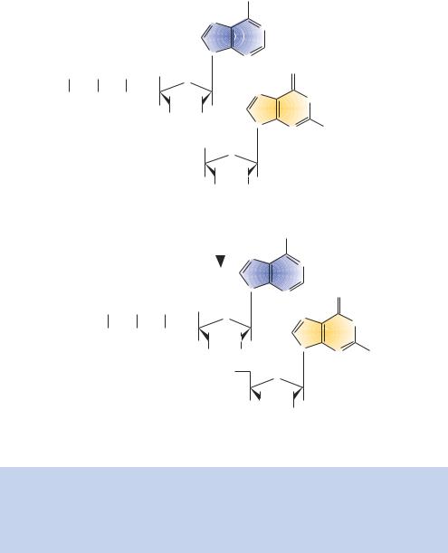
Figure 1.6. The joining of nucleotides. The joining of an adenine and a guanine nucleotide. The phosphates on the sugar ring of guanine are designated as α, β or γ . In the formation of the dinucleotide, pyrophosphate (representing the β and γ phosphates) is lost and the phosphodiester bond links the 3 hydroxyl to the phosphate on the 5 carbon atom of the sugar. DNA molecules invariably have a free 5 phosphate and 3 hydroxyl
firing a beam of X-rays at a regular array of molecules – either a crystal or a fibre. When the X-rays hit an atom in the array they will be diffracted, and the diffracted beams are detected as spots on X-ray film. Analysis of the diffraction patterns yields information about the structure and shape of the molecules in the array. As early as 1938 William Astbury applied the technique to fibres of DNA. By 1947, he had detected a periodicity (or repeating unit) within DNA of

1.3 THE DOUBLE HELIX |
11 |
|
|
Table 1.1. Chargaff’s rules. The ratios of individual nucleotides isolated from DNA of various sources. While the ratios of purine:purine and pyrimidine:pyrimidine vary widely, the ratio of purine:pyrimidine was found to be a constant unity
Organism |
A to G |
T to C |
A to T |
G to C |
Purines: |
|
|
|
|
|
pyrimidines |
|
|
|
|
|
|
Ox |
1.29 |
1.43 |
1.04 |
1 |
1.1 |
Human |
1.59 |
1.75 |
1 |
1 |
1 |
Hen |
1.45 |
1.29 |
1.06 |
0.91 |
0.99 |
Salmon |
1.43 |
1.43 |
1.02 |
1.02 |
1.02 |
Sea urchin |
1.83 |
1.80 |
1.02 |
1.00 |
1.01 |
Wheat |
1.22 |
1.18 |
1 |
0.97 |
0.99 |
Yeast |
1.67 |
1.92 |
1.03 |
1.2 |
1 |
Hemophilus influenzae |
1.75 |
1.54 |
1.06 |
0.93 |
1.01 |
Escherichia coli |
1.05 |
0.95 |
1.09 |
0.99 |
1 |
Serratia marcescens |
0.76 |
0.63 |
1.03 |
0.85 |
0.92 |
Bacillus schatz |
0.68 |
0.58 |
1.07 |
0.9 |
0.96 |
|
|
|
|
|
|
0.34 nm. Between 1950 and 1953, Rosalind Franklin obtained improved X-ray data from highly purified samples of DNA. Her work confirmed the 0.34 nm periodicity, and suggested that the structure of DNA was some sort of helix. Franklin, however, did not propose a model for the structure of DNA. Rather, Linus Pauling and Robert Corey used Franklin’s data, together with that of others, to propose that DNA was a triple helix with the phosphates near the centre of the axis and the bases on the outside (Pauling and Corey, 1953).
1.3The Double Helix
Franklin noted that DNA fibres could give two distinct types of diffraction pattern depending upon how the samples were prepared and stored. The first (termed Structure A) was composed of fibres that were relatively dehydrated, while the second (Structure B) was prevalent over a wide variety of conditions. She noted that the change from Structure A to Structure B was reversible, depending on the levels of sample hydration (Franklin and Gosling, 1953). It is thought that the B-form of DNA is the biologically significant conformation. Other forms of DNA (the right-handed A form and the left-handed Z form) certainly do exist under certain conditions, and may play significant roles in certain cellular processes. For example, a family of proteins that bind specifically to Z-DNA has recently been described (Schwartz et al., 2001). Here, however, we will concentrate on the properties and interactions of B-form DNA.
12 |
DNA: STRUCTURE AND FUNCTION 1 |
|
|
In 1953, James Watson and Francis Crick attempted to build molecular models of DNA and realized that the Pauling–Corey structure was incorrect, with some atoms having to be closer together than was possible. By combining Franklin’s X-ray diffraction patterns with Chargaff’s rules, Watson and Crick proposed the, now famous, double-helix model in 1953 (Watson and Crick, 1953a). This model, shown in Figure 1.7, has the following major features, some of which have been updated slightly from the original model in the light of high-resolution crystal X-ray diffraction data.
(a)Two long polynucleotide chains coiled around a central axis, forming a right-handed double helix – this means that the turns are clockwise when looking down the helical axis.
(b)The two chains are antiparallel; that is, each chain has a specific orientation, and these run in opposite directions.
(c)The bases of both chains are flat structures, lying perpendicular to the axis. They are ‘stacked’ on one another, 0.34 nm apart, and are located on the inside of the helix.
(d)The nitrogenous bases of opposite strands are paired to one another by hydrogen bonds.
(e)Each complete turn of the helix is 3.4 nm long. This means that just over ten bases from each strand (10.4 bp) form one complete turn of the helix.
(f)Along the molecule, alternating larger major grooves and smaller minor grooves are apparent.
(g)The double helix measures approximately 2 nm in diameter.
The pairing of the nitrogenous bases in the centre of the helix is the most significant feature of the model by Watson and Crick. However, several other features are also important to understand the double helix.
1.3.1The Antiparallel Helix
The antiparallel nature of the two polynucleotide chains is a key part of the double helix. Given the constraints of the bond angles of the bases and sugar
phosphates, the double helix could not be constructed easily if both chains ran parallel to each another. One chain of the helix runs in the 5 to 3 orientation, and the other chain runs in the 3 to 5 orientation. This is illustrated in Figure 1.8. The 5 and 3 nomenclature is derived from the numbering system of the sugar ring that we saw in Figure 1.5. By convention, DNA sequences are

1.3 THE DOUBLE HELIX |
13 |
|
|
Major groove ~2.2 nm
One turn of the helix 3.4 nm
Diameter
2.0 nm
Figure 1.7. The Watson and Crick model of DNA
Minor groove ~1.2 nm
0.34 nm
written in the 5 to 3 direction. This means that a single DNA chain begins with a free phosphate group on the 5 carbon of a deoxyribose ring. Additional nucleotides are joined to the chain through phosphodiester bonds, which link the hydroxyl group on the 3 carbon atom of one sugar with the phosphate on the 5 carbon atom of an adjoining sugar. The chain terminates in a free hydroxyl group on the 3 carbon atom of the last sugar.
14 |
DNA: STRUCTURE AND FUNCTION |
1 |
|
|
|
|
|
|
|
|||||
|
5' |
|
|
|
|
H |
|
|
|
|
|
3' |
|
|
|
|
|
|
|
|
|
|
|
|
|
|
|
||
|
|
H3C |
O H |
N |
|
|
|
|
|
|
|
|
||
|
|
|
C |
C |
|
|
C |
|
|
N |
|
|
|
|
|
|
|
|
|
|
|
|
|
|
|
|
|||
|
|
|
|
|
|
|
C |
|
|
CH |
|
|
|
|
|
|
|
HC T |
|
|
|
A |
|
|
|
|
|
||
|
P |
|
N |
H |
N |
|
|
|
|
|
|
|||
|
|
C |
|
N |
|
|
|
|
||||||
|
|
|
N |
C |
|
HC |
|
|
|
|
|
|
||
|
|
O |
|
N |
|
|
|
|
|
|
|
|||
|
|
|
|
|
|
|
|
|
|
|
|
|||
|
|
|
|
|
|
|
|
|
|
|
|
|
|
|
|
|
|
|
O |
|
|
|
|
|
|
|
|
O |
|
|
|
|
|
|
H |
|
|
|
|
|
|
|
|
|
|
|
|
|
|
H |
O |
|
|
|
|
|
|
|
|
|
|
|
|
|
N |
|
|
|
|
|
P |
|
||
|
|
|
|
H |
|
|
|
|
|
|
|
|
||
|
|
|
|
C |
|
|
|
|
|
N |
|
|
||
|
|
|
|
C |
|
|
C |
|
C |
|
|
|
|
|
|
|
|
|
|
|
|
|
|
|
CH |
|
|
||
|
|
|
|
C |
|
|
|
|
|
|
|
|
||
|
|
P |
HC |
N |
H |
N |
G |
|
|
|
|
|||
|
|
C |
|
|
|
|
||||||||
|
|
|
|
|
|
|
N |
|
|
|
||||
|
|
|
|
N |
C |
|
|
|
|
|
|
|
||
|
|
|
O |
|
C |
|
N |
|
|
|
|
|||
|
|
|
|
|
|
|
|
|
|
|
||||
|
|
|
|
|
|
|
|
|
|
|
|
|
||
|
|
|
|
|
O |
H |
N |
|
|
|
|
|
O |
|
|
|
|
|
|
|
|
H |
|
|
|
|
|
|
|
|
|
|
|
|
|
|
|
|
|
|
|
|
|
|
|
|
|
|
|
N |
|
|
H |
|
H |
O |
|
CH3 |
P |
|
|
|
P |
HC |
|
|
N |
|
|
|
||||
|
|
|
|
|
C |
C |
|
|
|
C |
C |
|
|
|
|
|
|
O |
|
N |
A |
|
|
|
|
|
T |
|
|
|
|
|
|
C |
N |
|
|
H |
N |
CH |
|
|||
|
|
|
|
|
|
|
|
|||||||
|
|
|
|
|
|
N |
C |
|
|
|
C |
N |
|
|
|
|
|
|
|
|
|
H |
|
|
|
|
|
|
|
|
|
|
|
|
|
|
|
|
|
O |
|
|
O |
|
|
|
|
|
|
|
N |
|
|
|
|
|
|
|
|
|
|
|
|
|
HC |
|
|
|
O |
H NH |
|
P |
||
|
|
|
P |
|
|
|
|
|
|
|||||
|
|
|
|
|
C |
C |
|
|
|
C |
C |
|||
|
|
|
|
|
|
|
|
|
|
|
|
|
H |
|
|
|
|
|
O |
N |
C |
G |
|
N |
H |
N |
C CH |
|
|
|
|
|
|
|
|
|
||||||||
|
|
|
|
|
|
|
|
|||||||
|
|
|
|
|
|
|
N |
C |
|
|
|
C |
N |
|
|
|
|
|
|
|
|
|
|
|
N |
H |
O |
|
O |
|
|
|
|
|
|
|
|
|
|
H |
|
|
|
|
|
|
|
|
|
|
|
|
|
|
|
|
|
|
|
P
5'
3'
Figure 1.8. DNA base pairing and complementation. The two chains of the helix, arrowed in the 5 to 3 direction, are antiparallel. The bases on one strand of the helix are complementary to those on the opposite strand, A always base pairs with T and G always base pairs with C
1.3.2Base Pairs and Stacking
The bases of both DNA chains are flat structures that lie approximately perpendicular to the helical axis. The bases themselves are stacked upon each other. The arrangement is best illustrated by inspection of a computergenerated model of high-resolution crystal X-ray diffraction data (Figure 1.9). It can be noted that the base pairs are not all perpendicular to the helical axis, and that some show propeller twist, where the purine and pyrimidine pair do not lie flat but are twisted with respect to each other, like the blades of a propeller (Dickerson, 1983). The pairing of a purine (A or G) with a pyrimidine (T or C) within the helix is important for the integrity of the helix.
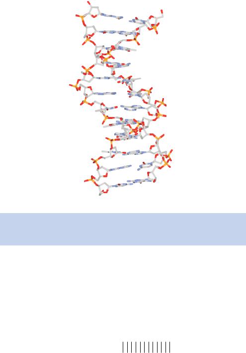
|
1.3 THE DOUBLE HELIX |
15 |
|
|
|
|
|
|
|
|
|
|
|
|
|
Figure 1.9. Computer generated model of DNA. The structure of double-stranded B- form DNA as derived from high-resolution X-ray diffraction of DNA crystals. Oxygen atoms are coloured red, phosphorus is orange, carbon is white and nitrogen is blue
The constant length of the purine – pyrimidine pairing would be disrupted if purine – purine (too large) or pyrimidine –pyrimidine (too small) pairings occurred. The purine –pyrimidine pairs are said to complement each other, and the two strands of a single DNA molecule are thus complementary to one another. Thus, if the sequence 5 -ATGATCAGTACG-3 occurs on one strand of the DNA, the other strand must have the sequence 5 -CGTACTGATCAT-3 . These two sequences are complementary to each other:
Strand one: 5′-ATGATCAGTACG-3′
Strand two: 3′-TACTAGTCATGC-5′
16 |
DNA: STRUCTURE AND FUNCTION 1 |
|
|
As we will we see in many of the subsequent chapters, the ideas of complementation between two strands of DNA form the basis of many genetic engineering experiments. The pairing of two DNA strands is very specific. Precise matches between two DNA strands, like those shown above, are highly stable and readily form helices. As we will see later, two DNA strands that are not precisely complementary to one another, but where there is still a high degree of complementation, retain the ability to interact with each other.
1.3.3Gaining Access to Information with the Double Helix without Breaking it Apart
How can the information held within the sequence of DNA be read without having to unravel the double helix? The invariant nature of the sugar –phosphate backbone would seem to provide an almost impenetrable barrier to ‘reading’ the DNA base sequence. The grooves along the helical axis do, however, provide a mechanism whereby the bases can be distinguished from one another. As we can see in Figure 1.7, DNA is composed of alternating major and minor grooves along its axis. This is a result of the glycosidic bonds that attach a base pair to its sugar rings not lying directly opposite each other across the helical axis. As a result, the two sugar – phosphate backbones of the double helix are not equally spaced along the helical axis, and the grooves that form between the backbones are not of equal size. The major groove is wide ( 0.22 nm) and shallow, while the minor groove is narrow ( 0.12 nm) and deep. The floor of the major group is composed mainly of nitrogen and oxygen atoms that belong to the unique portions of each base pair. In contrast, the floor of the minor groove is filled with nitrogen and oxygen atoms that are generally common to either the purines or to the pyrimidines. Thus, the potential of the major groove for interactions shows a much greater dependence on base sequence than that of the minor groove. This finding led to the speculation that DNA sequence-specific binding proteins recognize DNA by forming hydrogen bonds predominantly to specific groups positioned within and along the major groove. One of the most common ways in which proteins can recognize specific DNA sequences is by the insertion of a protein α-helix into the major groove of DNA. The α-helix, originally postulated by Pauling and Corey in 1951, is a protein secondary structure motif in which a right-handed helix is formed by amino acids on a polypeptide chain (Pauling et al., 1951). Each amino acid in the helix occupies a vertical distance of 0.15 nm, and there are 3.6 amino acid residues per turn of the helix (see Appendix 1). The diameter of the polypeptide backbone in an α-helix is approximately 0.5 nm; however, the amino acid side chains project away from the helical axis. This results in a protein α-helix
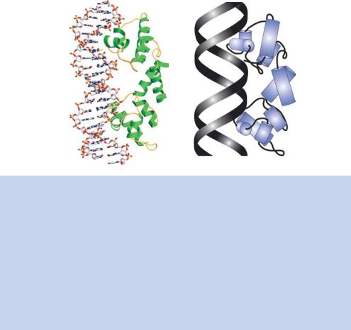
1.3 THE DOUBLE HELIX |
17 |
|
|
being able to fit almost exactly into the major groove of double-stranded DNA. The amino acid side chains that project away from the α-helix are able to form hydrogen bonds with the DNA bases in the major groove. The type of protein –DNA interaction is shown in Figure 1.10.
1.3.4Hydrogen Bonding
The Watson and Crick model of DNA structure predicts that the two polynucleotide chains are held together by non-covalent hydrogen bonds rather than by covalent interactions. This raises several important questions – what is a hydrogen bond, and is it sufficiently strong to maintain the integrity of the double helix?
To address the first question, a hydrogen bond is a weak electrostatic interaction between a covalently bonded hydrogen atom and an atom with
(a) |
(b) |
2 1
3
4
5
5
1 4
3
2
Figure 1.10. The interaction between the λ-repressor of bacteriophage λ and DNA.
(a)Computer generated model of the interaction between DNA, shown with the same colouring scheme as in Figure 1.9, and λ-repressor, whose α-helices are shown in green and are connected by amino acids that do not adopt secondary structure. Two molecules of the λ-repressor (a dimer) interact with a 17 bp segment of double-stranded DNA.
(b)The five helices of λ-repressor. Each monomer of the λ-repressor DNA binding domain has five helices, numbered 1–5 from the amino terminal end of the protein. Helix 3 lies in the major groove and the side chains (not shown) extend to the edges of the major groove and make contacts with the DNA bases. Helices 2 and 3 form a ‘helix–turn–helix’ DNA binding motif that is found in many DNA binding proteins. Helix 3 – the recognition helix – forms DNA sequence-specific contacts in the major groove, while helix 2 – the stabilization helix – interacts non-specifically with the DNA backbone to provide stability to the DNA–protein interaction