John Wiley & Sons - 2004 - Analysis of Genes and Genomes
.pdf168 |
POLYMERASE CHAIN REACTION |
4 |
|
|
|
|
|
|
|||||
|
|
|
|
|
Polystyrene derivertized with |
|
|
|
|
|
|||
|
|
|
|
|
|
phosphoramidite T |
|
|
|
|
|
||
|
|
|
|
|
|
OCH3 |
|
O |
|
|
|
|
|
|
|
|
|
|
|
|
|
|
|
|
|
|
|
|
|
|
|
|
|
|
|
NH |
|
|
|
|
|
|
|
|
|
|
|
|
|
N O |
|
|
|
|
|
|
|
|
|
H3CO |
|
O |
O |
|
|
|
|
|
|
|
|
|
|
|
|
|
|
|
|
|
|
|
|
|
|
|
|
|
|
|
O |
|
H+ |
|
|
|
|
|
|
|
|
|
|
|
|
|
(b) |
|
O |
|
|
|
|
|
|
|
|
Polystyrene |
|
|
|
|
|
||
|
|
|
|
O |
|
|
|
|
|
N |
|
||
|
|
|
|
|
|
|
|
|
|
|
|
||
|
OCH3 |
|
|
NH |
|
|
|
|
|
|
N |
|
|
|
|
|
|
|
(a) |
|
|
|
|
O |
|
||
|
|
|
|
|
|
|
|
|
|
|
|||
|
|
|
N |
|
|
|
|
|
|
|
|
|
|
|
|
|
N |
|
|
|
|
OH |
|
|
|
|
|
|
|
|
|
|
|
|
|
O |
|
|
|
||
|
|
|
|
|
|
|
|
|
|
|
|
|
|
|
|
|
N |
N |
|
|
|
|
|
|
|
|
|
|
|
|
|
|
|
|
|
|
|
|
|
|
|
H3CO |
O |
|
O |
O |
|
|
|
|
|
O |
|
|
|
|
|
|
|
|
|
|
|
|
|
||||
|
|
|
|
|
|
|
|
|
|
|
|
|
|
|
|
O |
|
NH |
|
|
|
|
|
Polystyrene |
|
|
|
|
|
|
|
|
|
|
|
|
|
|
|
||
|
O |
P |
O |
N |
O |
|
|
|
|
|
|
|
|
|
|
|
|
|
|
|
|
|
|
|
|||
|
|
O |
O |
|
|
|
|
|
|
|
|
|
|
|
|
|
|
|
|
|
|
|
|
|
|
|
|
|
N |
|
|
|
|
|
|
|
|
|
|
|
|
|
|
|
O |
|
|
|
|
O |
|
|
H |
|
|
|
|
|
|
|
|
|
|
|
|
N |
|
|
|
|
|
|
|
|
OCH3 |
|
|
NH |
|
|
N |
|
|
|
|
|
|
|
|
|
|
N |
N |
|
|
||
|
|
|
Polystyrene |
|
|
|
|
|
|
|
O |
||
|
|
|
|
|
|
N |
|
(c) |
|
|
|||
|
|
|
|
|
|
|
N |
|
|
|
|||
|
|
|
|
|
|
|
|
|
|
NH |
|||
|
|
|
|
|
|
|
|
|
|
OCH3 |
|
||
|
|
(d) |
|
|
|
N |
N |
|
|
|
N |
|
|
|
|
|
|
|
|
|
|
|
|
||||
|
|
|
|
|
|
|
|
|
|
N |
|||
|
|
|
H3CO |
|
O |
|
|
O |
|
|
|
|
|
|
I2, H2O |
|
|
O |
|
|
|
|
|
|
|||
|
|
|
|
|
|
|
|
|
|
||||
|
|
|
|
|
|
|
|
|
|
N |
N |
||
|
|
|
|
|
|
|
|
NH |
|
|
|
|
|
|
|
|
|
|
|
O |
|
|
H3CO |
|
O |
O |
|
|
|
|
|
|
|
|
|
N |
O |
|
|
|
|
|
|
|
|
|
|
P |
|
|
|
|
|
||
|
|
|
|
|
|
|
|
|
|
|
|
|
|
|
|
|
|
|
|
O |
|
|
|
|
|
|
|
|
|
|
|
|
|
O |
|
O |
|
|
|
O |
|
|
|
|
|
|
|
|
|
|
|
|
|
|
|
|
|
|
|
|
|
|
|
|
|
|
|
P |
|
|
|
|
|
|
N |
|
|
|
|
|
|
O |
|
N
O
N
Polystyrene
Phosphoramidite nucleoside
Figure 4.6. The synthetic production of oligonucleotide primers. The production of 5 - AT-3 . Phophoramidite nucleosides are modified with a dimethoxytrityl protecting group on the 5 -end (red) and a β-cyanoethyl protected 3 -phosphite group (blue). Additionally, other modifiers (green) protect primary amines occurring elsewhere in the molecule. See the text for details of the reaction cycle
The nucleoside condensation reaction is highly efficient, with less than 1 per cent of the 5 -hydroxyl groups not reacting with the incoming nucleoside. To prevent these unreacted molecules participating in subsequent reactions, and resulting in unwanted truncation deletions, the unreacted 5 -OH groups are blocked by acetylation (capping) with acetic anhydride before the oxidation step. The efficiency of coupling ensures that primers up to about 60 nucleotides in length can be manufactured routinely.
After synthesis is complete, the oligonucleotide is cleaved from the solid support with concentrated ammonium hydroxide at room temperature. Continued incubation in ammonia at elevated temperature will deprotect the phosphorus
4.5 PRIMER MISMATCHES |
169 |
|
|
via β-elimination of the cyanoethyl group and also removes the protecting groups from the heterocyclic bases. The finished oligonucleotide can be purified from contaminating chemicals by precipitation, and the full-length sequence is usually isolated by HPLC purification.
The great advantage to chemical primer synthesis is that any sequence can be manufactured rapidly and relatively cheaply (<$0.5 per base). The sequence of a primer may also be mixed. For example, with the primer outlined in Figure 4.6, if a 1:1 mixture of dT and dC monomers were added to the reaction at stage C, then the resulting primer could have the sequence of either 5 -TA-3 or 5 -CA-3 . The final product would be an equal mixture of the two species. By controlling the amount of each monomer added at the condensation step, the primer can be doped to any specified concentration at any position except the 3 -end. Additionally, modified bases (e.g. those containing phosphothioates, or labelled with biotin) can be added to the primer sequence.
4.5Primer Mismatches
The oligonucleotide primers that are used in a PCR experiments need not match the target sequence exactly. This is particularly relevant when trying to make mutations, or deliberate changes, in the amplified DNA sequence or when attempting to search for gene sequences that are homologous to one already known. We will discuss some of the more straightforward uses of PCR-based mutagenesis here, but will save some of the more elegant mutagenic strategies for Chapter 7. The only place within the primer sequence that must match the target sequence exactly is the extreme 3 -end of the primer. If the 3 -end of the primer does not precisely match the target sequence, then the polymerase will not efficiently extend the primer. A consequence of this is that the PCR will be inefficient, or will fail completely. This property has, however, been exploited in the diagnosis of point mutations within genes (see below).
To think about how PCR might be used to introduce mutations into amplified products, we need to think about the primers themselves. The primers initiate the DNA replication process, but are themselves incorporated into each strand of the final amplified product. Consequently, any base changes between the primer and the template DNA will be carried forward into the amplified product. Since we cannot introduce mutations at the 3 -end of the primer, a favourite location to introduce changes is the 5 -end of the primer (Figure 4.7).
In this case, we are amplifying the same sequence as shown in Figure 4.5. However, this time the oligonucleotide primers contain additional sequences at their 5 -ends. In the case of primer 1, it contains the recognition sequence for the EcoRI restriction enzyme at the 5 -end of the sequence used to recognize

170 |
POLYMERASE CHAIN REACTION 4 |
|
|
Primer 1
5′-GAATTCATGAAGCTACTGTCTTCT-3′
5′-...TGAAAGATGAAGCTACTGTCTTCT  GGATTATTTGTACAAGATAATGTG...-3′
′ GAL4 gene
3 -...ACTTTCTACTTCGATGACAGAAGA  CCTAATAAACATGTTCTATTACAC...-5′ 3′-CCTAATAAACATGTTCTACCTAGG-5′
Primer 2
PCR
EcoRI |
BamHI |
5′-GAATTCATGAAGCTACTGTCTTCT  GGATTATTTGTACAAGATGGATCC-3′ 3′-CTTAAGTACTTCGATGACAGAAGA GAL4 gene CCTAATAAACATGTTCTACCTAGG-5′
Figure 4.7. Primers to amplify part of the GAL4 gene from the Saccharomyces cerevisiae genome and to include restriction enzyme recognition sites. The final PCR product contains the sequences present in the primers
the GAL4 gene. This primer and the primer shown in Figure 4.5 will bind to the same template DNA sequence with approximately the same affinity. The EcoRI recognition sequence does not match the template sequence exactly, but the mismatches are not sufficient to prevent specific primer binding. The EcoRI recognition sequence will, however, be incorporated into the final PCR product as shown in Figure 4.7. Primer 2 in this figure contains the recognition site for the BamHI restriction enzyme, which will also be incorporated into the final product. Cloning of the final product now becomes straightforward after cutting with the two restriction enzymes – having first ensured that there are no EcoRI or BamHI restriction enzyme sites in the PCR product itself! Often restriction enzymes require more DNA than just their recognition site in order to cleave efficiently. Therefore three to six additional residues are usually added to the 5 -end of the primer before the restriction enzyme recognition site. These are often G or C (termed a GC clamp) to provide the maximum level of annealing between the two DNA stands and efficient cleavage by restriction enzymes.
Any mismatches between the primer and the template DNA will be carried forward into the final PCR product. Therefore, deliberate mutations may be introduced into the final PCR product by altering the sequence of the primer. This is particularly important if you want to alter the coding sequence of a gene to change the amino acid sequence of a protein, or if you want to alter the codon usage of a gene to, for example, introduce a restriction enzyme recognition site without altering the amino acid sequence of the resulting protein, or so that preferred codons are used if the resultant protein is to be expressed at high levels.
The second major use of mismatched primers is in the search for genes encoding a particular protein, and in the search for homologous genes. The
4.5 PRIMER MISMATCHES |
171 |
|
|
isolation and characterization of a protein is common in biochemistry. For example, you may isolate a protein and sequence its amino terminal end to find the following amino acids: Met – Ile –Trp –Pro –Phe. The degeneracy of the genetic code means that this amino acid sequence could be encoded for by the following DNA sequences:
Met-Ile-Trp-Pro-Phe
5 -ATG-ATA-TGG-CCA-TTC-3 C C T
TT G
With just the protein sequence at hand, it is impossible to tell which codon will be used in a particular gene to encode an individual amino acid. Therefore to PCR amplify the gene encoding this protein sequence, we must design a degenerate primer. The primer must be able to bind to all possible combinations that could encode the protein sequence. The primer below could be synthesized to perform this function:
5 -ATG-ATA-TGG-CCN-TTC-3 C T
T
where N is an equimolar mixture of A, T, C and G. The primer above would be produced as a mixture of 24 different primers. Only one of these 24 combinations will be a perfect match to the protein coding sequence, but others will differ by only a single nucleotide from the target sequence and may still promote efficient PCR. For the case shown here, an additional primer recognizing sequences 3 to those shown here is required for PCR to proceed.
An alternative to mixing all four nucleotides at one position in a primer is to use inosine. Inosine is a purine, which occurs naturally in tRNAs, that can form base pairs with C, T and A. The inosine –A pairing will not fit correctly in double-stranded DNA as a purine-purine pairing, so there will be an energetic penalty to pay when the helix bulges out at this pairing. Inosine can be used in primers at positions where any of the four bases might be required. Each use of inosine thus reduces the degeneracy of the primer pool fourfold. Inosine –G mismatches may occur, but precise base pairing at other positions in the primer may overcome such a problem. Using inosine in the primers requires that the DNA polymerase used in the PCR experiment is capable of synthesizing DNA over an inosine-containing template. Taq polymerase can do this, but some other polymerases (e.g. Vent and Pfu) are unable (Knittel and Picard, 1993).
Many genes occur in families with similar amino acid sequences (and consequently similar gene sequences) encoding a similar function within a different
172 |
POLYMERASE CHAIN REACTION 4 |
||
|
|
|
|
(a) |
|
|
|
|
POU-SPECIFIC BOX |
||
|
|
||
Pit-1: KSKLVEEPIDMDSPEIRELEKFANEFKVRRIKLGYTQTNVGEALAAVHG---SEFSQTTICRFENLQLSFKNACKLKAILSKWLEEAEQV |
Oct-1: DTPSLEEPSDLE-----ELEQFAKTFKQRRIKLGFTQGDVGLAMGKLYG---NDFSQTTISRFEALNLSFKNMCKLKPLLEKWLNDAENL |
Oct-2: PPSHPEEPSDLE-----ELEQFARTFKQRRIKLGFTQGDVGLAMGKLYG---NDFSQTTISRFEALNLSFKNMCKLKPLLEKWLNDAETM |
unc-86: RYPIAPPTSDMDT-DPRQLETFAEHFKQRRIKLGVTQADVGKALAHLKMPGVGSLSQSTICRFESLTLSHNNMVALKPILHSWLEKAEE- |
Consensus: .........D........LE.FA..FK.RRIKLG.TQ..VG.A............SQ.TI.RFE.L.LS..N...LK..L..WL..AE.. |
Sequence 1
POU HOMEO BOX
Pit-1: GALYNEK-----------VGAN-ERKRKRRTTISIAAKDALERHFGEQNKPSSQEIMRMAEELNLEKEVVRVWFCNRRQREKRVKTSLNQ |
Oct-1: SSDSSLSSPSALNSP--GIEGL-SRRRKKRTSIETNIRVALEKSFLENQKPTSEEITMIADQLNMEKEVIRVWFCNRRQKEKRINPPSSG |
Oct-2: SVDSSLPSPNQLSSPSLGFDGLPGRRRKKRTSIETNVRFALEKSFLANQKPTSEEILLIAEQLHMEKEVIRVWFCNRRQKEKRINPCSAA |
unc-86: -AMKQKDTIGDIN----GILPN-TDKKRKRTSIAAPEKRELEQFFKQQPRPSGERIASIADRLDLKKNVVRVWFCNQRQKQKRDFRSQFR |
Consensus: .............................RT.I.......LE..F.....P....I...A..L...K.V.RVWFCN.RQ..KR....... |
Sequence 2
(b) Sequence 1 Sequence 2
..PheLysValArgArgIleLysLeuGly.........ArgValTrpPheCysArgGlnArgGln..
Gln |
|
|
Arg |
|
5′-CCNAAAGTNCGNCGNATAAAACTNGGN-3′ |
5′-CGNGTNTGGTTCTGCCGNCGNCGNCAA-3′ |
|||
GCA A A |
C GT |
A |
T TA AA A |
G |
|
T |
|
|
|
(c)
Primer 1 (26 mer): 5′-CCNAA(AG)(GC)(TA)N(CA)GN(CA)GNAT(ACT)AA(AG)(CT)TNGG-3′ Primer 2 (26 mer): 5′-(TC)TGNC(GT)N(CT)(GT)NC(GT)(GA)CA(GA)AACCANACNC-3′
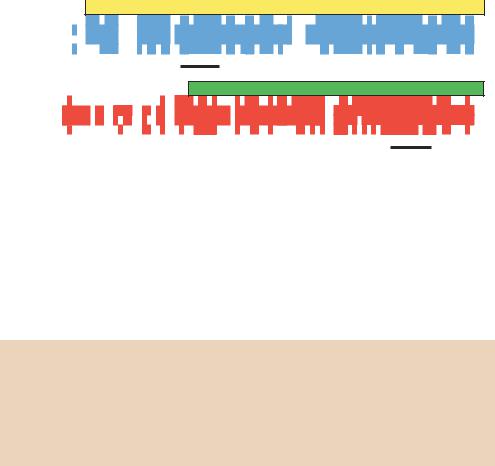
Figure 4.8. The use of degenerate PCR primers to identify novel POU genes. (a) Sequence alignment of the Pit-1, Oct-1, Oct-2 and unc-86 proteins. Sequences present in at least two of the proteins are coloured, while sequences present in all four proteins are written below as the consensus. (b) The nine amino acid sequences that are most highly conserved within the POU specific box and the POU homeo-box, and the DNA sequences that may encode them. (c) The sequence of primers used to identify additional POU-domain proteins
protein. PCR can be used to identify additional family members once some have already been characterized. To illustrate this, it is easiest to look at a specific example. The POU (pronounced ‘pow’, to rhyme with ‘how’) domain containing proteins are a family of transcription factors that share a highly conserved DNA binding domain (Figure 4.8(a)). The family derives its name from the first four members to be characterized – three mammalian proteins Pit-1, Oct-1 and Oct-2, and the unc-86 gene from the nematode Caenorhabditis elegans (Herr et al., 1988). There are two highly conserved portions of the POU domain – termed the POU specific domain and the POU homeodomain. The latter has a high degree of homology with Drosophila homeobox containing developmental control proteins. Mutations in these genes have dramatic effects on the development of an organism. For example, mutations in the mouse Pit-1 gene result in a failure in pituitary gland development and the production of dwarf mice (Li et al., 1990).
4.6 PCR IN THE DIAGNOSIS OF GENETIC DISEASE |
173 |
|
|
To identify other members of the POU-domain family, primers were designed to the most conserved regions of the POU specific region and of the POU homeobox (Figure 4.8(b)). These primers were highly degenerate, based on the degeneracy of the genetic code, but have been successfully used to identify additional POU domains from a variety of sources (Lillycrop et al., 1992). The two primers shown here (Figure 4.8(c)) were found to amplify an approximately 400 bp DNA fragment that was somewhat heterologous. The heterogeneity is due to differences in length of individual genes encoding POU-domain proteins that are amplified from DNA samples. This approach led to the identification of many new genes, including Brn-3 family members that are highly expressed in neuronal cells and plays a role in the differentiation and survival of sensory and motor neurons (Erkman et al., 1996).
4.6PCR in the Diagnosis of Genetic Disease
The power of PCR to amplify specific DNA sequences has made it a valuable tool in the diagnosis of genetic defects and mutations. Since PCR requires prior knowledge of the DNA sequence that is to be amplified, the site of the mutation must be known before PCR analysis can be attempted. The great advantage of PCR based methods is the small amount of DNA required to make a diagnosis. Samples of blood, or cells from the inside of the cheek, provide sufficient material to make adult diagnosis, while small samples of chorionic villi can be used to made a diagnosis in utero.
PCR is used to detect both insertion/deletion mutations and point mutations. A wide range of techniques have been developed to detect mutations by PCR. Here we will concentrate on a couple of examples, but the reader should be aware that many alternatives also exist.
•Insertion/deletion mutations. Waardenburg syndrome, an inherited autosomal dominant disease that is characterized by a combination of deafness and abnormal pigmentation. The disease is responsible for over 2 per cent of the cases of adult deafness, and is often associated with a frontal white blaze of hair and white eyelashes. Certain types of Waardenburg syndrome are caused by mutations in the PAX-3 gene, a transcription factor involved in regulating embryonic development. One of the first mutations found within PAX-3 from a Waardenburg syndrome patient was an 18 bp deletion in the DNA encoding the DNA binding domain of the transcription factor (Tassabehji et al., 1992). This deletion can be detected in other Waardenburg syndrome patients by PCR. Primers can be designed to flank the site of the deletion (Figure 4.9). Amplification of the wild-type sequence

174 |
POLYMERASE CHAIN REACTION 4 |
|
|
will yield a large DNA fragment (156 bp in Figure 4.9), while amplification of the mutant sequence will yield a smaller DNA fragment (138 bp in Figure 4.9). Bands of these sizes can be easily separated on polyacrylamide or agarose gels. Thus, the presence of the mutation can be determined by the size of the PCR product obtained. In this case, because the disease is dominant, most sufferers will be heterozygotes, having one copy of the wild-type gene and one copy of the mutant gene. PCR amplification of a heterozygote will yield two different sized DNA fragments (156 and 138 bp in Figure 4.9).
(a) Primer 1
5'-CAG GGC CGC GTC AAC CAG CTC GGC-3'
5'-GGC CAG GGC CGC GTC AAC CAG CTC GGC GGC GTT TTT ATC AAC GGC AGG CCG CTG
|
G |
Q |
G R V N |
Q |
L G |
|
G V F I N |
G R P L |
|||||||||||||
CCC AAC CAC ATC CGC CAC AAG ATC GTG GAG ATG GCC CAC CAC GGC ATC CGG CCC TGC |
|||||||||||||||||||||
P |
N |
H |
I R H K |
I |
V E |
|
M A H H G |
I R P C |
|||||||||||||
GTC ATC TCG CGC CAG CTG CGC GTG TCC CAC GGC TGC GTC TCC AAG ATC CTG-3' |
|||||||||||||||||||||
V |
I |
S |
R Q L R |
V |
S H |
|
G C V S K |
I L |
|||||||||||||
|
|
|
|
|
3'-AGG GTG CCG ACG CAG AGG TTC TAG-5' |
||||||||||||||||
|
|
|
|
|
|
|
|
|
|
|
|
|
|
|
|
Primer 2 |
|
||||
(b) |
|
|
|
|
Wild-type Mutant |
|
|||||||||||||||
|
|
|
|
|
|
|
|
|
|
|
|
|
|
|
|
|
|
|
|
156 bp |
|
|
|
|
|
|
|
|
|
|
|
|
|
|
|
|
|
|
|
|
|
|
|
|
|
|
|
|
|
|
|
|
|
|
|
|
|
|
|
|
|
|
|
|
|
|
|
|
|
|
|
|
|
|
|
|
|
|
|
|
|
|
|
|
|
|
|
|
|
|
|
|
|
|
|
|
|
|
|
|
|
|
|
|
|
|
|
|
|
|
|
|
|
|
|
|
|
|
|
|
|
|
|
|
|
|
|
|
138 bp |
|
|
|
|
|
|
|
|
|
|
|
|
|
|
|
|
|
|
|
|
|
|
||
Figure 4.9. PCR to detect a pathological deletion mutation. A. The wild-type DNA sequence of the sense strand of part of exon 2 from the human Pax-3 gene. The sequence shown represents the first part of the paired box, a highly conserved element that functions as a transcription factor. Certain patients suffering from Waardenburg syndrome contain a deletion of the sequence shown in bold. This results in the deletion of seven amino acids (MAHHGIR) and the formation of a new codon (AGG, which encodes arginine). The net result is the deletion of six amino acids (MAHHGI) from the protein. Primers are designed that flank the site of the deletion. (b) PCR amplification of a normal individual with primers 1 and 2 will yield a 156 bp DNA fragment. Amplification of the DNA from an individual suffering from this Waardenburg syndrome mutation will yield two DNA fragments – the 156 bp band and a smaller 138 bp band. The smaller band is derived from the chromosome that contains the 18 bp deletion. Since Waardenburg syndrome is a dominant trait, most sufferers are heterozygotes
4.7 CLONING PCR PRODUCTS |
175 |
|
|
•Point mutations. The method described above cannot be used to detect point mutations (base changes) within a gene. Flanking primers would simply produce the same sized DNA fragment for both mutant and wildtype individuals. The PCR products could be subjected to DNA sequence analysis (Chapter 8), but this would be time consuming. What is required is a PCR based system to identify single-base mutations. Allele-specific PCR
exploits the property that, in order to be efficiently extended by DNA polymerase, a primer needs to have a correctly base paired 3 -end (Newton et al., 1989; Wu et al., 1989). The allele-specific PCR amplification of a mutation within the β-globin gene causing sickle cell anaemia is outlined in Figure 4.10. To detect point mutations within a gene, three primers must be designed to take part in two PCR reactions. One primer (primer 3 in Figure 4.10) is common to both reactions, while the other primers (primers 1 and 2) detect the presence of either the wild-type or the mutated DNA sequence. The amplification of the wild-type DNA sequence with primers 1 and 3 will proceed. However, if primers 2 and 3 are used, the reaction
will fail because of the mismatch between the wild-type sequence and the extreme 3 -end of primer 2. Similarly, amplification of the mutant DNA sequence with primers 1 and 3 will fail, whereas PCR will proceed normally with primers 2 and 3 using the mutant sequence as the template. The results of such an analysis are shown diagrammatically in Figure 4.10(b). Here, we are comparing a wild-type DNA sequence with that of an individual who is homozygous for the mutation. If an individual were heterozygous, then both PCR reactions would proceed normally since both alleles would be present in the template DNA. To ensure that all PCR reactions are working correctly, a set of control primers – which amplify a region of an unrelated gene – are usually included in the PCR experiment so that one band should always be present, the control PCR, and the presence or absence of an additional band is searched for.
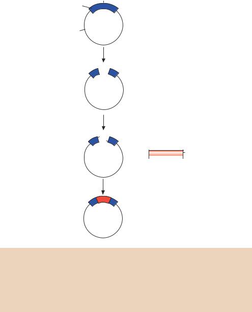
4.7Cloning PCR Products
As we have already seen, it is possible to clone PCR products by the insertion of extra sequences on the 5 -ends of primers such that restriction enzyme recognition sites are incorporated. It is also possible to clone PCR products directly, taking advantage of the terminal tranferase activity of Taq DNA polymerase to add a template-independent A residue to the 3 -end of PCR products. A consequence of this activity is that most of the DNA molecules amplified using Taq polymerase possess single 3 -A overhangs. In a process termed TA cloning, these can be ligated to a linearized ‘T-vector’, which has

176 |
POLYMERASE CHAIN REACTION |
4 |
|
|
|
|
|
|||||
|
|
|
|
|
|
|
|
|
|
|
|
|
(a) |
|
|
|
|
Human b-globin gene |
|
|
|
|
|||
|
|
|
|
Exon 1 |
|
Exon 2 |
Exon 3 |
|||||
|
5′ |
|
|
|
|
|
|
|
|
|
|
3′ |
|
|
|
|
|
|
|
|
|
|
|
||
|
|
|
|
|
|
|
|
|
|
|
||
|
|
|
|
|
|
|
|
|
|
|
|
|
|
5′-UTR |
Intron 1 |
|
Intron 2 |
|
|
3′-UTR |
|||||
Primer 1
Primer 2
|
|
|
5′-CAC CTG ACT CCT GA-3′ |
|
|
|
|
|
|
|
|
|
|||||||||
|
|
|
5′-CAC CTG ACT CCT GT-3′ |
|
|
|
|
|
|
|
|
|
|||||||||
5′-acc ATG GTG CAC CTG ACT CCT GAG GAG AAG TCT GCC GTT ACT GCC CTG |
|||||||||||||||||||||
(M) V |
H L |
T |
P |
E |
E K |
S |
A |
V |
T |
A |
L |
||||||||||
|
|
|
|
|
|
|
|
|
|
|
|
|
|
|
|
|
|
|
|
|
Exon 1 |
|
TGG GGC AAG GTG AAC GTG GAT GAA GTT GGT GGT GAG GCC CTG GGC |
||||||||||||||||||||
|
W G |
K V |
N V D |
E V |
|
G G E |
A L |
G |
|||||||||||||
|
|
|
|
||||||||||||||||||
|
|
AGgttggtatcaaggttacaagacaggtttaaggagaccaatagaaactgggcatgtgg |
|
||||||||||||||||||
|
R |
|
|
|
|
|
|
|
|
|
|
|
|
|
|
|
|
|
|
|
Intron 1 |
|
agacagagaagactcttgggtttctgataggcactgactctctctgcctattggtctat |
|
|||||||||||||||||||
|
|
|
|
|
|
|
|
|
|
|
|
|
|
|
|
|
|||||
|
|
|
|
|
|
|
|
|
|
|
|
|
|
|
3′-GACGGATAACCAGATA-5′ |
||||||
|
tttcccacccttag |
G CTG CTG GTG...-3′ |
|
|
|
|
|
Primer 3 |
|
|
|||||||||||
|
|
|
|
L |
|
L |
|
V ... |
|
|
|
|
|
|
|
|
|
||||
|
|
|
|
|
|
|
|
|
|
|
|
|
|
|
|||||||
|
|
|
|
|
Exon 2 |
|
|
|
|
|
|
|
|
|
|
|
|
|
|||
(b) |
|
|
|
|
Wild-type |
|
|
Mutant |
|
|
|
|
|
||||||||
|
|
|
|
|
|
|
|
|
|
|
|||||||||||
|
|
|
|
|
|
|
|
|
|
|
|
|
|||||||||
|
|
|
Primers: |
|
|
1&3 |
2&3 |
|
|
1&3 |
2&3 |
|
|
|
|
|
|||||
|
|
|
|
|
|
|
|
|
|
|
|
|
|
|
|
|
|
|
|
|
|
|
|
|
|
|
|
|
|
|
|
|
|
|
|
|
|
|
|
|
|
|
|
Control PCR
Test PCR
Figure 4.10. PCR to detect a point mutation. (a) Sickle cell anemia can be caused by a single base pair mutation, an A to T transversion converting Glu6 to Val, in the human
β-globin gene. In individuals that are homozygous for this mutation, the substitution in the
β-globin subunit of haemoglobin results in reduced solubility of deoxyheamoglobin and erythrocytes that assume irregular shapes. (b) The identification of a single base change in DNA using allele-specific PCR
single 3 -T overhangs on both ends to allow direct, high-efficiency cloning of PCR products (Zhou, Clark and Gomez –Sanchez, 1995; Marchuk et al., 1991). The complementarity between the PCR product 3 -A overhangs and vector 3 -T overhangs aids efficient ligation that does not occur with blunt-ended DNA molecules. The TA cloning strategy is shown diagrammatically in Figure 4.11.