

John Wiley & Sons - 2004 - Analysis of Genes and Genomes
.pdf48 |
DNA: STRUCTURE AND FUNCTION 1 |
||
|
|
|
|
|
|
|
|
Activator binding
ACT
DBD
Chromatin modification and remodelling
ACT


 DBD
DBD 


Recruitment of RNA polymerase II holoenzyme
DBD ACT
SRB |
TFIIH |
|
|
Pol II |
TFIIB |
|
|
|
Gal11 |
TAFs |
TFIIF |
|
||
|
TBP |
mRNA |
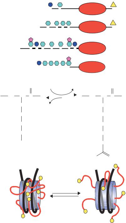
Figure 1.24. The multiple roles of transcriptional activators in switching on gene expression in eukaryotes. The binding of the activator protein through its DNA binding domain (DBD) can occur either in a nucleosome-free region, as shown, or on the DNA already bound within a nucleosome. The activation domain of the activator (ACT) recruits chromatin modifying and chromatin remodelling complexes to the gene. The activator then functions to recruit an RNA polymerase II holoenzyme complex to the gene so that transcriptional initiation can occur. The polymerase holoenzyme may exist as one or more discrete sub-complexes within the cell
An additional problem for the transcription of eukaryotic genes is that the majority of the DNA is wrapped into nucleosomes, which act as physical barriers to the assembly of the transcriptional complexes. Many activators are able to bind to their cognate DNA recognition sequences whether or not these are contained within nucleosomal DNA (Taylor et al., 1991), while others require that the nucleosomes are removed before they can bind DNA. After
1.13 TRANSCRIPTION |
49 |
|
|
DNA binding, the activator recruits one, or more, of the following complexes to the gene.
•Chromatin-modifying complexes. Histone proteins form the core of the nucleosome. However, as can be seen in Figure 1.15, the amino-terminal ends of the histones protrude from the complex. These ‘tails’ can be modified, by acteylation, phosphorylation or methylation of particular amino acids (Figure 1.25(a)), and the modifications play a significant role in the ability of nearby genes to be transcribed. Although these tails are not needed to maintain the structural integrity of the nucleosome, they do
have roles in higher-order chromatin structure and in interactions with nonhistone chromosomal proteins. Much attention has been focused recently on the role of histone acteylation in the process of transcription. It has been known for many years that increased levels of histone acetylation at a gene or chromosomal region are associated with transcriptional activity, whereas under-acetylation of histones is observed in non-transcriptionally active regions (Allfrey, Falkner and Mirsky, 1964), but the significance of this observation was not fully realized until it was discovered that transcriptional activators recruit chromatin modifying complexes to the promoters of genes (Bhaumik and Green, 2001; Larschan and Winston, 2001). The acetylation of histones is performed by enzymes called histone acetyltransferases (HATs) while the reverse reaction is catalysed by enzymes call histone deacetylases (H-DACs). The acetylation of lysine residues results in the elimination of positive charge from the protein (Figure 1.25(b)). It has been speculated that in under-acetylated histones (transcriptionally inactive) the histone tails wrap around the DNA with their positive charge being attracted to the negatively charged sugar –phosphate backbone. Acetylated histone tails, on the other hand, may interact with the DNA more loosely so that the histone –DNA complex is less rigid and may be more easily dissociated to allow transcription to occur.
•Chromatin remodelling complexes. These complexes, such as the yeast SWI/SNF proteins (pronounced ‘switch –sniff’, based on the nomenclature used in the original genetic screens that isolated the genes), use energy derived from ATP to move nucleosomes. Their precise mechanism of action is unclear, but they may function as ATP driven motors that move along DNA and disrupt protein –DNA interactions as they move (Pazin and Kadonaga, 1997).
•RNA polymerase II holoenzyme. RNA polymerases were first isolated in 1960 (Stevens, 1960; Weiss, 1960). Multiple forms of the enzyme in
50 |
DNA: STRUCTURE AND FUNCTION 1 |
|
|
(a) |
P |
Ac |
S |
K |
|
|
H2A |
1 |
5 |
|
|
|
Ac |
Ac |
Ac |
Ac |
|
K |
K |
K |
K |
H2B |
512 15 20
Me |
|
|
|
|
Me |
|
|
Ac |
P |
Ac |
Ac |
Ac |
Ac |
P |
|
K |
S |
K |
K |
K |
K |
S |
H3 |
9 |
10 |
14 |
18 |
23 |
27 |
28 |
|
|
|
|
|
|
Me |
|
|
|
P |
Ac |
Ac Ac |
Ac |
Ac |
|
|
S |
|
K |
|
K |
|
K |
|
|
K |
|
K |
|
|
|
|
|
|
||||||
1 |
5 |
8 |
12 |
|
16 |
20 |
|||||
(b) |
|
|
|
|
|
|
|
Acetylation |
|||
O |
Acetyl CoA |
|
|
|
CoA |
||||||
HN CH C |
|
|
|
|
|
||
|
CH2 |
Ac |
|
|
|
||
Lysine |
CH2 |
|
Deacetylation |
|
|
||
|
|
|
|
CH2
CH2
NH3+
(c)
|
+ |
|
|
N |
|
|
|
|
|
|
|
+ |
N |
+ |
|
+ |
HAT |
|
|
+ |
|||
+ |
|
|
+ |
Acetylation |
|
+ |
|
|
|
||
N |
+ N |
|
|||
+ |
|
|
+ |
|
|
|
N |
|
|
+ |
Deacetylation |
+ |
|
|
|
||
|
|
|
|
HDAC |
|
+ |
|
|
|
|
|
+ |
|
|
+ |
|
|
+ |
|
|
|
||
+ |
|
|
|
+ |
|
N |
|
+ |
|
|
|
|
|
|
|
||
H4
HN
H3C
Ac Ac
N Ac
Ac
Ac
Ac
Ac
Ac
N
Ub
K
119
Ub
K
120
O
CH C
CH2
CH2 Acetyl-lysine
CH2
CH2
NH
Positive charge is removed
O
Ac N Ac
Ac Ac
Ac
NAc
N
Ac
Ac
Ac
Ac
Ac
Ac
N

1.13 TRANSCRIPTION |
51 |
|
|
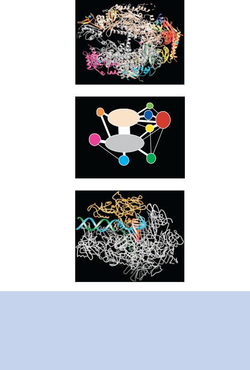
eukaryotic cells were identified by their sensitivity to the bicyclic octapeptide α-amanitin – RNA polymerase I is not affected by this compound; RNA polymerase II is rapidly inhibited at low concentrations of α-amanitin; RNA polymerase III from mammalian cells is inhibited by high levels of α-amanitin. Each of the polymerases is a large ( 500 kDa) multiprotein complex typically containing 8 –14 individual subunits. Here we will concentrate on RNA polymerase II (pol II) since it is responsible for the transcription of protein coding genes. Pol II is an enzyme comprising 12 subunits encoded, in yeast, by the RPB1 to RBP12 genes. The crystal structure for 10 of the subunits of RNA pol II has been solved recently (Cramer et al., 2000; Cramer, Bushnell and Kornberg, 2001) and is shown in Figure 1.26; it gives tantalizing insights into the structural mechanism of RNA production. The largest subunit Rpb1p contains a highly conserved carboxy-terminal repeat domain (CTD) with the consensus sequence Tyr – Ser –Pro –Thr –Ser –Pro –Ser. The yeast CTD includes 26 or 27 repeats and the human CTD has 52 repeats. This sequence can be extensively phosphorylated, which is an important step in converting RNA pol II from a form involved in promoter recognition to a form involved in transcriptional elongation (Hampsey, 1998). We will see later that, besides its role in transcription, the CTD is also critical for co-transcriptional RNA processing events such as capping, polyadenylation and splicing. As stated previously, pol II is incapable of binding DNA on its own. It requires other proteins – called general transcription factors – to achieve this function. There are six principal general transcription factors (TFIIA, IIB, IID, IIE, IIF and IIH) that are essential for gene-specific DNA binding and the formation of a polymerase complex capable of transcriptional initiation. Again, most of the general transcription factors are not single proteins, but are multi-protein complexes – for example TFIID is composed of 13 polypeptides – the TATA-box binding protein (TBP) and 12 TATA-box binding
Figure 1.25. Modifications of the histone tails. (a) The amino-terminal ‘tails’ of the histone proteins may be modified by phosphorylation (P), acetylation (Ac) or methylation (Me) at the serine (S) or lysine (K) residues indicated. The numbers refer to the amino acid positions within each protein. Additionally, histones H2A and H2B can be ubiquitinated (Ub) at their carboxy-terminal ends. The expression of genes contained within or close to nucleosomes containing these modified histones can be drastically altered. Reprinted from FEMS Microbiology Review, Vol 23, Perez´-Mart´ın, Chromatin and transcription in Saccharomyces cerevisiae, pp. 503–523, Copyright (1999), with permission from Elsevier. (b), (c) The acetylation of the histone tails alters their charge, and results in a ‘loosening’ of the histone–DNA complex. This allows transcription to occur more readily
52 |
DNA: STRUCTURE AND FUNCTION 1 |
|
|
|
(a) |
(b)
|
|
12 |
9 |
2 |
10 |
|
||
|
3 |
|
|
|
|
|
|
11 |
5 |
1 |
|
|
|
|
|
6 |
8 |
|
|
(c)
Figure 1.26. Architecture of yeast RNA polymerase II. (a) The backbone model for 10 subunits of RNA polymerase as determined by X-ray crystallography. The red sphere in the large cleft at the centre of the molecule represents the magnesium ion at the active site. A diagrammatic arrangement of the subunits of the protein present in the structure is shown in panel (b) and the protein complexed with both double-stranded DNA (blue and green) and RNA (red) is shown in (c). These figures were kindly provided by Patrick Cramer (University of Munich) and are reprinted with permission from Science (Cramer et al., 2000; Gnatt et al., 2001). Copyright American Association for the Advancement of Science
1.13 TRANSCRIPTION |
53 |
|
|
associated factors (TAFs) (Lee and Young, 2000). Pol II exists in the cell as a large complex of proteins, termed the holoenzyme. The holoenzyme is composed of SRB (suppressor of RNA polymerase B) proteins that interact with the CTD, Med (mediator) proteins and some of the general transcription factors. The precise composition of the general transcription factors involved with the holoenzyme is still unclear. The SRB and Med proteins appear to act as a regulatory ‘glue’ that stabilizes interactions between RNA pol II and general transcription factors and may also confer responsiveness to transcriptional activators (Koleske and Young, 1994; Malik and Roeder, 2000). The mediator complex is assumed to enter the initiation complex with RNA pol II and is released at the end of initiation or early in RNA chain elongation (Svejstrup et al., 1997). Transcriptional activators interact with a number of the components of the Pol II holoenzyme, including TBP, TAFs, TFIIB and some of the SRB and Med proteins. Whatever interactions occur within the cell, the end result appears to be simply the recruitment of the pol II complex to a promoter that has been freed from nucleosomes. Once the polymerase is bound to the promoter, transcription will be initiated.
The interactions between activators and the proteins described above lead to a model for gene activation in which the activator must first bind to DNA. The activator then recruits chromatin modifying and remodelling complexes to remove nucleosomes from the promoter, and finally pol II is brought to the promoter so that transcription can begin. The entire gene need not be devoid of nucleosomes for full transcription to occur. If the promoter is accessible to RNA polymerase and transcription factors, the presence of nucleosomes will not inhibit the elongation of the message. The structure of the nucleosome is such that the positive charges (lysines and arginines) of the histones are placed adjacent to the negative DNA helix. As the polymerase advances, it displaces DNA from the nucleosome and forms a closed loop, while the torsion ahead of the RNA polymerase generates supercoiling of the DNA. This displaces the histone octamer, which keeps contact with the DNA behind the RNA polymerase. In this way, the nucleosome need never lose contact with DNA as the RNA polymerase passes by (Studitsky, Clark and Felgerfeld, 1995). So once the RNA polymerase is transcribing the gene, nucleosomes do not stop it. The important event for transcription-level gene expression is to get the RNA polymerase bound and functioning at the promoter.
The molecular processes involved in transcriptional termination in eukaryotes are relatively poorly defined. Some transcripts terminate over 1000 bp downstream of the 3 end of the mature mRNA, and appear to end at termination regions rather than at specific sites. The transcript is cleaved from the extending polymerase before the polymerase itself terminates transcription
54 |
DNA: STRUCTURE AND FUNCTION |
1 |
|
|
|
|
||||||||||||||||||||||||||||
|
|
|
|
|
|
|
|
|
|
|
|
|
|
|
|
|
|
|
|
|
|
|
|
|
|
|
|
|
|
|
|
|
|
|
|
|
OH OH |
|
|
|
|
|
|
|
|
|
|
|
|
|
|
|
|
|
|
|
|
|
|
|
|
|
|
|
|
|
|
|
|
|
|
|
|
|
|
O |
O |
|
O |
|
|
Base |
|
|
|
|
||||||||||||||||||
|
|
O |
|
|
|
|
|
|
|
|
|
|
|
|
|
|
|
|
|
|
|
|
|
|
|
|
||||||||
|
|
|
|
O |
|
P O |
|
P |
|
O |
|
P |
|
O |
O |
|
|
|
|
|
|
|
|
|
|
|
|
|
||||||
|
|
|
|
|
|
|
|
|
O− |
|
|
|
|
|
|
|
|
|
|
|
|
|
|
|
|
|
|
|
|
|
||||
H2N N |
N |
|
|
O− |
|
|
O− |
|
|
|
|
|
|
|
|
|
|
|
|
|
|
|||||||||||||
5′ HN |
N + |
|
|
|
|
|
|
|
|
|
|
|
|
|
|
|
O |
|
O CH3 |
|
Base |
|
|
|
|
|||||||||
|
|
|
|
|
|
|
|
|
|
|
|
|
|
O |
|
P |
|
O |
|
|
|
|
O |
|
|
|
|
|
||||||
O |
|
CH3 |
|
|
|
|
|
|
|
|
|
|
|
|
|
|
|
|
|
|
|
|
|
|
|
|
|
|||||||
|
|
|
|
|
|
|
|
|
|
|
|
|
|
|
|
O− |
|
|
|
|
|
|
|
|
|
|
|
|||||||
|
|
|
|
|
|
|
|
|
|
|
|
|
|
|
|
|
|
|
|
|
|
|
|
|
|
|
|
|
|
|||||
7-methyl guanosine |
|
|
|
|
|
|
|
|
|
|
|
|
|
|
|
|
|
|
|
|
|
|
|
|
OH |
Coding region |
|
|
||||||
|
|
|
|
|
|
|
|
|
|
|
|
|
|
|
|
|
|
|
O |
|
A100-200 3′ |
|||||||||||||
|
|
|
|
|
|
|
|
|
|
|
|
|
|
|
|
|
|
|
|
|
|
|
|
|
|
|
|
|
|
|
|
STOP |
|
|
|
|
|
|
|
|
|
|
|
|
|
|
|
|
|
|
|
|
|
|
|
|
O |
|
P |
|
O |
|
|
AUG |
|
|
|||
|
|
|
|
|
|
|
|
|
|
|
|
|
|
|
|
|
|
|
|
|
|
|
O− |
|
5′-UTR |
|
|
|
|
|||||
|
|
|
|
|
|
|
|
|
|
|
|
|
|
|
|
|
|
|
|
|
|
|
|
3′-UTR |
||||||||||
|
|
|
|
|
|
|
|
|
|
|
|
|
|
|
|
|
|
|
|
|
|
|
|
|
|
|
|
|
|
|
||||
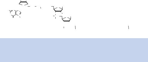
Figure 1.27. The structure of a mature mRNA molecule in eukaryotes. After transcription, a 7-methyl guanosine cap is added to the 5 -end of the message, and the ribose sugar of the first, and sometimes second, nucleotide is methylated at the 2 -position. The 3 -end of the transcript is polyadenylated with the addition of 100–200 A residues
(Figure 1.27). The function of the RNA polymerase complex is not finished once the transcript has been cleaved. The polymerase also functions to direct processing of the transcript, such as splicing and polyadenylation (McCracken et al., 1997; Hirose and Manley, 1998). Indeed, recent evidence suggests the enzymes and cellular machinery involved in transcription, RNA processing and translation may be extensively coupled to form ‘gene expression factories’ that maximize the efficiency and specificity of each stage of gene expression (Maniatis and Reed, 2002).
1.14RNA Processing
Perhaps the most obvious difference between the genes of prokaryotes and eukaryotes is that genes of the later are split into exons (coding regions) and introns (non-coding regions). In the mid-1970s, Philip Sharp and Richard Roberts independently hybridized a messenger RNA to the DNA from which it had been transcribed, and viewed the resulting hybrids using electron microscopy. They found that RNA hybridization occurred at discontinuous sequences within the DNA. That is, the binding of mRNA to the DNA sequence from which it was derived would result in the formation of loops in the DNA, corresponding to sequences in the DNA that were not present in the mRNA molecule (Chow et al., 1977; Berget, Moore and Sharp, 1977). Walter Gilbert gave names to these various regions. The parts of the gene that are represented in the mRNA, and are therefore part of the expressed region, he called exons. The parts of the gene that are the intervening sequences he called introns. The mechanism by which the precursor RNA (pre-mRNA) is converted into the final mRNA – in other words, how introns are removed – has been the subject of intense scrutiny since their discovery. Before we address this issue, we need to see what happens to an mRNA molecule as it is produced.
1.14 RNA PROCESSING |
55 |
|
|
Once a gene has been transcribed, the mRNA produced is extensively modified. Almost as soon as mRNA synthesis is initiated, the 5 -end of the message is capped by the addition of a 7-methyl guanosine residue. The cap is added to the 5 -end of the mRNA via a 5 –5 condensation of guanosine to the mRNA to form a structure consisting of 3 –G– 5 ppp5 –N-3 p. After cap formation, a methyl group is added to the guanine residue and to the first and/or second adjacent nucleotide (Figure 1.27). The function of the cap is not entirely clear. It has been suggested that the cap, and proteins that bind to it, direct ribosome binding and correct translational initiation. In addition to the cap, the 3 -end of the mRNA is cleaved from the extending chain and then polyadenylated.
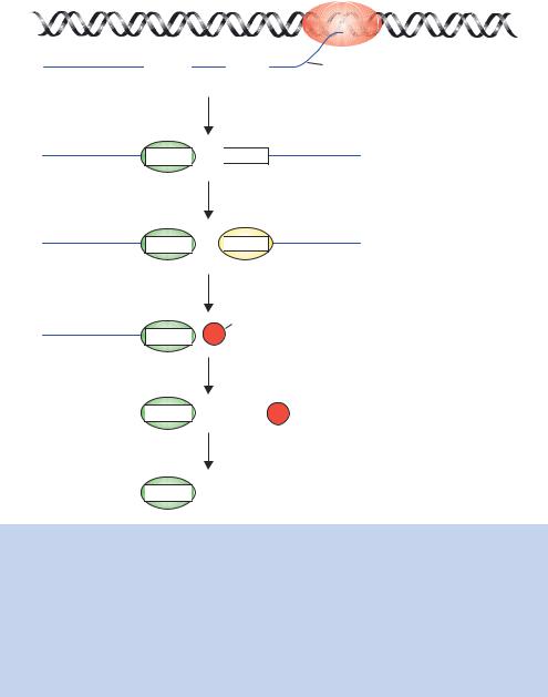
RNA transcript cleavage and polyadenylation are directed by a polyA signal within the RNA (Figure 1.28). The core polyA signal for vertebrate pre-mRNAs consists of two recognition elements flanking a cleavage –polyadenylation site. Typically, an almost invariant 5 -AAUAAA-3 hexamer is found 20 –50 nucleotides upstream of a more variable element rich in U or GU residues. Cleavage of the newly formed transcript occurs between these two elements and is coupled to the addition of approximately 200 adenosines to the 3 -end of the 5 cleavage product. Two protein factors required for this process are the cleavage and polyadenylation specificity factor (CPSF), which binds to the AAUAAA motif, and the cleavage stimulation factor (CStF), which binds the downstream GU-rich element. As the polyA signal is extruded from the polymerase, it is recognized by a subset of cleavage and polyadenylation factors that have already been recruited to the RNA polymerase CTD. Then assembly continues as more factors join the complex. Ultimately cleavage occurs, followed by polyadenylation in which a stretch of 200 A residues is added to the 3 -end of the message. Polyadenylation only occurs to mRNAs, but not all mRNAs are polyadenylated; e.g. the mRNA species that encode histone proteins are not polyadenylated. The process of polyadenylation is carried out by an enzyme called polyA polymerase (PAP), and not only confers stability on the transcript, but is also required for the movement of the mature mRNA out of the nucleus into the cytoplasm, where translation occurs. As we will see in later chapters, the addition of polyA to mRNAs has important practical consequences. The hybridization of polyT sequences to only mRNA, and not other RNAs, is a vital tool to the genetic engineer.
1.14.1RNA Splicing
There are several different mechanisms of RNA splicing, which function on different RNA species. Here, however, we will concentrate on the mechanism
56 |
DNA: STRUCTURE AND FUNCTION 1 |
|
|
5'
5'
5'
5'
5'
5'
RNA polymerase II
PolyA site
|
|
|
|
|
AAUAAA |
|
|
G/U |
Newly synthesised transcript |
|
|
|||
|
|
|
|
|
PolyA signal
CPSF
AAUAAA 
 G/U
G/U
CPSF
CStF, CFI, CFII, PAP
AAUAAA
 G/U
G/U
CStF
Cleavage
PAP
AAUAAA  3'
3'
 AAUAAA
  AAAAAAAAAA
PABII
 AAUAAA
  AAAAAAAAAAAA200
Figure 1.28. Cleavage and polyadenylation of the mRNA transcript. The polyA site within the newly formed transcript is shown, together with the signal AAUAAA and the downstream U/GU region. A complex of cleavage and polyadenylation specificity factor (CPSF), cleavage factors I and II (CFI and II) and cleavage stimulation factor (CStF) bind to these sequences. PolyA polymerase (PAP) also joins the complex at this stage. RNA cleavage occurs, and CPSF remains bound to the 5 -end of the cleaved message. The 3 -end is rapidly degraded. PAP begins the synthesis of the polyA tail, resulting in the addition of the first 10 A residues. Finally, polyA binding protein II (PABII) joins the reaction, stimulating the synthesis of polyA and extending the tail to about 200 A residues
of mRNA splicing since understanding it, and its consequences, are most applicable to the genetic engineer. The capped and polyadenylated mRNA is spliced to remove the introns and fuse the exons together into a single unit that can be translated. The need to produce precisely spliced products is obvious. Any slippage in the fusing of exons together would have disastrous
|
|
|
|
1.14 |
RNA PROCESSING |
57 |
|
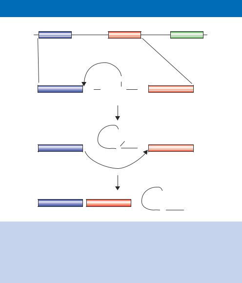
5' |
Exon 1 |
Intron 1 |
Exon 2 |
Intron 2 |
Exon 3 |
3' |
|
|
|
|
|||||
|
OH |
|
|
Exon 1 |
pGU YNCURAY |
AGp |
Exon 2 |
|
Branch point |
|
|
|
sequence |
|
|
Step1
Lariat
U
G Branch point p
Exon 1 |
OH |
A |
AGp |
Exon 2 |
|
|
|
Step 2 |
|
|
|
|
|
|
U |
|
Exon 1 |
p |
Exon 2 |
+ |
G Lariat |
|
|
p |
|
|||
|
|
|
|
A |
AG-OH |
Figure 1.29. The splicing of exons to form a mature mRNA. Nuclear pre-mRNA splicing takes place in two distinct mechanistic steps. Step 1 involves a nucleophilic attack by the 2 -OH group of the branch point A residue on the phosphodiester bond of the 5 exon–intron boundary, displacing the 5 exon (shown in blue) as the leaving group and giving the lariat structure shown. The second step is a nucleophilic attack of the 3 -end of the 5 exon on the phosphodiester bond of the 3 intron–exon boundary, displacing the intron (as a lariat form) and sealing the two exons together (the blue and red)
consequences for the protein that is to be eventually made. How are the boundaries between an intron and an exon marked, and by what molecular mechanism does splicing actually occur? Perhaps surprisingly, there is relatively little RNA sequence conservation at the exon –intron boundary. Most introns begin with the dinucleotide sequence 5 -GU-3 and end with the dinucleotide sequence 5 -AG-3 (Figure 1.29). There are, however, other less conserved sequences present within the intron that act as binding sites for complexes that are essential for splicing. These complexes, collectively called the spliceosome, contain both RNA and protein components, and are found exclusively in the nucleus. The RNA molecules found within the spliceosome are small (100 –200 nucleotides) and are complexed with proteins to form small nuclear