

John Wiley & Sons - 2004 - Analysis of Genes and Genomes
.pdf28 |
DNA: STRUCTURE AND FUNCTION 1 |
|
|
Double-stranded DNA
2 nm
“Beads on a string”
Cross-section of 30 nm fibre
10 nm
30 nm
Loops of 30 nm fibre
Folded and coiled loops
Protein scaffold
Mitotic chromosome
Figure 1.16. Packaging of DNA into the eukaryotic chromosome. The 2 nm DNA double helix is wrapped into nucleosomes as illustrated in Figure 1.15. At some point histone H1 enters the complex, possibly at the DNA entry/exit point on the nucleosome. The nucleosomes can form extended 10 nm fibres, which are long arrays of ordered nucleosomes. The nucleosomes can further condense to form a 30 nm fibre. This may be a ‘zig-zag’ array of nucleosomes. The 30 nm fibre is then wrapped onto a protein scaffold, which can be additionally folded and coiled to form the mitotic chromosome that is observed under the electron microscope
1.7The Replication of DNA
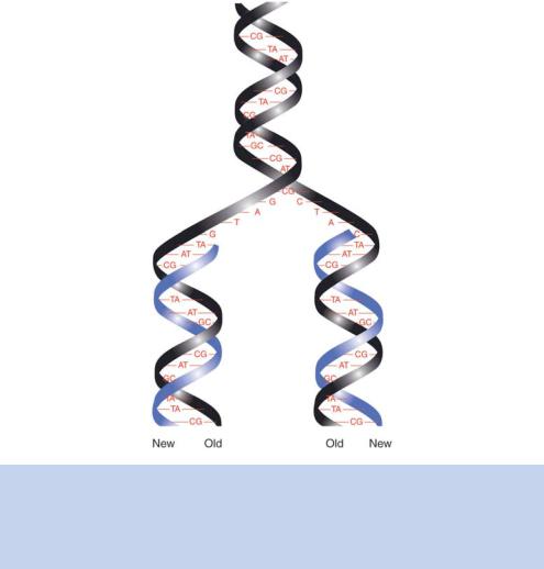
Watson and Crick finished their famous 1953 paper with the statement ‘It has not escaped our notice that the specific pairing we have postulated immediately suggests a possible copying mechanism for the genetic material’. It was apparent to them from the arrangement and nature of the bases that each strand
1.7 THE REPLICATION OF DNA |
29 |
|
|
of the DNA double helix could serve as a template for the synthesis of a complementary strand (Figure 1.17). They proposed that if the double helix were unwound, each nucleotide on the parental strands would have an affinity for its complementary nucleotide (Watson and Crick, 1953b). Thus, each of the parental DNA strands could act as a template for the synthesis of a new DNA strand that would result in the production of two identical double-stranded DNA duplexes. In this model, each of the newly synthesized DNA molecules consists of one parental and one newly synthesized DNA strand; hence, the process is known as semi-conservative replication.
Two other modes of replication that also rely on the parental strands serving as templates for new DNA synthesis were also considered. In conservative
Figure 1.17. The replication of double-stranded DNA as suggested by Watson and Crick. The separation of the DNA strands of the parent helix (black) means that each strand can act as a template of the synthesis of a new strand of DNA (blue). This is termed semi-conservative replication, as the daughter DNA molecules contain one old strand and one new strand

30 |
DNA: STRUCTURE AND FUNCTION 1 |
|
|
replication, the parental helix would be opened to reveal the base sequence, and a newly synthesized helix would be produced. The two newly synthesized DNA strands would come together to form a helix and the parental helix would reform. In the second alternative mode, called dispersive replication, the parental DNA from one strand could be dispersed into either of the newly synthesized strands, which would then be a mixture of new and old DNA.
Matthew Meselson and Franklin Stahl provided the experimental evidence to suggest that semi-conservative replication was used by bacteria to produce new DNA molecules (Meselson and Stahl, 1958). The results of their classic experiment are depicted in Figure 1.18. They grew E. coli cells for many generations in a medium in which the sole source of nitrogen was a heavy – but
E.coli grown in 15N media |
|
Many generations |
|
One replication |
One replication |
in 14N-media |
in 14N-media |
|
|
|
|
|
|
|
|
|
|
|
|
|
|
|
|
|
|
|
|
|
|
|
|
|
|
|
|
15N/15N |
15N/14N |
|
|
14N/14N 15N/14N |
|||
|
|
|
||||||
Figure 1.18. The Meselson–Stahl experiment. Bacterial cells were grown for many generations in a medium containing the heavy isotope of nitrogen, 15N. The DNA isolated from such bacteria was isolated and centrifuged through a density gradient. The mobility of the DNA band was noted. The bacteria were then transferred to media containing the normal isotope of nitrogen (14N). After one generation, all the DNA isolated from the bacteria ran through the density gradient with a mobility different to that seen previously. After two generations, two DNA species were observed. One had the same mobility as that seen after one generation, and the other ran with the same mobility as DNA isolated from bacteria grown on unlabelled media. In subsequent generations, the intensity of the latter band increased and the band of intermediate density decreased. These data are most readily explained by DNA replication occurring through a semi-conservative mechanism
1.8 DNA POLYMERASES |
31 |
|
|
non-radioactive – derivative of ammonium chloride (15NH4Cl). This meant that the DNA of these E. coli cells, into which the 15N was incorporated as part of the bases, was heavier than the DNA from cells grown on normal ammonium chloride (14NH4Cl). The normal and heavy forms of DNA could be distinguished after centrifugation through a density gradient of caesium chloride. The heavier DNA will travel further under such conditions than the lighter normal DNA. This provided a means of monitoring the levels of each nitrogen isotope within the bacterial replicated DNA as it was replicated. Meselson and Stahl took their bacteria that contained heavy DNA and transferred them into media containing normal ammonium chloride and then isolated DNA after each successive generation. After a single round of DNA replication on the normal media, they found that the bacterial DNA now possessed an intermediate density between the expected positions for wholly 15N-containing and wholly 14N-containing DNA. This result is consistent with semi-conservative replication, but not with conservative replication, where two distinct bands would have been expected. After two cell divisions, Meselson and Stahl did observe the presence of two bands of different densities – the first corresponding to the position of a mixed 14N/15N DNA species and the second, of equal intensity, corresponding to the expected position of unlabelled 14N/14N DNA. Subsequent rounds of DNA replication resulted in an increased proportion of the 14N/14N DNA band.
The experiments described above suggest that conservative replication does not occur, but could not rule out dispersive replication. Meselson and Stahl, however, dismissed this mode of replication by the analysis of individual single strands of replicated DNA. They heated their DNA (Figure 1.11) and found that individual strands possessed either the density of 15N- or 14N-containing single-stranded DNA, but not that of an intermediate density – which would have been expected if a newly synthesized DNA strand contained a mixture of the parental and newly synthesized sequences. Semi-conservative replication has also been shown to occur in eukaryotes (Taylor, Woods and Hughes, 1957), and is now widely accepted as the universal mechanism for DNA replication.
1.8DNA Polymerases
Both bacteria and eukaryotic cells possess multiple DNA polymerase enzymes. E. coli contain three such enzymes (Table 1.2). The first to be isolated, now called DNA polymerase I, is primarily involved in DNA repair rather than DNA replication. DNA polymerase II is also involved in DNA repair, while DNA polymerase III is the main replicating enzyme (Kornberg and Baker, 1992). DNA polymerase III is a multisubunit enzyme that functions as a dimer of these

32 |
DNA: STRUCTURE AND FUNCTION 1 |
|
|
Table 1.2. The properties of DNA polymerases I, II and III from E. coli
|
Pol I |
Pol II |
Pol III |
|
|
|
|
|
|
Main function |
DNA repair and |
DNA repair |
DNA replication |
|
|
primer removal |
|
|
|
Size, kDa |
109 |
120 |
140 |
|
Structural genes |
polA |
polB |
DnaE, N, Q, X, Z |
|
Molecules per cell |
400 |
+ |
10 – 20 |
|
5 to 3 polymerization |
+ |
+ |
||
3 to 5 exonuclease |
+ |
+ |
+ |
|
+ |
− |
− |
||
5 to 3 exonuclease |
||||
|
|
|
multiple subunits. Many DNA polymerase enzymes have been studied at the structural level, which, combined with extensive biochemical analysis, has led to the elucidation of their molecular mechanism of action.
DNA polymerase enzymes can possess three distinct enzymatic activities.
•All DNA polymerases direct the synthesis of DNA fragments in a 5 to 3 direction.
•Many DNA polymerases also possess 3 to 5 exonuclease activity. This enables the enzyme to remove nucleotides at the 3 - end of the newly synthesized chain. This is often considered a ‘proof-reading activity’, which can be used to excise incorrectly incorporated nucleotides before their replacement with the correct bases.
•Some DNA polymerases also have a 5 to 3 exonuclease activity. This allows the enzyme to remove sequences that have already been synthesized and, as we will see below, is involved in joining up discontinuous DNA fragments produced on the lagging strand during DNA replication.
DNA polymerases are not able to initiate DNA synthesis on their own. They have an absolute requirement for a free 3 -end onto which the enzyme can add new nucleotides. This means that two primers are required to initiate DNA replication in each direction from the origin – one complementary to each DNA strand to be replicated. As stated above, DNA synthesis only proceeds in a 5 to 3 direction. That is, new nucleotides are added on to the 3 end of an existing polynucleotide chain. This leads to a problem. Both strands of the parent DNA molecule must be replicated, but only one of the strands is orientated in a 5 to 3 direction from the origin of replication. This strand, called the
1.9 THE REPLICATION PROCESS |
33 |
|
|
RNA primer |
DNA polymerase III dimer |
|
Leading strand |
Okazaki fragments
Helicase
Lagging strand
Direction of DNA replication
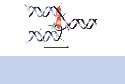
Figure 1.19. DNA replication by DNA polymerase III. The strands of the parent DNA molecule (black) are unwound by a helicase at the replication fork. The leading strand and the lagging strand are then fed into a dimer of DNA polymerase III. DNA replication on the leading strand occurs in a 5 to 3 direction by extending a single RNA primer. Many primers are required to produce the lagging strand in short sections (Okazaki fragments), which are later joined using DNA ligase
leading strand, can be copied in a continuous manner – DNA replication can occur without stopping as the DNA polymerase is working in its favoured orientation. Replication of the other strand, the lagging strand, would appear to have to occur in the 3 to 5 direction, which is not possible. The lagging strand is replicated in a discontinuous fashion (Figure 1.19). For discontinuous strand synthesis, a series of short DNA segments are produced in a 5 to 3 direction (Okazaki et al., 1968) that are then ligated together to produce the intact newly synthesized strand.
An enzyme that we will return to in later chapters is the RNA-dependent DNA polymerase called reverse transcriptase. This enzyme is found in certain viruses (called retroviruses) that contain RNA, rather than DNA, as their genetic material. Following infection of the host cell, the viral RNA serves as a template for the synthesis of a complementary DNA (cDNA) molecule. The DNA may then be incorporated into the host’s genome where, if the DNA is expressed, the retroviral RNA genome will be produced. The ability to use the reverse transcriptase enzyme in vitro to produce DNA from RNA will be discussed in Chapter 5.
1.9The Replication Process
How does DNA replication start and finish? As we have already seen, the DNA molecules inside cells are big and the need to tightly control the replication of
34 |
DNA: STRUCTURE AND FUNCTION 1 |
|
|
DNA sequences is obvious. We can consider the replication process in three phases – initiation, elongation and termination.
•Initiation of replication. The initiation of DNA replication does not occur at random sites around the genome, but rather it is initiated at specific points called origins of replication. Once DNA synthesis has been initiated, two replication forks, extending in either direction from the origin of replication, proceed to allow the full replication of the genome. Bacteria, such as E. coli, have a single origin of replication (called OriC). Eukaryotic cells, on the other hand, have multiple origins of replication which are different from OriC – the yeast Saccharomyces cerevisiae has been estimated to have about 300 replication origins, whilst human cells utilize over 20 000 origins during the replication of the genome. OriC is a 245 bp DNA sequence that acts as the binding site for a number of proteins (namely DnaA, B and C). The binding of these proteins promotes the melting (opening) of the DNA helix, a process that is essential so that DNA replicating enzymes can read the base sequence. Replication origins in both prokaryotes and eukaryotes probably serve the same overall function, but the replication origins of prokaryotic cells will not substitute for their eukaryotic counterparts and vice versa. Once the helix has been unwound, the DNA polymerase can access the base sequences by ‘reading’ the base pair hydrogen bonds such that a complementary DNA chain may be
synthesized. However, as we noted above, the polymerase can only function if a free 3 hydroxyl group is present. This hydroxyl group is provided by an RNA primer (which is complementary to the DNA) that is 5 –15
nucleotides long. The synthesis of the primer is directed by a form of RNA polymerase (called primase) that does not require a free 3 - end to initiate synthesis. DNA replication then proceeds simultaneously on both strands. Other proteins are also required for DNA replication to occur. The sections of single-stranded DNA produced during replication are stabilized through the binding of single-stranded binding proteins (SSBs), and the DNA is unwound into the polymerase complex with the help of DNA helicases (Figure 1.19). Additionally, topoisomerase enzymes (e.g. DNA gyrase) are required to relieve tension in the helix that results as a consequence of the unwinding process.
•Elongation. Given that DNA polymerase III can only replicate in a 5 to 3 direction, how can both DNA strands be simultaneously replicated? A mechanism for leading and lagging strand DNA synthesis is shown in Figure 1.19. In this model, DNA polymerase III, which is a dimer, is
1.9 THE REPLICATION PROCESS |
35 |
|
|
able to replicate the leading strand in a continuous fashion. The lagging strand, however, forms a loop so that nucleotide polymerization can occur on both template strands in a 5 to 3 direction. Looping will invert the orientation of the template with respect to the enzyme but not the direction of actual synthesis on a lagging strand. After the synthesis of approximately 1000 –2000 base pairs, the monomer of the enzyme on a lagging strand encounters a completed Okazaki fragment, at which point it releases the lagging strand. A new loop is then formed with the lagging strand and the process is repeated. The 5 to 3 exonuclease activity of DNA polymerases may aid the complete formation of Okazaki fragments. DNA ligase is then required to join the phosphodiester backbone of the Okazaki fragments to form a complete newly synthesized lagging strand. We will discuss the mechanism of action of DNA ligase and its use in genetic engineering in Chapter 2.
•Termination. Just as important as the correct initiation of DNA synthesis is its correct termination. For bacteria, such as E. coli, which contain a circular genome, if replication initiates bidirectionally from a single point, then termination should occur at a point halfway round the genome. Termination is not a passive process. It occurs at defined DNA sequences (called terminator sequences) that act as binding sites for a protein called Tus. Tus binding to the terminator sequences is highly asymmetric, which allows replication forks travelling in one direction to pass, but blocks DNA replication in the opposite direction (Kamada et al., 1996). Little is known about the termination of DNA replication in eukaryotes.
In eukaryotic cells, genome replication must be coordinated with the cell cycle so that two copies of the entire genome are available when the cell divides. The cell cycle is a four-stage process that is based upon microscopic observations of dividing cells (Figure 1.20). These observations showed that dividing cells pass through repeated cycles of metaphase, when nuclear and cell division occurs, and interphase, where few changes can be detected using a microscope. DNA replication occurs during interphase. The four stages of the cell cycle are the following.
•M-phase (mitosis) – the period when the nucleus and the cell divides.
•G1-phase (gap 1) – a growth phase where transcription, translation and other general cellular activities occur.
•S-phase (synthesis) – where DNA synthesis occurs and the genome is replicated.

36 |
DNA: STRUCTURE AND FUNCTION 1 |
|
p |
ter |
|
In |
|
hase
G1
se a h p
c i t o
t
i M
M
Cell division |
|
Mitosis |
DNA synthesis |
cyclin |
S |
Chromosome |
|
separation |
|
G2
Figure 1.20. The eukaryotic cell cycle is split into cell division (mitosis) and the period between divisions (interphase). After a division is complete, the cell grows during the first gap phase (G1). DNA synthesis then occurs (S) before a second gap phase (G2). Finally, the duplicated chromosomes separate and cell division occurs (M). The decisions to proceed with particular phases of the cycle are controlled by proteins – cyclins and cyclin-dependent kinases (CDKs). These are termed checkpoint controls
•G2-phase (Gap 2) – the second interval period. Another growth phase when final preparations for cell division take place.
A human cell in culture takes about 20 hours to progress through one complete cell cycle. Of this, over 9 h will be spent in G1, while S-phase takes about 8 h to complete, and G2 lasts about 2 h. The actual time the cell spends dividing (i.e. in M-phase) is only about 45 min. Clearly, it is important that the S- and M-phases are coordinated so that the genome is completely replicated once, but only once, before cell division occurs. The periods immediately before entry into S- and M-phases are the key cell-cycle checkpoints. Proteins, cyclins and cyclin-dependent kinases (CDKs) act to control entry into each phase of the cell cycle. Cyclins are a diverse family of proteins that bind and activate some members of the CDK family. Most cyclins display dramatic changes in concentration during the cell cycle, which help to generate the oscillations in CDK activity that form the foundation of the cell-cycle control. Other checkpoints also exist, primarily within S-phase, to ensure that, for example, DNA damage is repaired before replication can be completed. Lee Hartwell, Paul Nurse and Tim Hunt were awarded the 2001 Nobel Prize in Physiology or Medicine for their discoveries of key regulators of the eukaryotic cell cycle.
1.10 RECOMBINATION |
37 |
|
|
1.10Recombination
DNA should never be considered as a static molecule. It is constantly changing, and one mechanism by which this change is brought about is recombination. Recombination is the large-scale rearrangement of a DNA molecule. This type of rearrangement occurs as a consequence of two DNA molecules sharing either extensive regions of similar sequence (homologous recombination) or very short regions of homology (site-specific recombination). Cells containing a diploid set of chromosomes have plenty of opportunities to find a homologous partner for recombination to occur. Recombination in a bacterial system was first demonstrated independently by Alfred Hershey and Max Delbruck¨ in 1947. They studied the infection of E. coli with bacteriophages. If an E. coli cell was infected at the same time with two genetically different T2 bacteriophages, the resulting phage population included recombinant phage types as well as the original parental phage types (Hershey, 1947).
Homologous recombination occurs between two DNA molecules that have essentially the same sequence. The genetic information is swapped and mixed up between the two DNA versions such that the recombined DNA molecules are a mixture of the starting ones. Several mechanisms have been proposed to explain the molecular basis of these events. The key to understanding the molecular processes involved in recombination was first articulated in 1964 by Robin Holliday (Holliday, 1964). His model for recombination required that a nick (cleavage at the phosphodiester bond between two nucleotides) occurred at the same site in two homologous DNA molecules (called the donor and recipient DNA). A strand exchange reaction then occurred, followed by sealing of the nicks using DNA ligase. The exchange of donor and recipient DNA strands leads to the formation of a Holliday junction – a structure in which the crossed DNA strands connect the donor and recipient DNA molecules. The Holliday junction can undergo branch migration, leading to more recombination between strands from different DNA molecules. Finally, resolution of the junction gives rise to the recombined DNA molecules. Here, however, we will concentrate on the modified mechanism proposed by Meselson and Radding (1975) since the event initiating recombination can be brought about by a single nick in one of the DNA strands rather than earlier models, which relied on two DNA nicks before recombination could occur. The basic model is shown in Figure 1.21(a). The formation of the Holliday junction and its resolution, together with some of the E. coli proteins known to be involved in the process, are outlined below.
•The nick is created by the RecBCD endonuclease, which cleaves DNA strands at sequences called chi (χ ) sites (5 -GCTGGTGG-3 ).