

John Wiley & Sons - 2004 - Analysis of Genes and Genomes
.pdfxviii ABBREVIATIONS AND ACRONYMS
HSV |
herpes simplex virus |
IMAC |
immobilized metal ion affinity chromatography |
IMPACT |
intein mediated purification with an affinity chitin binding tag |
ITR |
inverted terminal repeat |
LTR |
long terminal repeat |
MBP |
maltose binding protein |
mRNA |
messenger RNA |
MCS |
multiple cloning site |
MLP |
major late promoter |
MSV |
maize streak virus |
NLS |
nuclear localization signal |
OD |
optical density |
ORF |
open reading frame |
PABII |
polyA binding protein II |
PAC |
P1 artificial chromosome |
PAP |
polyA polymerase |
PCR |
polymerase chain reaction |
PFGE |
pulsed-field gel electrophoresis |
RdRp |
RNA-dependent RNA polymerase |
RF |
release factor |
|
replicative form |
RFLP |
restriction fragment length polymorphism |
RIP |
ribosome inactivating protein |
RISC |
RNA induced silencing complex |
RNAi |
RNA interference |
rRNA |
ribosomal RNA |
RT |
reverse transcription |
|
reverse transcriptase |
RT-PCR |
reverse transcription-polymerase chain reaction |
SAM |
S-adenosylmethionine |
SDS |
sodium dodecyl sulphate |
siRNAs |
small inhibiting RNAs |
SNP |
single-nucleotide polymorphism |
snRNP |
small nuclear ribonucleoprotein |
SRB |
suppressor of RNA polymerase B |
STS |
sequence tagged site |
SV40 |
simian virus 40 |
TAF |
TATA-box binding associated factor |
ABBREVIATIONS AND ACRONYMS |
xix |
|
|
TBP |
TATA-box binding protein |
TdT |
terminal deoxynucleotidal transferase |
TGMV |
tomato golden mosaic virus |
TK |
thymidine kinase |
tRNA |
transfer RNA |
VA RNAs |
viral associated RNAs |
VNTR |
variable number tandem repeat |
YAC |
yeast artificial chromosome |

DNA: Structure 1 and function
Key concepts
The genetic information is contained within nucleic acids
DNA is a double-stranded antiparallel helix
Base pairing (A to T and G to C) holds the two strands of the helix together
DNA replication occurs through the unwinding of the DNA strands and copying each strand
The central dogma of molecular biology:
◦DNA makes RNA makes protein
Transcription is the production of an RNA copy of one of the DNA strands
Translation is decoding of an RNA molecule to produce protein
Every organism possesses the information required to construct and maintain a living copy of itself. The basic concepts of heredity and, as a consequence, genes can be traced back to 1865 and the studies of Gregor Mendel – discussed by Orel (1995). From the results of his breeding experiments with peas, Mendel concluded that each pea plant possessed two alleles for each gene, but only displayed a single phenotype. Perhaps the most remarkable achievement of Mendel was his ability to correctly identify a complex phenomenon with no knowledge of the molecular processes involved in the formation of that phenomenon. Hereditary transmission through sperm and egg became known about the same time and Ernst Haeckel, noting that sperm consists largely of nuclear material, postulated that the nucleus was responsible for heredity.
Analysis of Genes and Genomes |
Richard J. Reece |
2004 John Wiley & Sons, Ltd |
ISBNs: 0-470-84379-9 (HB); 0-470-84380-2 (PB) |

2 |
DNA: STRUCTURE AND FUNCTION 1 |
|
|
1.1Nucleic Acid is the Material of Heredity
The idea that genetic material is physically transmitted from parent to offspring has been accepted for as long as the concept of inheritance has existed. Both proteins and nucleic acid were considered as likely candidates for the role of the genetic material. Until the 1940s, however, many scientists favoured proteins. There were two main reasons for this. Firstly, proteins are abundant in cells; although the amount of an individual protein varies considerably from one cell type to another, the overall protein content of most cells accounts for over 50% of the dry weight. Secondly, nucleic acids appeared to be too simple to convey the complex information presumed to be required to convey the characteristics of heredity. DNA (deoxyribonucleic acid) was first isolated in 1869 by the Swiss chemist Johann Frederick Miescher. He separated nuclei from the cytoplasm of cells, and then isolated an acidic substance from these nuclei that he called nuclein. Miescher showed that nuclein contained large amounts of phosphorus and no sulphur, characteristics that differentiated it from proteins. In what proved to be a remarkable insight, he suggested that ‘if one wants to assume that a single substance. . . is the specific cause of fertilization then one should undoubtedly first of all think of nuclein’.
In 1926, based on the idea that DNA contained approximately equal amounts of four different groups, called nucleotides, and by determining the type of linkage that joined the nucleotides together, Levene and Simms proposed a tetranucleotide structure (Figure 1.1) to explain the chemical arrangement of nucleotides within nucleic acids (Levene and Simms, 1926). They proposed a very simple four-nucleotide unit that was repeated many times to form long nucleic acid molecules. Because the tetranucleotide structure was relatively simple, it was widely believed that nucleic acids could not provide the chemical variation expected of the genetic material. Proteins, on the
OH
HO PO Sugar Adenine
HO PO Sugar Uracil
HO PO Sugar Guanine
HO PO Sugar Cytidine
Figure 1.1. The tetranucleotide model for nucleic acid structure proposed by Levene and Simms in 1926. At the time that this model was proposed, it was thought that plant and animal nucleic acid might be different, and the differences between DNA and RNA were not fully understood
1.1 NUCLEIC ACID IS THE MATERIAL OF HEREDITY |
3 |
|
|
other hand, containing 20 different amino acids, could provide the basis for substantial variation.
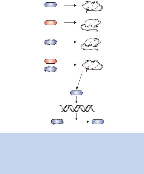
In 1928, Frederick Griffith performed experiments using several different strains of the bacterium Streptococcus pneumoniae (Griffith, 1928). Some of the strains used were termed virulent, meaning that they caused pneumonia in both humans and mice. Other strains were avirulent, and did not cause illness. Virulent and avirulent strains are morphologically distinct in that the virulent strains have a polysaccharide capsule surrounding the bacterium and form smooth, shiny-surfaced colonies when grown on agar plates. Avirulent bacteria lack the capsule and produce rough colonies on the same plates. The smooth bacteria are virulent because the polysaccharide capsule means that they are not easily engulfed by the immune system of an infected animal, and thus are able to multiply and cause pneumonia. The rough bacteria that lack the polysaccharide capsule do not have this protection and are consequently readily engulfed and destroyed by the host immune system.
Griffith knew that only living virulent bacteria would produce pneumonia when injected into mice. If heat-killed virulent bacteria were injected into mice, no pneumonia would result, just as living avirulent bacteria failed to produce the disease when similarly injected. Griffith’s critical experiment (Figure 1.2) involved the injection into mice of living rough bacteria (avirulent) combined with heat-treated smooth bacteria. Neither cell type caused death in mice when they were injected alone, but all mice receiving the combined injections died. The analysis of blood of the dead mice revealed a large number of living smooth bacteria when grown on agar plates. Griffith concluded that the heat-killed smooth bacteria were somehow responsible for converting the live avirulent rough bacteria into virulent smooth ones. He called the phenomenon transformation, and suggested that the transforming principle might be some part of the polysaccharide capsule or some compound required for capsule synthesis, although he noted that the capsule alone did not cause pneumonia.
In 1944, Oswald Avery, Colin MacLeod and Maclyn McCarty published their work to show that the molecule responsible for the transforming principle was DNA (Avery, MacLeod and McCarty, 1944). They began by culturing large quantities of smooth Streptococcus pneumoniae cells. The cells were harvested from cultures and then heat-killed. Following homogenization and several extractions with detergent, they obtained an extract that, when tested by co-injection with live rough bacteria, still contained the transforming principle. Protein was removed from the extract by several chloroform extractions and polysaccharides were enzymatically digested and removed. Finally, precipitation of the resultant fraction with ethanol yielded a fibrous mass that still retained the ability to induce transformation of the rough avirulent cells. From the
4 |
DNA: STRUCTURE AND FUNCTION 1 |
|
|
Bacteria
Injection
Live smooth
Mouse dies
Heat-killed smooth
Mouse lives
Live rough
Mouse lives
Heat-killed smooth
+
 Mouse dies Live rough
Dead mouse has live smooth bacteria
Extract DNA
Rough |
Transformation |
Smooth |
Figure 1.2. The key features of Griffith’s experiments, combined with data of Avery, MacLeod and McCarty to identify DNA as the transforming principle from Streptococcus pneumoniae. Griffith noted that injecting live smooth bacteria into mice led to the formation of pneumonia and eventually to the death of the mouse. Heat-treating the bacteria before injection did not result in the formation of the disease. Non-virulent bacterial strains, which did not cause the disease on their own, could be transformed by co-injection with heat-treated virulent bacteria. Avery, MacLeod and McCarty identified DNA as the transforming principle
original 75 L culture of bacterial cells, the procedure yielded 10 –25 mg of the ‘active factor’. Further testing established beyond a reasonable doubt that the transforming principle was DNA. The fibrous mass was analysed for its nitrogen/phosphorus ratio, which was shown to coincide with the ratio expected for DNA. In order to eliminate all probable contaminants from their final extract, they treated it with the proteolytic enzymes trypsin and chyomtrypsin, and then digested it with an RNA digesting enzyme called
1.1 NUCLEIC ACID IS THE MATERIAL OF HEREDITY |
5 |
|
|
ribonuclease. Such treatments destroyed any remaining activity of proteins and RNA, but still retained the transforming activity. The final confirmation that DNA was transforming principle came by digesting the extract with deoxyribonuclease, which destroyed the transforming activity.
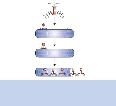
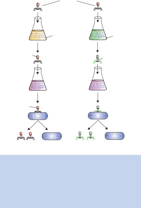
The second major piece of evidence supporting DNA as the genetic material was provided by the study of the infection of the bacterium Escherichia coli by one of its viruses, bacteriophage T2. Often simply referred to as phage, the virus consists of a protein coat surrounding a core of DNA (see Figure 1.3). In the early 1950s, little was known about the early steps of phage infection. The phage was known to be adsorbed to the surface of the bacteria, after which there was a latent period of approximately ten minutes before infectious virus particles started to be made, ultimately leading to host cell lysis and phage release. Alfred Hershey and Martha Chase reasoned that if they knew the fate of the phage protein and the nucleic acid at the beginning of the infection process, they would understand more about the nature of those early steps.
Protein coat  Phage DNA
Tail fibers
Infection
Bacterial cell
Ghost |
Insertion of genetic material
Production of new phage particles
Figure 1.3. The life cycle of T2 phage. During the course of infection, the bacteriophage adheres to the surface of the Escherichia coli cell. The genetic information, but not the whole phage particle, is inserted into the bacterium, where it is replicated. The phage ‘ghost’, which lacks the genetic material, remains at the bacterial surface. Once the newly synthesized phage particles are produced, bacterial cell lysis occurs and the phage particles are released into the surrounding medium
6 |
DNA: STRUCTURE AND FUNCTION 1 |
|
|
T2 phage
Infect bacteria in labelled media
32P |
35S |
Isolate phage |
|
DNA |
Protein |
labelled |
|
|
labelled |
Infect bacteria in |
|
unlabelled media |
|
Ghost |
Separate phage ghosts from bacterial cells
Ghosts |
Bacteria labelled |
Ghosts labelled |
Bacteria |
unlabelled |
with 32P |
with 35S |
unlabelled |
Figure 1.4. The Hershey– Chase blender experiment to show that nucleic acid was the genetic material. Hershey and Chase grew T2 bacteriophages on bacteria whose media contained either 32P (to label the phosphorus of nucleic acid) or 35S (to label the sulphur of proteins – the side chains of the amino acids methionine and cysteine both contain sulphur). They used their radio-labelled bacteriophages to infect a new culture of unlabelled bacteria. After a brief incubation, the bacteria were harvested by centrifugation and put into a blender to shear the bacteria away from the phage particles attached to their surface. They found that, when the DNA was labelled, the label was transferred to the bacterial cell, while the labelled protein remained with the phage ghosts. They concluded, therefore, that the material of heredity – i.e. the material passed on to make new offspring – was nucleic acid
1.2 STRUCTURE OF NUCLEIC ACIDS |
7 |
|
|
They used the radioisotopes 32P and 35S to follow the molecular components of the phages during infection (Figure 1.4). Because DNA contains phosphorus but not sulphur, 32P effectively labels DNA, and because proteins contain sulphur but not phosphorus, 35S labels protein. If E. coli cells are grown in the presence of 32P or 35S and then infected with T2 virus, the newly synthesized phages will have either a radioactively labelled DNA core or a radioactively labelled protein coat, respectively. These labelled phages can be isolated from the medium of infected cultures and used to infect other unlabelled bacteria. Hershey and Chase labelled the T2 phages with either 35S or 32P, and allowed them to adsorb to unlabelled bacteria. The cells were then separated from the unadsorbed material by centrifugation. The cells were resuspended and the suspension was blended to separate the phage ‘ghosts’ from the infected bacteria. The ghosts should not contain the genetic material, which needs to be replicated inside the host bacteria. Hershey and Chase found that 80% of the 35S label was removed from the bacteria whereas only 20% of the 32P label was removed. They concluded that ‘most of the phage DNA enters the cell, and a residue containing at least 80% of the sulphur-containing protein of the phage remains at the cell surface’. This work, together with that of Avery, MacLeod and McCarty, provided overwhelming evidence that DNA was the molecule responsible for heredity. Curiously, however, Hershey and Chase seemed somewhat skeptical about their findings, concluding their paper by saying that the ‘. . . protein has no function in phage multiplication, and that DNA has some function’ (Hershey and Chase, 1952).
1.2Structure of Nucleic Acids
As we have seen in the above section, the material of heredity is DNA, or more correctly nucleic acids. In most organisms, the hereditary material is DNA. However, a number of viruses use RNA (ribonucleic acid) as the building block for their genome. DNA and RNA are polymeric molecules made up of linear chains of subunits called nucleotides. Each nucleotide has three parts: a nitrogenous base, a five-carbon-atom sugar and a phosphate group (Figure 1.5). The combination of base and sugar is termed a nucleoside, while the base –sugar –phosphate is called a nucleotide. Since they contain the sugar 2 -deoxyribose, the nucleotides of DNA are termed deoxyribonucleotides, while those of RNA, which contain the sugar ribose, are known as ribonucleotides. The nucleotide bases can be either a double-ringed purine or a single-ringed pyrimidine. DNA and RNA are both built up from two purine containing nucleotides and two pyrimidine containing nucleotides. The purines of both DNA and RNA are the same – adenine (A) and guanine (G). The pyrimidine