John Wiley & Sons - 2004 - Analysis of Genes and Genomes
.pdf
38 |
DNA: STRUCTURE AND FUNCTION 1 |
|
|
(a)5′ 
 3′
3′
3′ 
 5′
5′
Nick
Stabilisation of single-strand
Strand invasion - formation of D loop
|
Nick and |
|
strand exchange |
|
Seal nicks to form |
|
Holliday junction |
5′ |
3′ |
3′ |
5′ |
3′ |
5′ |
5′ |
3′ |
Branch migration
Resolution
(b)
l phage DNA
attP
E. coli genome
attB
Int, IHF 
 Int, Xis, IHF
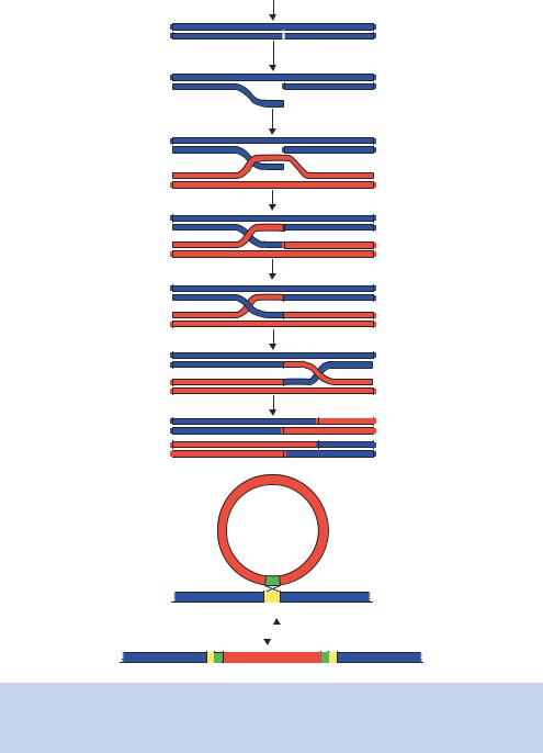
Figure 1.21. Homologous and site-specific recombination. (a) The Meselson–Radding model for homologous recombination. (b) The site-specific integration of λ DNA into the E. coli genome. See text for details
1.11 GENES AND GENOMES |
39 |
|
|
•Strand invasion is catalysed by the RecA protein, along with single-strand DNA binding protein (SSB), which stabilizes the free single DNA strand.
•The ‘D-loop’ that is formed after strand invasion is cut, again by the RecBCD endonuclease.
•Strand exchange occurs and the ends are sealed using DNA ligase. This forms the Holliday junction.
•The formation of the Holliday junction is followed by branch migration, catalysed by the proteins RuvA and RuvB.
•The resolution of the Holliday junction requires the RuvC protein, which nicks two of the DNA strands. DNA ligase then seals the strands after they have been cut.
At the end of these processes, the cell will have two molecules that have exchanged DNA. The same process outlined above also takes place in eukaryotes, using similar kinds of enzymatic activity. Recently, the high-resolution structure of the Holliday junction stabilized by the RuvA protein has been solved (Hargreaves et al., 1998).
Unlike homologous recombination, site-specific recombination occurs when a particular DNA, that is not at all homologous to another DNA, has a sequence that acts as a target for the recombination of that other DNA. In prokaryotes, this is the case when bacteriophage λ integrates its DNA into the E. coli chromosome, where it will reside in the lysogenic state. We will look at the life cycle of λ in more detail in Chapter 3. This recombination event occurs between sites in the bacteriophage DNA (attP) that have a sequence homology with sites in the bacterial DNA (attB), even though the two DNA molecules are completely different in all other sequences. This is shown in Figure 1.21(b). The enzyme that catalyses this event is called integrase (Int), along with a host protein called integration host factor (IHF). When the bacteriophage DNA is excised from the E. coli genome to begin the formation of new phage particles, a phage protein called Xis is required. This site-specific recombination still requires the base pairing of a short homologous region (the attP with the attB sites), but does not involve any of the proteins of the homologous recombination pathways.
1.11Genes and Genomes
The genetic information contained within the DNA base sequence directs the production of the proteins and enzymes that build the cell. The nucleic
40 |
DNA: STRUCTURE AND FUNCTION 1 |
|
|
acids are arranged into units called genes, and the whole collection of genes constitutes the genome. Most genes are used as templates to produce proteins. The complement of proteins within a particular cell type is distinctive – a hair follicle will produce keratin and a pancreatic β-cell will produce insulin – but the protein content of a cell can also change dramatically depending upon, for example, the availability of nutrients. This means that in certain cell types, certain genes will be expressed and others will not, and the cell must have the ability to activate certain genes in response to external signals. This raises a number of issues. How is the cell able to discern within its genome what is a gene and what is not, and how is the cell able to turn a particular gene on while at the same time not affecting the expression of other genes?
1.12Genes within a Genome
What is a gene? Surprisingly, there is not a universally accepted answer to this question. In its broadest terms a gene may be considered as a unit of heredity composed of nucleic acid. In classical genetics, a gene is described as a discrete part of a chromosome that determines a particular characteristic. Here, we will use the term gene to describe an open reading frame (ORF – the region of the gene that will be transcribed and translated into a protein sequence) together with its transcriptional control elements (promoter and terminator). Thus a gene is not just the coding sequence itself, but contains the information required for the expression of that coding sequence. Most protein coding ORFs have the same overall format (Figure 1.22). They start with a particular triplet of DNA bases (ATG) and end at stop signal, again a triplet of DNA bases (either TGA, TTA or TAG). The sequence in between will either directly code for the protein (prokaryotes) or be split into a series of exons and introns (eukaryotes). We will discuss exons and intron later, but with the advent of fully sequenced genomes (Chapter 9), a gene is usually defined as a DNA sequence that has at least 100 codons between the start and the stop signals. This sequence alone, is however insufficient to direct protein production. Each gene requires other DNA sequences surrounding it that act as binding sites for proteins such as the RNA polymerase and for those involved in transcriptional and translational initiation and termination. When combined, the coding portions of the gene and these promoter and terminator sequences form the functional gene, sometimes referred to as a ‘transcriptional unit’.
How many genes are present within a genome, how many genes are essential to the cell and how many are expressed at one time? The advent of completely sequenced genomes (Chapter 9) has allowed us to address, at least in part, some of these questions.

1.12 GENES WITHIN A GENOME |
41 |
|
|
Repressor
Prokaryotic
Stop
Start
Polymerase
 Terminator
Gene(s)
|
Promoter |
Operator |
|
||
|
||
|
binding site |
|
|
||
|
CAP |
|
|
||
|
Protein |
Met Thr Met... |
mRNA |
|
|
|
|
|
−10
−35
operon:lac
5′-CAACGCAATTAATGTGAGTTAGCTCACTCATTAGGCACCCCAGGCTTTACACTTTATGCTTCCGGCTCGTATGTTGTGTGGAATTGTGAGCGGATAACAATTTCACACAGGAAACAGCT ATG ACC ATG...-3′
3′-GTTGCGTTAATTACACTCAATCGAGTGAGTAATCCGTGGGGTCCGAAATGTGAAATACGAAGGCCGAGCATACAACACACCTTAACACTCGCCTATTGTTAAAGTGTGTCCTTTGTCGA TAC TGG TAC...-5′
mRNA
lacrepressorbinding |
|
Stop |
|
|
|
|
|
|
Terminator |
|
|
|
|
|
|
|
|
|
|
||||
|
|
|
|
|
|
|
|
||||
|
|
|
|
|
|
|
|
||||
binding |
|
|
|
|
|
|
|
Exon |
|
||
|
|
|
|
|
|
|
|||||
|
|
|
|
|
|
|
|
||||
|
|
|
|
|
|
|
|||||
|
|
|
|
|
|
|
|
|
|
|
|
polymerase |
|
|
|
|
|
|
|
|
|
|
|
|
|
|
|
|
|
Gene |
|
|
|
||
RNA |
|
Start |
|
|
|
|
|
|
|
Intron |
Mouseb-globingene: |
|
|
|
|
|
|
|
|
||||
|
|
|
|
|
|
|
|
|
EnhancerActivator TATA binding box sites |
||
|
|
|
|
|
|
|
|
|
|||
CAPbindingsites |
Eukaryotic |
Repressor bindingsite |
|
|
|
|
|||||
|
|
|
|
||||||||
|
|
|
|
||||||||
|
|
|
|||||||||
|
|
|
|||||||||
|
|
|
|
||||||||
|
|
|
|
||||||||
|
|
|
|||||||||
|
|
|
|
||||||||
|
|
|
|
|
|
|
|
||||
|
|
|
|
|
|
|
|
||||
|
|
|
|
|
|
||||||
|
|
|
|
|
|
|
|||||
|
|
|
|
|
|
|
|
|
|||
|
|
|
|
|
|
|
|
|
|
||
GGTGAGGTAGGATCAGTTGCTCCTCACATTTGCTTCTGACATAGT...-3′ |
CCACTCCATCCTAGTCAACGAGGAGTGTAAACGAAGACTGTATCA...-5′ |
5′-CGTAGAGCCACACCCTGGTAAGGGCCAATCTGCTCACACAGGATAGAGAGGGCAGGAGCCAGGGCAGAGCATATAA |
3′-GCATCTCGGTGTGGGACCATTCCCGGTTAGACGAGTGTGTCCTATCTCTCCCGTCCTCGGTCCCGTCTCGTATATT |
TATA-box
CCAAT-box
Figure 1.22. The architecture of a typical prokaryotic and a typical eukaryotic protein coding gene. In prokaryotes, families of genes
required, for instance, to produce all the enzymes of a pathway in prokaryotes are often transcribed together as a polycistronic message.
The operon contains the binding sites for the RNA polymerase (promoter) and enhancer elements, such as the cyclic AMP activator protein
(CAP). The binding of these proteins triggers transcription. Additionally, the operon may contain the binding sites for repressor proteins,
which, when bound to DNA, occlude polymerase binding and thus stop transcription. The transcript ends at terminator sequences at the
producedare
eukaryotes,In genessingle
operon. lacthe
controlthe ofregion
theshown ofsequence
operon.the isBelow
ofend - 3
enhancercontain mayalso gene.They theof end-
5the
bindingrepressor atsites
and individually.regulated containThey andactivator
theassembly |
ofthis.Again, |
expression.fullitsforrequiredare Theactivatorpromotes |
shortabeginstranscriptionand distancetothe3 |
|
side |
|
|
ofthegenewhich |
attheTATAbox, |
bacepairsupstream |
holoenzymecomplex |
elementsseveralthousand |
oftheRNApolymeraseII |
and prokaryotes theis
differencemajor eukaryotesbetween
gene.the thePerhaps
- ofend
3the
specificat atsites
occurstermination
(exons).Theintronsareremovedbysplicingbeforethetranscriptistranslated.The |
β-globingeneisalsoshown |
sequences |
themouse |
presenceofintronsthatsplitthecoding |
DNAsequenceofthepromoterregionof |
42 |
DNA: STRUCTURE AND FUNCTION 1 |
|
|
•How many genes are contained within a genome? The E. coli genome (4.6 Mb in length) contains about 4200 genes. The average length of a gene in E. coli is 950 bp, and the average separation between genes is 118 bp, so the majority of the genome codes for RNA or protein. The yeast Saccharomyces cerevisiae, a single-cell eukaryote, has a larger genome size (13.5 Mb) and some 6000 genes. Humans, on the other hand have a genome that is almost 250-fold larger that of yeast (at 3300 Mb) but may have as few as 30 000 genes – representing an increase of only fivefold.
•How many genes are essential to the organism? To determine whether a gene is essential for the organism, the function of the gene must be impaired in some way. This is usually achieved by gene knockouts, where the gene is removed from the genome and its effects on cell viability determined. Analyses such as these suggest that about half the E. coli and yeast genes are required for viability. Many apparently non-essential genes may play specialist roles under conditions not examined in this type of experiment (nutrient starvation for example). The apparently low percentage of essential genes, however, may reflect a functional degeneracy among certain sets of genes.
•How many genes are expressed at one time? The genes that are expressed in a particular cell define that cell. Some of the genes expressed in a liver cell must be different to those expressed in a skin cell. Genome-wide analysis of the expression of all genes within fully sequenced organisms such as yeast suggests that about 90% of genes are expressed at any one time, but 80% of these are expressed with very low abundance levels – in the order of 0.1 –2 transcripts per cell (Causton et al., 2001). The level of expression of individual genes will vary widely. Highly abundant proteins are produced from highly expressed genes, whilst other proteins that may be present at a much lower level (e.g. one copy per cell) are often produced from genes that are expressed at a very low level.
The intermediary between DNA and its encoded protein is RNA. The process of converting DNA sequences into proteins occurs in two steps. The DNA is first transcribed into RNA which is subsequently translated into the polypeptide sequence of the protein. As we have already seen (Figure 1.5), RNA differs from DNA in that it contains a ribose sugar rather than a deoxyribose sugar. Additionally, uracil replaces thymine as the complementary base to adenine in RNA. Transcription is similar to DNA replication except that only one of the two DNA strands is copied. There are two distinct classes of RNA that are produced by transcription, messenger RNA (mRNA) and structural RNAs
1.13 TRANSCRIPTION |
43 |
|
|
(ribosomal RNA (rRNA) and transfer RNA (tRNA)). mRNA, although by far the least abundant form of RNA – in most cells representing a few percentage points of the total RNA – is the carrier of the genetic information.
1.13Transcription
Transcription is the process by which an RNA copy of one of the strands in the DNA double helix is made. The antisense strand of the DNA directs the synthesis of a complementary RNA molecule. The RNA molecule produced is therefore identical to the sense strand of the DNA – except that it contains U instead of T. There are fundamental differences in the ways in which genes are transcribed in prokaryotes and eukaryotes. Here, it is important to understand the processes involved in each case. Many of the experiments we will look at in later chapters involve the use of eukaryotic cells, but the bacterium E. coli still plays a vital role in almost all genetic engineering experiments.
Transcription begins at specific DNA sequences called promoters. Like DNA replication, transcription occurs in three phases – initiation, elongation and termination. Initiation of transcription usually occurs to the 3 side of the promoter, and termination occurs at specific sites downstream of the coding sequence of the gene. At first glance, the overall architecture of a typical prokaryotic gene and a typical eukaryotic gene may appear to be similar (Figure 1.22). However, the controlling region for eukaryotic genes will not function in a prokaryotic cell, and vice versa.
Most protein coding genes in prokaryotes are transcriptionally active by default. That is to say, in the absence of other factors, the RNA polymerase can recognize the promoter of a gene, bind to it and produce RNA. Transcriptional control is brought to bear on the gene by repressor proteins that bind to DNA sequences adjacent to the RNA polymerase binding site. DNA binding by the repressor either occludes RNA polymerase binding and/or prevents a bound polymerase from transcribing. The eukaryotic RNA polymerase involved in the production of protein coding genes (pol II) is unable to recognize promoter sequences on its own. Therefore, eukaryotic genes are transcriptionally inactive in the absence of other factors. In both prokaryotes and eukaryotes, transcription is a highly regulated process. Proper timing and levels of gene expression are essential to almost all cellular processes.
1.13.1Transcription in Prokaryotes
Francois¸ Jacob and Jacques Monod were the first to elucidate a transcriptionally regulated system (Jacob and Monod, 1961). They worked on the lactose
44 |
DNA: STRUCTURE AND FUNCTION 1 |
|
|
metabolism system in E. coli. Lactose is a disaccharide of galactose and glucose. When the bacterium is in an environment that contains lactose as the sugar source it expresses the following structural genes.
•β-galactosidase – the enzyme hydrolyses the bond between the two sugars, glucose and galactose. It is coded for by the gene lacZ.
•Lactose permease – the enzyme spans the cell membrane and brings lactose into the cell from the outside environment. The membrane is otherwise essentially impermeable to lactose. It is coded for by the gene lacY.
•Thiogalactoside transacetylase – the function of this enzyme is not known. It is coded for by the gene lacA.
These three enzymes are located adjacent to each other in the E. coli genome (Figure 1.23) and a region of DNA that is responsible for their transcriptional regulation is located just to the 5 side of these structural genes. This assortment of genes and their regulatory regions is called the lac operon. In order to metabolize lactose, the structural genes must be expressed and the protein products produced. One could imagine, therefore, a simple genetic switch in which the lac structural genes are transcriptionally inert in the absence of lactose and on in its presence. There are, however, other factors that influence the transcriptional activity of the lac structural genes. The favoured sugar source for E. coli is glucose, since it does not have to be modified to enter the respiratory pathway. So, if both glucose and lactose are available, the bacterium turns off lactose metabolism in favour of glucose metabolism. The lac operon has the following DNA sequence elements.
(a)Operator (lacO) – the binding site for repressor.
(b)Promoter (lacP) – the binding site for RNA polymerase.
(c)Repressor (lacI) gene – which encodes the Lac repressor protein. This protein binds to DNA at the operator and blocks transcription of the structural genes by RNA polymerase bound at the promoter.
(d)Pi the promoter for lacI.
(e)CAP binding site for cAMP/CAP complex.
In the absence of lactose, RNA polymerase binds to the promoter but is prevented from transcribing the structural genes by the Lac repressor which is bound to the operator site (Lee and Goldfarb, 1991). Since no RNA can be made, no protein is produced. The repressor itself is produced from the lacI

|
|
|
|
|
|
1.13 |
TRANSCRIPTION |
45 |
||||
|
|
|
|
|
|
|
|
|
|
|
|
|
|
Pi lacI CAP P O |
lacZ |
|
lacY |
|
lacA |
|
|||||
|
|
|
|
|
|
|
|
|
|
|
|
|
|
|
|
|
|
|
|
|
|
|
|
|
|
Transcription
 Repressor
Induction by lactose
Translation
Pi lacI CAP P O |
lacZ |
|
lacY |
lacA |
||||||
|
|
|
|
|
|
|
|
|
|
|
|
|
|
|
|
|
|
|
|
|
|
Transcription of the
structural genes
b-galactosidase Lactose Thiogalactoside
permease transacetylase
Figure 1.23. The activation of the lac operon in E. coli. In the absence of lactose, the lac repressor binds to the operator (O) and prevents RNA polymerase bound to the promoter
(P) from transcribing the structural genes. When lactose enters the cell, it binds to the lac repressor and causes a conformational change that inhibits its ability to bind DNA. Consequently, the polymerase bound at P will transcribe the lac structural genes (lacZ, lacA and lacY). Full activation of the lac genes only occurs in the absence of glucose when the catabolite activator protein (CAP) binds to the operon and aids the binding of RNA polymerase to P
gene by an RNA polymerase bound to another promoter (Pi). When lactose is present within the cell, it acts as an inducer of the operon. It binds to the lac repressor, and induces a conformational change that results in the repressor dissociating from DNA. Now the RNA polymerase bound to the promoter is free to move along the DNA and RNA can be made from the three genes. Lactose can now be metabolized.
A pertinent question to ask here is how the lac genetic switch actually gets turned on initially. As stated above, the lac structural genes, including the permease required to get lactose into the cell, are switched off in the absence of lactose. It therefore seems impossible to achieve activation, since activation is required to get lactose into the cell. This is, however, an over-simplification. Each of the lac structural genes is transcribed at a low, basal, level (approximately five
46 |
DNA: STRUCTURE AND FUNCTION 1 |
|
|
copies per cell) in the absence of lactose. This means that when the bacterium encounters a source of lactose it is able to transport a few molecules into the cell so that full induction of the lac structural genes can occur. When fully induced, approximately 5000 copies of each protein product are present in the cell.
Regulatory control of the lac operon is more complicated since it needs to be turned off (repressed) if glucose is present, no matter whether lactose is present or not. The repression of the lac operon by glucose is mediated through the CAP site. When levels of glucose (a catabolite) in the cell are high, the production of cyclic AMP (cAMP) is inhibited, so when glucose levels drop, more cAMP forms. cAMP binds to a protein called CAP (catabolite activator protein), which then binds DNA at the CAP site in the lac operon. This activates transcription of the lac structural genes by increasing the affinity of the promoter for RNA polymerase. This phenomenon is called catabolite repression, since glucose inhibits the transcription of the lac operon by not allowing its full activation.
Like many operons in prokaryotes, the three lac structural genes are transcribed as a single mRNA. This polycistronic message is translated as it is produced since there is no physical separation of the DNA from the cytoplasm. Each of the ORFs contains its own ribosome binding site (see below) within the encoded mRNA and is translated independently of the other lac genes.
We have seen above how the transcript is initiated, but how does transcription end? Once RNA polymerase has started transcription, the enzyme moves along a DNA template, synthesising RNA, until it meets a terminator sequence. Here, the polymerase stops adding nucleotides to the growing RNA chain, releases the completed product and dissociates from the DNA. Termination can be brought about in one of two ways.
•Rho-dependent termination. The protein rho binds to the newly synthesized RNA chain and appears to cause termination when the polymerase pauses at certain DNA sequences.
•Rho-independent termination. Intrinsic termination sequences may form hair-pin loops in the RNA that promote polymerase dissociation. This type of termination is independent of additional factors and is the most common form of termination in E. coli.
1.13.2Transcription in Eukaryotes
The process of activating gene expression in eukaryotes is far more complex than in prokaryotes. There are several differences between eukaryotes and prokaryotes that impinge upon gene activation and RNA processing.

1.13 TRANSCRIPTION |
47 |
|
|
•Eukaryotes possess multiple RNA polymerase enzymes.
•The wrapping of DNA into nucleosomes represents a barrier to transcription.
•The physical separation of the nucleus (where transcription occurs) and the cytoplasm (where translation occurs) in eukaryotes means that mechanisms must exist to protect the mRNAs from degradation in the cytoplasm before they have been translated.
•Eukaryotic genes are monocistronic, with each gene being produced as a separate transcript from its own promoter.
•The genes of eukaryotes are not continuous and are split into coding regions and non-coding regions.
We will touch upon each of these issues as we consider how a typical eukaryotic gene may be transcribed, and the subsequent fate of the RNA that is produced.
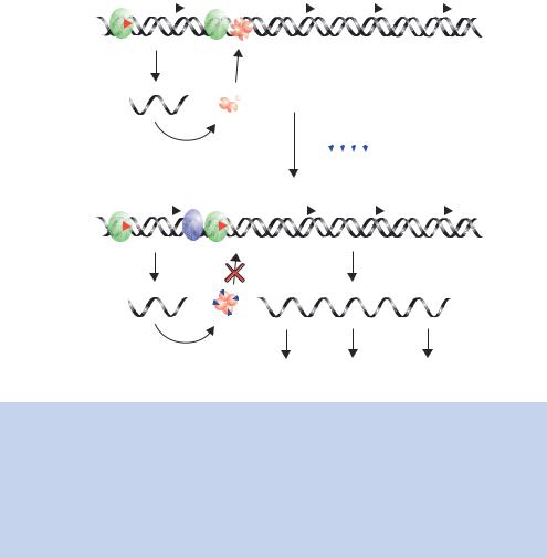
Eukaryotes contain three different RNA polymerases, each responsible for the transcription for a certain class of genes (Table 1.3). Each of the polymerases is a large complex of proteins that are involved in, amongst other things, regulating polymerase activity. Activation of RNA polymerase II genes (those coding for proteins) is brought about by transcriptional activator proteins binding to DNA sequences within the promoter of a gene (Figure 1.24). In the absence of these activators, the genes are not transcribed. The role of the activator has been the focus of much speculation. However, it appears to act simply as a way of recruiting various protein complexes to a particular promoter. Control of gene expression is therefore targeted through the activator. Activators have at least two separate functions. First, they must be able to bind to DNA so that they can find their target gene within the mass of genomic DNA. Second, they participate in protein –protein contacts necessary to recruit the RNA polymerase and other complexes to the gene through an activation domain. Either of these functions can be modulated to regulate gene expression in response to specific signals.
Table 1.3. Properties of eukaryotic RNA polymerases. In older papers, these are sometimes referred to as polymerases A, B and C
RNA polymerase |
Location |
Products |
|
|
|
I |
Nucleolus |
28S, 18S and 5.8S rRNA |
II |
Nucleus |
mRNA, and some snRNA |
III |
Nucleus |
tRNA, 5S rRNA, and some snRNA |
|
|
|
