

John Wiley & Sons - 2004 - Analysis of Genes and Genomes
.pdf108 |
BASIC TECHNIQUES IN GENE ANALYSIS 2 |
|
|
bacterial cells (1 –5 mL of bacterial culture) and can yield up to 10 µg of purified plasmid DNA in a typical mini-prep procedure. The basic method for plasmid purification is as follows (Figure 2.25).
•The bacterial cells are treated with the detergent sodium dodecyl sulphate (SDS) in the presence of sodium hydroxide to ensure an alkaline pH. The SDS disrupts the cell membranes and denatures proteins. During this process, the chromosomal DNA becomes denatured, but plasmid DNA molecules will remain in solution.
•The mixture is then neutralized using acidic potassium acetate (pH 5.2). The chromosomal DNA renatures upon neutralization, but aggregates to form an insoluble network. The high concentration of potassium acetate also causes precipitation of the protein –SDS complexes and of the high-molecular-weight RNA. Again, plasmid DNA molecules will remain in solution.
•The precipitate is then removed by centrifugation and the plasmid DNA can be further purified from the supernatant.
•The plasmid DNA in solution is then purified from any contaminants (metabolites, RNA, fragments of genomic DNA etc.) by binding the DNA to either silica or a positively charged (anionic) ion exchange matrix. For many small-scale mini-prep procedures, the plasmid DNA is bound to a silica matrix in buffer containing high levels of salt. Under these conditions, other contaminants will not bind to the matrix and can be washed away. Subsequently, the DNA can be eluted from the matrix using a low-salt buffer (or water). Larger-scale plasmid preparations procedures will often bind the plasmid DNA to a positively charged ion-exchange resin, such as DEAE (diethylaminoethanol), in buffer containing, for example, 1 M NaCl. This level of salt allows the DNA to bind to the matrix, but not the contaminants. After washing, the salt concentration is raised to 1.6 M to elute the DNA. Procedures such as this require that the DNA is precipitated (with ethanol or isopropanol) and resuspended in a low-salt buffer before it can be used in many molecular biology protocols.

3 Vectors
Key concepts
Vectors are autonomously replicating DNA molecules that can be used to carry foreign DNA fragments
DNA fragments cloned into vectors can be isolated in quantities sufficient for most laboratory manipulations
Vectors are based on naturally occurring DNA sequences that have been modified and combined to serve particular functions
The choice of vector depends chiefly upon the size of the DNA molecules that must be inserted into it
Vectors have been developed for a variety of specific purposes, including the production of single-stranded DNA, the high-level expression of protein encoding genes, and the production of RNA
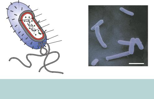
The vast majority of molecular cloning experiments utilize the bacterium Escherichia coli for the propagation of cloned DNA fragments. Even if the final destination of a cloned DNA fragment is a eukaryotic cell, DNA constructs are invariably produced in E. coli prior to being shuttled into their ultimate host. Cloning is possible in other organisms, but the advantages of E. coli have led to its widespread acceptance as the genetic engineering organism of choice. Escherichia coli, named for the German physician Theodor Escherich (1857 –1911), is a gram-negative, rod shaped bacterium propelled by long, rapidly rotating flagella (Figure 3.1). It is part of the normal flora of the human mouth and gut, helping to protect the intestinal tract from bacterial infection, aiding digestion and producing small amounts of vitamins B12 and K. The bacterium, which is also found in soil and water, is widely used in laboratory research and is probably the most thoroughly studied life form. As a laboratory organism, E. coli has a number of distinct advantages.
Analysis of Genes and Genomes |
Richard J. Reece |
2004 John Wiley & Sons, Ltd |
ISBNs: 0-470-84379-9 (HB); 0-470-84380-2 (PB) |
110 |
VECTORS 3 |
|
|
Outer membrane
Plasma membrane
Perplasmic space
DNA
Ribosome
Pilus
Flagellum
2 m
Figure 3.1. Structure of the bacterium Escherichia coli. A cut-away model of the bacterium showing some of the cellular layers and components, and E. coli cells viewed using an electron microscope
•It is easy to grow in simple, inexpensive growth medium.
•The organism has a rapid doubling time of about 20 –30 minutes during log-phase growth.
•Its genetics are well understood.
•Laboratory strains of E. coli are generally safe and contain mutations that do not allow them to escape the laboratory environment.
•It has a fully mapped and sequenced genome.
•Extra-chromosomal copies of DNA (plasmids and bacteriophage DNA) can be exploited to carry foreign DNA fragments.
E. coli cells generally reproduce asexually but, in order to increase diversity and share the gene pool, they have mechanisms for the transfer of genetic material from one bacterium to another. As far back as the mid-1940s it was known that bacterial cells were able to exchange genetic material with each other in a semisexual manner. The experiments of Lederberg and Tatum clearly demonstrated the transfer of genetic information through bacterial conjugation (Lederberg and Tatum, 1946). The ability to perform this transfer is conferred by a set of genes called F (for fertility). These genes can exist on a circular piece of DNA that replicates independently from the bacterial chromosome, or they can be integrated into the chromosome. A bacterium containing these genes (often

VECTORS 111
referred to as the ‘male’ bacterium) uses a pilus to attach to a neighbouring bacterium. The two cells then are drawn together, and DNA is transferred from one bacterium to another. There are three manifestations of the fertility factor.
•F – also called the F episome. This is a large circular double-stranded DNA molecule (99 159 bp) that carries only the fertility genes. It is maintained in the bacterium as an extra-chromosomal plasmid.
•Hfr – the F element has become integrated into the E. coli genome. When conjugation occurs, the F genes start travelling across the pilus, dragging the rest of the genome behind them. Eventually, the pilus breaks, so most often the entire genome is not transferred. The bacterial genome can be measured, in minutes, from the origin of transfer with the amount of time it takes for a particular gene to be transferred from one bacterium to another indicating how far it is from the origin of replication.
•F – this is a large circular double-stranded DNA molecule that contains the fertility genes and a few other genes. These other genes are transferred very efficiently from one bacterium to the next because the length of the transferred DNA is short enough that it can move across the connection between the two bacterial cells before the pilus breaks.
The ability of the F episome to replicate and be maintained as a separate entity from the bacterial chromosomes would make it seem an ideal candidate to carry foreign DNA sequences. As we will see later, engineered versions of the F episome have indeed been modified to carry foreign DNA fragments. However, the size of the F episome precludes easy analysis and manipulation and its gene transfer properties can make it unstable.
In general, foreign DNA fragments need to be carried in a vector to ensure propagation and replication within a host cell. A vector is probably best described as autonomously replicating DNA sequences that can be used to carry foreign DNA fragments. All vectors are based on naturally occurring DNA sequences that can be replicated under particular circumstances. Most commonly used vectors are based either upon plasmids or bacteriophage lambda (λ). In general, vectors can be thought of as a series of discrete modules that provide requirements essential for efficient molecular cloning. A vector that is used predominantly for reproducing the DNA fragment is often referred to as a cloning vector, while if it is used for expressing a gene contained within the cloned DNA, it is called an expression vector.
A vector must possess the following characteristics to make it useful for molecular cloning:
112 |
VECTORS 3 |
|
|
|
|
|
|
|
|
|
|||
|
|
|
|
|||
|
|
Table 3.1. The general properties of commonly used vectors |
|
|||
|
|
|
|
|
|
|
|
|
|
|
|
|
|
Vector |
|
Main uses |
Maximum |
Example |
|
|
|
|
|
insert size |
|
|
|
|
|
|
(kb) |
|
|
|
|
|
|
|
|
|
|
Plasmid |
|
General DNA manipulation |
10 |
– 20 |
pBR322, pUC18 |
|
|
|
Numerous specialized derivatives |
10 |
|
|
|
λ (insertion) |
Construction of cDNA libraries |
λgt11 |
|
|||
λ (replacement) Construction of genomic libraries |
23 |
λZAP, EMBL4 |
|
|||
Cosmid |
|
Construction of genomic libraries |
44 |
pJB8 |
|
|
M13 |
|
In vitro mutagenesis |
8 |
– 9 |
M13mp18 |
|
|
|
DNA sequencing |
|
|
|
|
Phagemid |
|
General DNA manipulation In vitro |
10 |
– 20 |
pBluescript |
|
|
|
mutagenesis |
|
|
|
|
YAC |
|
Construction of genomic libraries |
1000 |
– 2000 pYAC4 |
|
|
PAC |
|
Construction of genomic libraries |
75 |
– 90 |
pAd10SacBII |
|
BAC |
|
Construction of genomic libraries |
130 |
– 150 |
pBAC108L |
|
|
|
|
|
|
|
|
•the ability to self-replicate
•a selectable characteristic so that transformed cells may be recognized from untransformed cells.
Additionally, most vectors will contain at least one, and often multiple, restriction enzyme recognition sites so that DNA fragments can be cloned into the vector relatively easily. A huge array of different types of vector is available today, with many being highly specialized and designed to perform a specific function. In this chapter, I will discuss some of the general points of vector design, but will concentrate on vectors that are commonly used in cloning experiments (Table 3.1).
3.1Plasmids
Plasmids are naturally occurring extra-chromosomal DNA fragments that are stably inherited from one generation to another in the extra-chromosomal state. Plasmids are widely distributed throughout prokaryotes and range in size from approximately 1500 bp to over 300 kbp. Most plasmids exist as closed-circular double-stranded DNA molecules that often confer a particular phenotype onto the bacterial cell in which they are replicated. That is, the plasmid will often

3.1 PLASMIDS |
113 |
|
|
carry a gene that encodes resistance to either antibiotics or heavy metals, or that produces DNA restriction and modification enzymes, that the bacterium would not normally possess. The replication of the plasmid is often coupled to that of the host cell in which it is maintained, with plasmid replication occurring at the same time as the host genome is replicated. Plasmids are often described as being either relaxed or stringent on the basis of the number of copies of the plasmid that are maintained within the cell. Relaxed plasmids (not to be confused with relaxed DNA, Chapter 1) are maintained at multiple copies per cell (10 –200), while stringent plasmids are present at a single copy, or a low number of copies (1 –2) per cell. At least part of the basis of this difference is the different mechanisms employed by plasmids in order to replicate themselves. In general, relaxed plasmids replicate using host derived proteins, while stringent plasmids encode protein factors that are necessary for their own replication.
Box 3.1. Naming genes and DNA
The names given to plasmids, genes and other DNA fragments may, at first glance, appear to be nothing more than a jumbled collection of letters and numbers. The process of naming genes and segments of DNA began by geneticists describing genes associated with visible phenotypes, e.g. in mouse coat colour c and A for the albino and agouti loci, respectively. Other gene names reflect biological function, for example, Hbb for the haemoglobin β-chain, and Adh for alcohol dehydrogenase enzymatic activity. Drosophila geneticists have brought the most whimsical approach gene naming – with names like fushi tarazu (ftz, from the Japanese words meaning ‘segment deficient’), spatzle¨ (spz, a type of German noodle), dunce (dnc), forkhead (fkh), hedgehog (hh) and ether-a-go-go (eag). The assignment of chromosomal locations for genes of unknown function developed soon after the establishment of successful metaphase spreads, chromosome banding methodologies, somatic cell hybrids, isozyme separation and the ability to associate genes and phenotypes with a particular site on the chromosome. It has also become common to use the same name for the gene as for the enzyme or other protein that it encodes. Often, the gene name is italicized whereas the gene product is not to distinguish between the two. This can, however, be a source of confusion, since there is not necessarily a one-to-one relationship between the two entities. Additionally, nomenclature used for one organism or species may be different in another. This non-systematic approach to gene naming can result not only in the same gene function in different organisms being designated differently, but also in the same gene

114 |
VECTORS 3 |
|
|
from the same organism having several different names depending upon which research group is describing it.
Traditionally, recombinant plasmids tend to bear the initials of their creator(s) followed by a number that may indicate the numerical order in which the plasmids were produced, or perhaps has some deeper meaning. For example, the name of the plasmid pBR322 can be dissected into the following components:
•p – plasmid,
•BR – named by Paco Bolivar and Ray Rodrigues, who developed the plasmid (Bolivar et al., 1977) and
•322 – the number of the plasmid within their stock collection.
Plasmids constructed within my own laboratory have the nomenclature pRJRXXX, where XXX is a three-digit number indicating the order in which plasmids have been constructed so they can be easily identified in a laboratory plasmid list. Other plasmids are named for specific, but still comparatively obscure, reasons, e.g. the pUC plasmids are named after The University of California, while others have names that more readily indicate their predominant function, e.g. pYAC4. Although many attempts have been made to standardize nomenclature (White, Mattais and Nebert, 1998), historical names tend to be maintained. Indeed, the naming of DNA by its constructor does allow for wide variations that may not otherwise be seen.
Most plasmids in common use today are based upon the replication origin of the naturally occurring E. coli plasmid ColE1, or its very close relative pMB1 – see Box 3.1 for a description of plasmid names. ColE1 is a 6646 bp closed-circular DNA molecule that encodes a bacteriocin, colicin E1 (the product of the cea gene), and a resistance gene that allows the host bacterium to escape the effects of the bacteriocin (the product of the imm gene). Colicin E1 is a transmembrane protein that causes lethal membrane depolarization in bacteria (Konisky and Tokuda, 1979). The drug resistance gene codes for a protein that interferes with the action of colicin by inhibiting its ability to form a channel through the bacterial membrane (Zhang and Cramer, 1993). Bacteria harbouring the ColE1 plasmid can be distinguished from their counterparts that do not possess the plasmid by their ability to grow on plates containing colicin E1. However, screening for this type of growth is technically difficult
3.1 PLASMIDS |
115 |
|
|
and has long since been abandoned in favour of simpler antibiotic screening methods (see below).
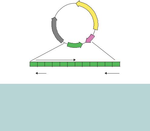
The ColE1 plasmid is replicated in a relaxed fashion using the DNA polymerase provided from the host cell. Unlike the bacterial origin of replication (OriC), however, the replication of ColE1 proceeds in one direction only. The plasmid does not encode proteins that initiate the replication process, but it does code for several non-translated RNA molecules that are involved in the initiation of replication and one protein that plays a role in regulating the RNA molecules. The ColE1 DNA replication origin is in a region of DNA from which two RNA molecules (RNAI and RNAII) are constitutively transcribed from their own promoters (Figure 3.2). RNAII is complementary to the ColE1 origin of replication and binds to it to form a DNA –RNA hybrid (Cesareni et al., 1991). The bound RNA II molecule is cleaved at the origin, by the host encoded enzyme RNase H, and serves as a primer from which DNA replication occurs. Through complementary base pairing, RNAII can bind on one DNA strand only, and hence the replication of the ColE1 plasmid is unidirectional.
|
|
|
cea |
|
|
|
ColE1 |
|
|
|
|
6646 bp |
|
|
|
imm |
|
|
|
|
|
ori |
rop |
|
|
RNAII |
|
|
|
−500 |
−300 |
−100 ori 100 |
300 |
500 bp |
RNAI |
|
|
|
rop |
Figure 3.2. The ColE1 plasmid and origin of replication. The ColE1 plasmid is a doublestranded closed-circular DNA molecule, 6646 bp in length. It codes for, amongst other things, the bacteriocin colicin E1 (the product of the cea gene) and an immunity protein (the product of the imm gene) that prevents the toxic effects of the bacteriocin in cells harbouring the plasmid. DNA replication occurs in one direction only, as indicated by the direction of the ori arrow. Two non-translated RNA molecules, termed RNAI and RNAII, and the protein product of the rop gene, control the replication process – see the text for details. The relative positions of these replication control elements to the origin (ori) are shown. The arrows on the genes indicate the direction of transcription
116 |
VECTORS 3 |
|
|
Control of the replication process is achieved by another non-translated RNA molecule. RNAI is complementary to the 5 -end of RNAII, and the RNA duplex formed between RNAII and RNAI cannot serve as a replication primer. RNAI is a relatively short-lived species, and consequently the ColE1 plasmid is maintained at a level of about 15 –20 copies per cell. The interaction between RNAI and RNAII is stabilized by the ROP protein (Helmer-Citterich et al., 1988).
The mechanism of DNA replication employed by ColE1 has several important consequences. The arrangement of regulators results in plasmid incompatibility – that is, two plasmids with the same origin of replication cannot coexist in the same cell. RNAI of the resident plasmid prevents RNAII of the incoming plasmid from forming a primer, so it is not replicated. This is important for cloning experiments when a mixed population of plasmids, which is often the result of a ligation reaction, is transformed into bacteria. Individual transformants produced in this way will contain a single plasmid and not a mixed population. Plasmids with different replication origins (e.g. ColE1 and p15A) are able to co-exist within the same cell. An additional consequence of the ColE1 replication mechanism is that fresh protein synthesis is not required to initiate DNA replication. In the presence of antibiotics that block protein synthesis (e.g. chloramphenicol), chromosomal DNA replication is halted, but ColE1 based plasmids continue to be replicated and accumulate at high levels (1000 –2000 copies per cell) (Clewell, 1972).
3.1.1pBR322
ColE1, and its very close relative pMB1, have the potential to be useful cloning vectors, but they suffer from a number of disadvantages. Primarily, the difficulty in the identification of recombinant plasmids (those in which the plasmid DNA has been ligated to an insert) meant that they did not gain widespread usage. What was required was a plasmid that could be replicated in the same way as ColE1, but in which recombinants could be easily recognized.
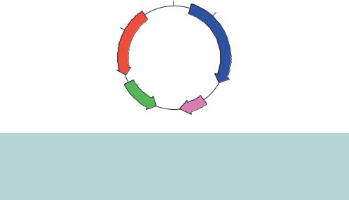
The plasmid pBR322 (Figure 3.3) was the first widely used plasmid vector. It is a small plasmid (4363 bp) that was constructed using components from naturally occurring plasmids and other DNA fragments (Bolivar et al., 1977). pBR322 contains the following components.
•Origin of replication. pBR322 carries the ColE1 replication origin and rop gene to ensure reasonably high plasmid copy number (15 –20 copies per cell), which can be increased 200-fold by chloramphenicol amplification.
3.1 PLASMIDS |
117 |
|
|
|
EcoRI |
|
BamHI |
PstI |
|
AMPR |
TETR |
|
pBR322 |
|
4361 bp |
ori
rop
Figure 3.3. The plasmid pBR322. This plasmid contains the ColE1 origin of replication (ori and rop), together with two antibiotic resistance genes. The ampicillin resistance gene (AMPR, or bla encoding β-lactamase) and the tetracycline resistance gene (TETR) each contain a number of restriction enzyme recognition sites that occur only once within the plasmid sequence
•Antibiotic resistance genes. pBR322 carries two genes that can be used as selectable markers. The ampicillin resistance gene (termed bla or, more commonly, AMPR) was cloned into the plasmid from the Tn3 transposon, and the tetracycline resistance gene (termed tet or TETR) was cloned from the plasmid pSC101 (Bernardi and Bernardi, 1984).
•Cloning sites. The plasmid carries a number of unique restriction enzyme recognition sites. Some of these are located in one or other of the antibiotic resistance genes. For example, sites for PstI, PvuI and SacI are found within AMPR, and sites for BamHI and HindIII are located within TETR.
The antibiotic resistance genes in pBR322 allow for the direct selection of recombinants in a process called insertional inactivation. For example, if we want to clone a DNA fragment into the BamHI site of pBR322, then the insert DNA will interrupt the gene responsible for tetracycline resistance, but the gene for ampicillin resistance will not be altered. Transformed cells are first grown on bacterial plates containing ampicillin to kill all the cells that do not contain a plasmid. Those cells that grow on ampicillin are then replica plated onto medium containing both ampicillin and tetracycline. Those cells that grow in the presence of the ampicillin, but die under tetracycline selection, contain plasmids that have foreign DNA inserts (Figure 3.4). In other words, the insertion of a foreign DNA fragment into an antibiotic resistance gene inactivates the gene product and leads to antibiotic sensitivity.