

новая папка / [libribook.com] 50 Cases in Clinical Cardiology_ A Problem Solving Approach 1st Edition
.PdfCase 1 Ventricular Septal Defect |
|
5 |
|
|
|
Figure 1.3: Various locations of ventricular septal defect (VSD)
RA: Right atrium; RV: Right ventricle
Table 1.1: Types of ventricular septal defect
• Perimembranous VSD • Subpulmonic VSD
• Supracristal VSD • Muscular VSD
A small VSD (Maladie de Roger) generates a loud pansystolic murmur in a localized area on the precordium. The murmur is located in the upper parasternal area in outlet VSD and in the mid-portion in perimembranous VSD. A muscular VSD produces a short systolic murmur since the defect shuts off during muscle contraction in later systole. This murmur is located over the lower parasternal area. A large VSD with elevated right ventricular pressure that equals left ventricular pressure (bidirectional shunt) is also associated with an early systolic murmur. Therefore, there is no correlation between the length or intensity of the murmur and the size of the VSD.
A large shunt may be accompanied by a diastolic flow murmur and a S3 sound, due to torrential flow across the mitral valve. The S2 is widely split due to early aortic valve closure. On ECHO, signal drop-out is not observed if the VSD is too small (<3 mm size) or muscular in location. The width of the colour flow map approximates the VSD size. On Doppler, high flow velocity indicates a small VSD. The flow velocity is low if the VSD is large and the shunt is bidirectional.
VSDisthecommonestformofcongenitalacyanoticheartdiseaseandaccounts for 25% of all cardiac malformations. VSD may occur in isolation or as part of a complex constellation of congenital cardiac abnormalities. Aortic regurgitation may be associated due to lack of support to the aortic valve in perimembranous

6 |
|
Section 1 Congenital Heart Diseases |
|
|
|
VSD. Complications of VSD in childhood are growth retardation and repeated chest infections. Reversal of shunt can occur later in life when pulmonary pressure exceeds the systemic pressure. Endocarditis can follow any non-cardiac surgical procedure.
MANAGEMENT ISSUES
Large sized VSDs allow large volumes of left-to-right shunt and usually present in childhood with failure to thrive, breathlessness and recurrent respiratory infections. They can lead to pulmonary hypertension, right heart failure and ultimately reversal of shunt (right-to-left). This is designated as the Eisenmenger’s syndrome. Such VSDs are usually closed in childhood to avoid complications and before the Eisenmenger’s syndrome has developed.
Medium sized VSDs are associated with a moderate sized shunt. The shunt is large enough to cause breathlessness, but not enough to cause pulmonary hypertension and shunt reversal. Such patients do reasonably well during childhood, but may become progressively symptomatic as left ventricular compliance declines with age and pulmonary venous congestion develops. Such VSDs are usually closed in adulthood, to avoid the development of heart failure.
Small sized VSDs do not cause significant shunting and are often asymptomatic. Some of them may close as the child grows older. Those that do not close spontaneously are closed by intervention for reasons other than the shunt. These reasons are development of endocarditis or associated significant aortic regurgitation (Table 1.2).
Table 1.2: Indications for surgical closure of VSD
• Large-sized VSD with volume overload
(pulmonary to systemic flow ratio >2:1)
• Medium-sized VSD with congestive symptoms without pulmonary hypertension
• Small-sized VSD without congestive symptoms with endocarditis or aortic regurgitation
RECENT ADVANCES
The last decade or two have witnessed remarkable progress in the percutaneous techniques for closure of ventricular septal defects, thus avoiding the risks associated with open heart surgery. Although transesophageal echocardiography (TEE)generallysufficestoguide thedeploymentofthe closuredevice,intracardiac ultrasound provides more accurate assessment. Sonography can provide vital information pertaining to the location and size of the defect and the rim around it, so as to facilitate proper device selection and placement.

|
|
C A S E |
|
|
|
|
|
|
|
|
|
||
|
|
|
|
|
||
|
|
2 |
|
Atrial |
|
|
|
|
|
|
|
|
|
|
|
|
|
Septal Defect |
|
|
|
|
|
|
|
|
|
|
|
|
|
|
|
|
Case Presentation
A 36-year old woman was referred to a physician by a gynecologist, for preoperative assessment prior to elective hysterectomy. The patient had multiple uterine fibroids on ultrasonography and complained of excessive bleeding during menstruation. For the past 6 months, she had been complaining of exertional dyspnea and fatigue, which were attributed to anemia as a result of blood loss. She denied complaints of chest pain, palpitations or dizziness. There was no history of cyanotic spells, joint pains or recurrent respiratory infections during her childhood. The patient was married, had 2 sons aged 11 and 9 years and she had never been hospitalized for any major illness or surgical procedure.
On examination there was mild anemia but no cyanosis, icterus or sign of congestive heart failure. The pulse was 90 beats/min. regular, with a BP of 136/80 mm Hg in the right arm. The apex beat was normal in location with a sustained left parasternal heave on palpation. The S1 was normal with a loud P2; no S3 or S4 sound was heard. The S2 components namely A2 and P2 were widely spaced and the time gap between them did not increase further during inspiration. A short systolic murmur was heard over the upper left sternal border. The murmur was not preceded by an ejection click or accompanied by a palpable thrill and did not radiate to the neck. The lung fields were clear on auscultation.
CLINICAL DISCUSSION
From the history and physical examination, this young woman had effort intolerance with an ejection murmur in the pulmonary area. Typical causes of such a murmur are innocent hemic murmur (Still’s murmur), pulmonary valve stenosis, pulmonary hypertension and atrial septal defect. The murmur of pulmonary stenosis may be preceded by an ejection click and accompanied by a palpable thrill. The P2 component of S2 is muffled and the splitting between A2 and P2 is wide, but widens further during inspiration. An innocent hemic murmur is not associated with a loud P2 or wide splitting of S2. Pulmonary hypertension of any etiology can produce a systolic murmur with loud P2 but wide fixed splitting of S2 is only a feature of atrial septal defect.
ECG of the patient showed sinus rhythm with incomplete right bundle branch block and a rightward QRS axis. X-ray chest showed enlarged right-sided

8 |
|
Section 1 Congenital Heart Diseases |
|
|
|
Figure 2.1: Color flow map extending from left atrium to right atrium
chambers with dilated main pulmonary artery, prominent hila and pulmonary plethora. On ECHO, the right atrium and right ventricle were dilated and a signal drop-out was noticed in the interatrial septum. On colour Doppler, an abnormal flow map was observed extending across the area of echo drop-out, from the left atrium to the right atrium (Fig. 2.1). There were no abnormalities of the cardiac valves and the estimated pulmonary artery pressure was normal. Therefore, the definite diagnosis in this case is atrial septal defect (ASD).
Figure 2.2: Atrial septal defect
In ASD, breach in the continuity of the interatrial septum creates a left-to- right shunt between the atria (Fig. 2.2). The septal defect occurs due to complexity of its embryological development. Most (75%) ASDs occur in the mid-portion of the septum, in the region of the foramen ovale and are termed as ostium secundum ASD. Some ASDs occur lower down the inter-atrial septum and are termed as ostium primum ASD (Fig. 2.3). Ostium primum ASDs are associated with cleft leaflets, regurgitation of the atrioventricular valves and are also known as endocardial cushion defect. An uncommon variety of ASD in the upper portion

Case 2 Atrial Septal Defect |
|
9 |
|
|
|
Figure 2.3: Various locations of atrial septal defect (ASD)
SVC: Superior vena cava; IVC: Inferior vena cava
Table 2.1: Types of atrial septal defect
• Ostium secundum ASD • Ostium primum ASD • Sinus venosus defect • Vena caval defect
is sinus venosus defect, which is accompanied by anomalous pulmonary venous connections (Table 2.1). Inferior vena caval defects are very rare. An ASD may be associated with trisomy 21 (Down’s syndrome) or abnormalities of the hand (Holt Oram syndrome).
The systolic murmur of ASD is due to increased flow across the pulmonary valve and not due to the shunt. The intensity of murmur does not correlate with the size of the ASD. However, a large ASD is associated with a diastolic flow murmur and a right-sided S3, due to torrential flow across the tricuspid valve. An accompanying pansystolic murmur due to mitral and/or tricuspid regurgitation is a feature of ostium primum ASD. In ASD, the splitting of S2 is wide and fixed. It is wide because of increased pulmonary ejection time, which delays the P2.
Other reasons for wide splitting of S2 are right bundle branch block or pulmonary stenosis (delayed P2) and mitral regurgitation or ventricular septal defect (premature A2). The splitting of S2 is also wide in WPW syndrome Type A, in which there is pre-excitation of the left ventricle. The splitting of S2 is fixed in ASD because the shunt equalizes atrial pressures throughout the respiratory cycle and there is no inspiratory augmentation of right ventricular filling.
On ECHO, since the signal from the interatrial septum is weak, false echo drop-out may be seen even in normal persons. The subcostal window may be a better option to diagnose an ASD but transesophageal echocardiography (TEE) provides excellent visualization particularly in endocardial cushion defects and sinus venosus ASD. Sometimes, contrast echo is needed to visualize the shunt using agitated saline, which contains air bubbles that cross over the septal defect.
ASD is the commonest congenital heart disease diagnosed in adulthood, with either absent or mild symptoms. It is 7 times more common in females than

10 |
|
Section 1 Congenital Heart Diseases |
|
|
|
in males. Complications of ASD in adults are effort intolerance and pulmonary hypertension. Reversal of shunt and right heart failure are rare compared to ventricular septal defect. Atrial tachyarrhythmias including atrial fibrillation are common. Typically, sinus arrhythmia is never observed, because the shunt negates the effect of inspiration on venous return. Systemic thrombo-embolism can occur due to emboli from peripheral or pelvic veins, passing across the septal defect (paradoxical embolism).
MANAGEMENT ISSUES
Most ostium secundum atrial septal defects are amenable to percutaneous device closure. Large ASDs allow large volumes of left-to-right shunt and usually persent with exertional breathlessness and fatigue. They can lead to pulmonary hypertension and right heart failure, although reversal of shunt (right-to-left) is less common than in case of ventricular septal defect. Therefore, ASDs larger than 10 mm in size should ideally be closed before significant pulmonary hypertension develops.
Smaller ASDs with small volumes of shunt may become progressively more symptomatic as left ventricular compliance declines with age and the degree of shunting increases. Such ASDs that lead to right ventricular dilatation, should be closed during adulthood. Patients with ASD may develop paradoxical emboli which arise in the venous system and cross the septal defect to reach the systemic circulation. ASDs with history of thrombo-embolism should be closed, irrespective of their size (Table 2.2).
Table 2.2: Indications for surgical closure of ASD
• |
Large-sized ASD more than 10 mm in size |
|
with pulmonary to systemic flow ratio > 1.5:1 |
• |
Medium-sized ASD with dilated right ventricle |
|
without significant pulmonary hypertension |
• |
Small-sized ASD without dilated right ventricle |
|
with history of systemic thrombo-embolism |
Ostium primum atrial septal defects with atrioventricular valvular abnormalities and sinus venosus defects with anomalous pulmonary venous drainage are not amenable to percutaneous device closure because of their complexity. They require a definitive surgical procedure for their correction.
RECENT ADVANCES
Percutaneous deployment of a closure device for atrial septal defect has been standard practice for several decades. Transesophageal echocardiography (TEE) is widely used to guide the deployment. Recently, intracardiac ultrasound has been used to accurately assess the anatomy, for better selection and placement of the device. Vital anatomical information includes size of the defect, the rim around the defect and proximity to the mitral and tricuspid valves.

|
|
C A S E |
|
|
|
|
|
|
|
|
|
||
|
|
|
|
|
||
|
|
3 |
|
Fallot’s |
|
|
|
|
|
|
|
|
|
|
|
|
|
Tetralogy |
|
|
|
|
|
|
|
|
|
|
|
|
|
|
|
|
CASE PRESENTATION
A 14-year old girl was admitted to a tertiary-care hospital of a metropolitan city, with the complaint of progressive shortness of breath since 2 months, more so for the last 5 days. There was no history of fever, productive cough, chest pain or hemoptysis. The girl’s mother also noticed an increase in the child’s abdominal girth and swelling around both her ankles. The patient was born after a Caesarian section and was noticed to be cyanosed at birth. At the age of 3 years, the girl had undergone a surgical operation for congenital heart disease, in this very hospital. There was no history of repeated chest infection or childhood asthma, but the patient’s growth milestones of early childhood were delayed.
On examination, the patient was slightly breathless at rest but was not distressed. The pulse rate was 84 beats/min., with a BP of 96/66 mm Hg. There was cyanosis over the tongue and lips and the finger-tips and toes were clubbed. Pitting ankle edema was present and the neck veins were engorged. Per abdomen findings were a 5 cm hepatomegaly with mild ascites. The breath sounds were vesicular in character without any rhonchi or crepts. On precordial examination, the apex beat was normal in location with a left parasternal heave. Auscultation revealed a normal S1 and S2 with an early-diastolic murmur over the pulmonary area and a soft pansystolic murmur over the lower left parasternal area.
CLINICAL DISCUSSION
From the history and physical examination, this young girl had congenital cyanotic heart disease that was operated upon during her early childhood. At present she was in right heart failure with pulmonary and tricuspid valve regurgitation and had right ventricular enlargement. Right ventricular enlargement is associated with a palpable left parasternal heave. Common causes of right ventricular enlargement are pulmonary valve stenosis and pulmonary arterial hypertension. The commonest cause of pulmonary regurgitation is pulmonary hypertension, but it can also follow a surgical procedure on the pulmonary valve. Rare causes of pulmonary regurgitation are subvalvular pulmonary stenosis and carcinoid syndrome (Table 3.1). Functional tricuspid regurgitation (dilated annulus) is a consequence of right ventricular dilatation due to any cause. Tricuspid regurgitation is associated with raised JVP with prominent v waves and rapid
12 |
|
Section 1 Congenital Heart Diseases |
|
|
|
Table 3.1: Causes of pulmonary regurgitation
• Primary: Carcinoid syndrome
• Congenital: Subvalvular stenosis • Iatrogenic: Pulmonary valvotomy
• Secondary: Pulmonary hypertension
y descent. An enlarged pulsatile liver with a pansystolic murmur over the lower left parasternal area are also observed.
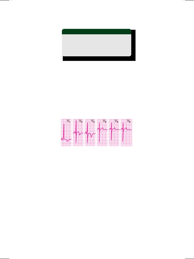
ECG showed sinus rhythm with tall R waves in right precordial leads and T wave inversion, suggestive of right ventricular hypertrophy with strain (Fig. 3.1).
P. pulmonale and right axis deviation of the QRS were also seen. X-ray chest findings were increased cardio-thoracic ratio with reduced pulmonary vascular markings and a right-sided aortic arch. On ECHO, the left ventricle was normal in size with an ejection fraction of 55% but the right ventricle was significantly dilated. The mitral and aortic valves were normal in structure but there was moderate pulmonary regurgitation and significant tricuspid regurgitation.
Figure 3.1: ECG showing tall R waves in leads V1 to V3.
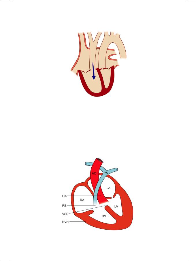
This patient was operated upon for a cardiac defect, when she was 3 years old. The most common congenital cyanotic heart disease that is associated with survival until adolescence and sometimes even into adulthood is tetralogy of Fallot. Therefore, in all probability, this patient was operated upon for Fallot’s tetralogy and had now developed pulmonary regurgitation (Fig. 3.2), as a complication of the surgical procedure on the pulmonary valve. The right ventricular enlargement that ensued, led to secondary tricuspid regurgitation because of annular dilatation.
Tetralogy of Fallot is the most common congenital cyanotic heart disease that survives into adolescence. It is associated with a right-to-left shunt since birth unlike isolated septal defects which are left-to-right shunts and undergo reversal only after the development of pulmonary hypertension. The four components (Fig. 3.3) of Fallot’s tetralogy are:
•Pulmonary stenosis (PS)
•Overriding of aorta (OA)
•Ventricular septal defect (VSD)
•Right ventricular hypertrophy (RVH)
Case 3 Fallot’s Tetralogy |
|
13 |
|
|
|
Figure 3.2: Pulmonary regurgitation
The primary developmental abnormality is of the pulmonary subvalvular or infundibulararealeadingtopulmonarystenosis(PS)andrightventricularoutflow tract (RVOT) obstruction. Rarely, the pulmonary valve is absent (pulmonary atresia). The ventricular septal defect (VSD) is membranous in location. The aortaisdisplacedrightwardandoverridestheseptum,theoverridingaorta(OA).
Therefore, the septum is not in line with the anterior aortic wall but with the aortic valve closure point. The right ventricular hypertrophy (RVH) is secondary to RVOT obstruction. Rarely, an atrial septal defect (ASD) may be associated, in which case the constellation is designated as pentalogy of Fallot.
Figure 3.3: The four components of Fallot’s tetralogy. AO: Aorta; OA: Overriding aorta; RA: Right atrium; LA: Left atrium; RV: Right ventricle; LV: Left ventricle PS: Pulmonary stenosis; PA: Pulmonary artery; VSD: Ventricular septal defect; RVH: Right ventricular hypertrophy

14 |
|
Section 1 Congenital Heart Diseases |
|
|
|
In a typical unrepaired case of Fallot’s tetralogy, auscultatory findings are a loud, single S2 and a parasternal systolic murmur. The S2 is single because the P2 is muffled and the A2 is loud because the aorta is anteriorly placed. The systolic murmur originates from the subvalvular pulmonary stenosis and not from the ventricular septal defect. The classical clinical features of Fallot’s tetralogy are central cyanosis, finger clubbing, anoxic spells, growth retardation and exercise intolerance. Congestive heart failure is rare because the septal defect balances the right and left ventricular pressures. If left unrepaired, catastrophic complications in adolescence are arterial thrombo-embolism and cerebral abscess.
MANAGEMENT ISSUES
Until the 1970s and even early-1980s, surgical interventions in very early childhood were largely palliative. These shunt procedures were performed to bypass the RVOT obstruction and to enhance pulmonary blood flow. These shunts were Blalock-Taussig shunt (subclavian artery to pulmonary artery) and
Waterstonshunt(ascendingaortatorightpulmonaryartery).However,evenafter these procedures, patients remained symptomatic and complications occurred unabated. Nowadays, total surgical correction is undertaken to close the shunt and to enhance pulmonary blood flow. This includes patch closure of the VSD with pulmonary subvalvular muscle resection and valvotomy.
Cardiac surgeons are increasingly encountering complications of prior surgical correction, as these children survive into their teens. Complications after surgery include residual shunt, residual stenosis or, post-valvotomy pulmonary regurgitation and right ventricular enlargement as in our case. Pulmonary valve replacement with tricuspid annular repair would be the best course of action in this case.
RECENT ADVANCES
Prior cardiac surgery often distorts the anatomy of the heart to an extent that the information obtained from transthoracic echocardiography is generally skewed and inconclusive. Modern cardiac imaging techniques of computed tomography (CT) and magnetic resonance imaging (MRI) are particularly useful to evaluate postoperative patients.
Percutaneous techniques are currently being evaluated in the management of Fallot’s tetralogy. Pulmonary balloon angioplasty and artificial valve deployment by non-surgical intervention have been recently described.