

новая папка / [libribook.com] 50 Cases in Clinical Cardiology_ A Problem Solving Approach 1st Edition
.Pdf
|
|
|
|
|
Case 27 Rheumatic Fever |
|
125 |
|
|
|
|
|
|
|
|
|
|
|
|
|
|
|
|
|
|
|
|
|
Table 27.1: Jone’s criteria for the diagnosis of rheumatic fever |
|
|
|
|||
|
Major criteria |
Minor criteria |
Streptococcal infection |
|
|
|
||
|
• |
Carditis |
• |
Fever |
• History of scarlet fever |
|
|
|
|
• |
Arthritis |
• |
Arthralgia |
• Throat swab culture + ve |
|
|
|
|
• |
Chorea |
• |
High ESR |
• Streptococcal antigen + ve |
|
|
|
|
• |
Eryth. marginatum |
• |
Raised CRP |
• High anti-Streptolysin O titre |
|
|
|
|
• |
Subcut. nodules |
• |
P-R interval |
|
|
|
|
|
|
|
|
|
|
|
|
|
|
|
|
|
|
|
|
|
|
A migratory or “fleeting” type of polyarthritis with fever and extreme weakness is the commonest manifestation of rheumatic fever. The arthritis typically involves the medium-sized joints such as the elbows, ankles and wrists. Chorea is usually observed in young children as an isolated entity, with rheumatic heart disease occuring a few days later. Erythema marginatum, which is hardly seen, is a pink rash on the trunk which blanches on pressure and is neither painful nor indurated. Rheumatic carditis is a pancarditis. Endocarditis manifests as a new murmur due to inflamed valve leaflets. Myocarditis presents as newly developed myocardial dysfunction. Evidence of pericarditis is a pericardial friction rub or presence of an effusion.
The major sequel of acute rheumatic fever is chronic valvular heart disease. Antibodies against streptococcal polysaccharide (carbohydrate cell wall) crossreact with cardiac tissues (myosin and laminin) to cause valve damage by molecular mimicry. During the acute stage, the mitral valve leaflets are inflamed resulting in a diastolic Carey-Coomb’s murmur. Later on, due to smouldering disease, the valves are chronically damaged. Mitral stenosis, aortic regurgitation, mitral regurgitation and aortic stenosis, follow in that order of decreasing incidence.
MANAGEMENT ISSUES
The two pillars in the management of acute rheumatic fever are aspirin and penicillin. Aspirin can be given in a dose of 325 mg 4 times a day, during the stage of acute inflammation. The dose is gradually brought down, as the fever and pain subside. Care should be taken to co-prescribe a proton pump inhibitor like pantoprazole and to advise to take aspirin soon after a meal. If the patient is sensitive to aspirin, an alternative agent like ibuprofen or diclofenac may be used. Crystalline penicillin is administered in full doses parenterally, during the stage of acute inflammation. If the patient is hypersensitive to penicillin, a macrolide antibiotic such as erythromycin or azithromycin is chosen and the dose is 500 mg 6 hourly or 300 mg daily, respectively. Once the acute flare has settled down, long-term prophylaxis is suggested, particular to women, until they attain the age of 40 years. Antibiotic prophylaxis against infective endocarditis before a dental or surgical procedure, depends upon the nature and degree of chronic valvular involvement.

126 |
|
Section 8 Myocardial Infections |
|
|
|
RECENT ADVANCES
As per the revised Jone’s criteria for rheumatic activity in developing countries, erythema marginatum has been dropped as it is hardly ever seen. Polyarthralgia with raised ESR and high ASLO titre is no less common than objective arthritis and has been included as a major criteria.

|
|
C A S E |
|
|
|
|
|
|
|
|
|
|
|
|
|
|
|
|
|
28 |
Acute |
|
|
|
|
|
|
|
|
|
|
|
Myocarditis |
|
|
|
|
|
|
|
|
CASE PRESENTATION
A 32-year old man presented to the emergency department with worsening breathlessness, over the preceding 3 days. Since the previous night, he was finding it difficult to lie flat in bed and had to sit up frequently to breathe. The patient was quite well until 5 days prior to his hospital visit, when he developed a“flu-like”illness. The low grade fever, headache, myalgia and mild sore throat had now settled down. There was no history of productive cough, chest pain or hemoptysis. There was also no history of skin rash, petechial spots or painful joints. He had not undergone any recent dental or endoscopic procedure. He did not suffer from recurrent pharyngotonsillitis during his childhood.
On examination, the patient was ill-looking and tachypneic. Pulse was fast and feeble and his extremities were sweaty. His heart rate was 116 beats/min with a BP of 104/68 mm Hg and the temperature was 98.80 F. There were no petechial hemorrhages under the finger-nails or in the conjunctiva and his joints were not swollen. The apex beat was displaced outwards and the S1 and S2 were normal. A soft S3 was audible in early diastole, but there was no murmur. No pericardial friction rub was heard. There were few basilar rales over the lung fields.
CLINICAL DISCUSSION
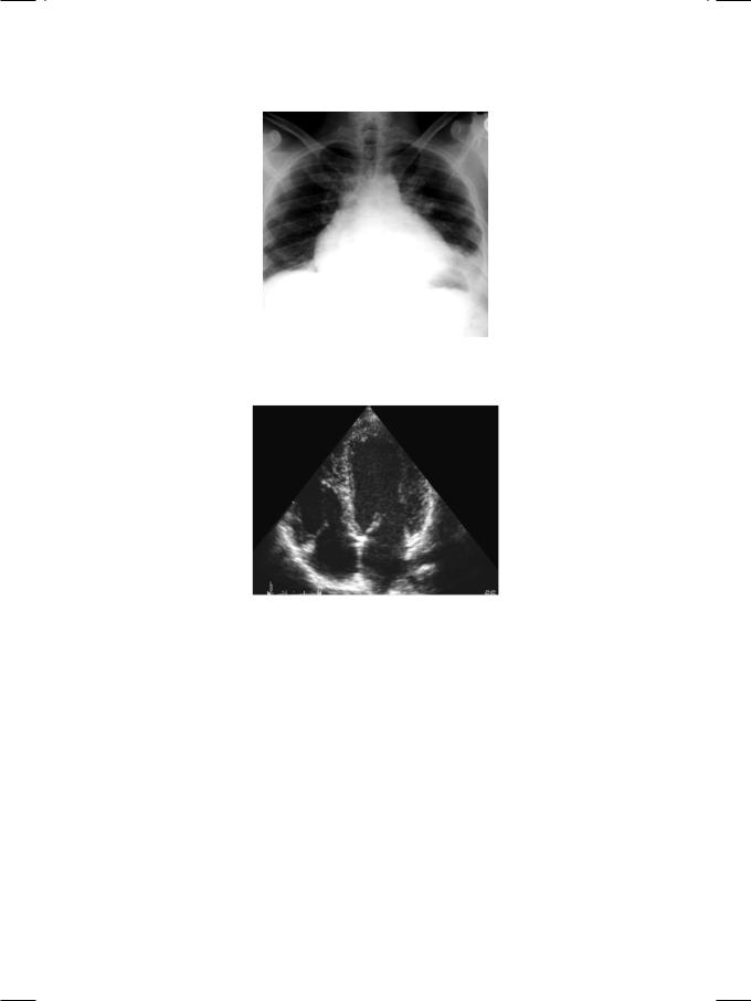
From the history and physical examination, it is obvious that the patient had a short febrile illness, culminating in left ventricular dysfunction within a week. ECG showed sinus tachycardia, low QRS amplitude and non-specific T wave inversion. X-ray chest findings were enlarged cardiac silhouette, hilar congestion (Fig. 28.1), Kerley B lines and pulmonary edema. ECHO revealed a dilated left ventricle with global hypokinesia (Fig. 28.2). There was no abnormality of the cardiac valves, septal defect or presence of pericardial effusion.
When a patient presents with a febrile illness and ECG abnormalities, there are four main diagnostic possibilities. These are:
•Rheumatic fever
•Acute pericarditis
•Acute myocarditis
•Infective endocarditis.
128 |
|
Section 8 Myocardial Infections |
|
|
|
Figure 28.1: X-ray showing cardiomegaly with left pleural effusion
Figure 28.2: ECHO showing an enlarged globular left ventricle
Acute pericarditis is unlikely because the patient had no chest pain or S-T segment elevation on the ECG or pericardial friction rub on clinical examination. Moreover, left ventricular dysfunction is unusual in pericarditis, unless there is an associated component of myocarditis. Infective endocarditis is also unlikely because the ECHO did not show any abnormality of the cardiac valves or presence of a septal defect. Rheumatic fever is a pancarditis where myocardial dysfunction is accompanied by pericarditis (friction rub) and endocarditis (inflammed valve leaflets). Moreover, rheumatic fever generally presents with a migratory polyarthritis with or without cutaneous nodules and an evanescent skin rash. Therefore, the most probable diagnosis in this case is acute myocarditis.
Acute myocarditis is an inflammatory disease afflicting the heart muscle and causing myocardial dysfunction over a short period of time. Although global hypokinesia is the hallmark of myocarditis, regional wall motion abnormalities can occur, if myocardial inflammation is patchy in distribution. The myocardial damage and necrosis is immune-mediated and the cardiac troponin levels are often elevated. The etiology is most often viral and Coxsackie B virus or Echovirus

Case 28 Acute Myocarditis |
|
129 |
|
|
|
Table 28.1: Pathogens implicated in acute myocarditis
• Viral |
Coxsackie B, Echovirus, HIV |
|
|
• Bacterial |
C. diphtheriae, Mycoplasma |
|
|
• Spirochetal |
Lyme’s disease, Leptospirosis |
|
|
• Protozoal |
Chaga’s disease |
|
|
are commonly implicated. Myocarditis may be bacterial as in Mycoplasma infection and Diphtheria. Rarely, it may be caused by a spirochete as in Leptospirosis or protozoa as in Chaga’s disease (Table 28.1).
PERTINENT INVESTIGATIONS
Thepatientofacutemyocarditismustbeinvestigatedtoestablishtheinflammatory nature of the disease and the type of inflammation. The total leucocyte count (TLC), erythrocyte sedimentation rate (ESR) and C-reactive protein (CRP) helps to establish inflammation. Anti-streptolysin O (ASLO) titre and throat swab culture for beta-hemolytic Streptococcus are indicated, if rheumatic fever is suspected. Suitable blood cultures are obtained, if the possibility of infective endocarditis is being entertained.
Viral serology for the diagnosis of viral myocarditis is generally unhelpful, because the implicated viruses are ubiquitous in nature and antiviral drugs have no role in the treatment. Endomyocardial biopsy is rarely performed because of its low diagnostic yield, except in situations where a specific etiology is suspected on clinical grounds and needs to be confirmed on tissue analysis.
These conditions are Loeffler’s endocarditis, hemochromatosis and amyloidosis.
Besides inflammation, electrolyte imbalance and nutritional deficiency are sometimes responsible for acute left ventricular dysfunction. These need to be excluded by appropriate tests such as serum potassium, calcium and magnesium levels. Thyroid hormone assay and urinary catecholamine levels are measured if thyrotoxicosis or pheochromocytoma are suspected.
MANAGEMENT ISSUES
The clinical features of myocarditis are similar to those of dilated cardiomyopathy, except that the onset of symptoms and signs is rapid. Therefore, the management of acute myocarditis is similar to that of dilated cardiomyopathy and follows the principles of standard heart failure treatment. The clinical manifestations not only evolve rapidly but also resolve within a short period of time. Loop diuretics are the mainstay of therapy to relieve pulmonary edema. An angiotensin converting enzyme(ACE)inhibitororangiotensinreceptorblocker(ARB)isaddedtoreduce cardiac afterload and to improve ventricular performance. An aldosterone antagonist (spironolactone or eplerenone) may be added if symptoms persist on the above treatment. Digoxin has no role in the acute setting since atrial fibrillation is unusual. Antiviral drugs and immunomodulatory agents do not improve clinical outcomes.

130 |
|
Section 8 Myocardial Infections |
|
|
|
RECENT ADVANCES
The role of auto-immunity in the pathophysiology of myocarditis and subsequent onset of dilated cardiomyopathy is being extensively investigated. However, so far neither anti-inflammatory drugs nor immunomodulatory therapy have been found to be successful. Auto-antibodies against beta-adrenergic receptors may also be relevant and patients who test positive for autoantibodies may respond more favourably to beta-blocker treatment.

S E C T I O N
9
Endocardial
Infections


C A S E
29 Aortic Valve
Endocarditis
CASE PRESENTATION
A 64-year old gentleman presented to the hospital emergency service with fever for the past 3 days. The fever was high grade and associated with chills but there were no rigors. The patient was discharged 2 weeks back from this very hospital, having undergone an aortic valve replacement with a St. Jude’s prosthesis for severe valve stenosis. Besides fever, he complained of headache, malaise, arthralgias and anorexia. He had not felt pain or noticed any redness at the sternal wound or at the site of intravenous cannula insertion. There was no history of productive cough, burning micturition, vomiting, pain abdomen or loose stools. He did not experience any significant chest pain or breathlessness. The patient denied having undergone any dental extraction or endoscopic procedure, after his surgery. He admitted having been noncompliant with the medication prescribed to him, at the time of discharge from the hospital.
On examination, he was ill-looking, toxic, febrile (101.80 F) and mildly tachypneic. Pulse was 110 beats/min with a BP of 130/60 mm Hg and his extremities were warm and sweaty. There was no sign of inflammation at the sternotomy wound or the site of intravenous line. The S1 was loud, S2 was sharp but no S3 or S4 was heard. There was an ejection systolic murmur along the left sternal border, but no pericardial friction rub was audible. Breath sounds were normal without any rhonchi or crepts. The abdomen was soft without hepato-splenomegaly or ascites. Additionally, there were petechiae under the finger nails (splinter hemorrhages) and sub-conjunctival spots (Roth spots) as well as tender nodules on the finger tips (Osler nodes).
The hemoglobin was 10.8 g/dL with a leucocyte count of 14,800/cu.mm; 86% were neutrophils and the ESR was 42 mm in 1st hour. Urine analysis showed albumin +1 with few RBCs but no WBC. ASLO titre was 180 IU with a CRP value of 48 mg/L. The biochemical parameters were Glucose 88 mg/dl, Creatinine 1.1 mg/dl, Bilirubin 2.2 mg/dl SGOT 24, SGPT 30 and Cholesterol 178 mg/dl. Urine culture did not yield any bacterial growth and throat swab culture was negative for beta-haemolytic Streptococcus. No pus was obtainable from the sternotomy wound for culture. Three sets of blood cultures were obtained, using aseptic skin precautions and all of them grew Staphylococcus epidermidis, after 72 hours of incubation.
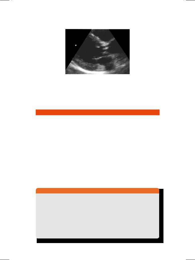
ECG showed sinus tachycardia with few ventricular premature beats but there was no conduction block. The X-ray chest did not show any cardiomegaly, parenchymal lung lesion (pneumonitis) or mediastinal widening (mediastinitis). ECHO revealed normal left ventricular size and ejection fraction. There were irregular echo-reflective masses attached to the aortic valve leaflets with mild aortic regurgitation (Fig. 29.1).
134 |
|
Section 9 Endocardial Infections |
|
|
|
Figure 29.1: ECHO showing nodular masses attached to aortic valve leaflets
Keeping in mind the history of a febrile illness with a prosthetic heart valve, positive blood cultures and demonstrable vegetations with aortic regurgitation, the most likely diagnosis in this case is aortic valve endocarditis.
CLINICAL DISCUSSION
Infective endocarditis may be caused by typical organisms, atypical bacteria and sometimes by fungi (Table 29.1). Non-infective endocarditis is observed in malignant disease (marantic), collagen disorder (Libman-Sacks) and rheumatic fever (pancarditis). High-risk lesions for endocarditis are regurgitant left-side valves (MR, AR), intracardiac shunts (VSD, PDA) and a prosthetic valve. Moderaterisk lesions are stenotic valves (MS, AS), MVP with MR and right-sided valves (TR and PS). Low-risk lesions are ASD, HOCM and MVP without MR. Modified Duke’s criteria are popularly used for the diagnosis of endocarditis (Table 29.2). Fever is associated with various vascular and immunological phenomena. Blood cultures may be negative due to prior antibiotic therapy, fastidious or atypical pathogen and in case of fungal endocarditis. ECHO is used to detect vegetations, diagnose the underlying lesion, to look for complications and to evaluate the response to therapy. Transesophageal echocardiography (TEE) is a better approach for detecting complications, such as prosthetic valve dehiscence or aortic root abscess and for the timing of surgical intervention.
Table 29.1: Pathogens implicated in infective endocarditis
BACTERIA |
• |
Typical |
Staphylococcus, Streptococcus |
|
|
|
|
|
|
|
HACEK * group bacteria |
|
|
|
|
|
• |
Fastidious |
Legionella, Brucella |
|
|
|
|
|
|
|
Neisseria, Nocardia |
|
|
|
|
|
• |
Intracellular |
Chlamydia, Coxiella |
|
|
|
|
|
|
|
Mycoplasma, Bartonella |
|
|
|
|
FUNGI |
• |
Typical |
Candida, Aspergillus |
|
|
|
|
*HACEK group: Haemophilus, Actinobacillus, Cardiobacterium, Eikenella, Kingella