новая папка / [libribook.com] 50 Cases in Clinical Cardiology_ A Problem Solving Approach 1st Edition
.Pdf
|
|
C A S E |
|
|
|
|
|
|
|
|
|
||
|
|
|
|
|
||
|
|
4 |
|
Ebstein’s |
|
|
|
|
|
|
|
|
|
|
|
|
|
Anomaly |
|
|
|
|
|
|
|
|
|
|
|
|
|
|
|
|
CASE PRESENTATION
A 26-year old married woman visited a cardiologist’s chamber, with the complaint of occasional fluttering sensation in the chest of 2 years duration. The episodes of palpitation were associated with some light-headedness, but she had never fainted. Her palpitation was at times related to some emotional upset or undue physical exercise, but there was no history of exertional fatigue, chest pain or breathlessness. There was also no history of tremor of the hands or weight loss. Her childhood had been uneventful with normal growth milestones and there was no history of cyanotic spells during sports activities. She also denied having had recurrent sorethroat, joint pains or any prolonged febrile illness during her school days.
On examination, the patient was comfortable, relaxed and not dyspneic. There was no tremor of the fingers, visible goiter or eye-signs of Grave’s disease. The JVP was raised 5 cm above the angle of Louis and showed large v waves with a prominent y descent. The pulse was regular, fair in volume, at a rate of 84 beats/min. and the BP was 130/80 mm Hg. On examining the abdomen, there were visible epigastric pulsations, with the liver edge 6 cm below the right costal margin and pulsatile; no ascites was demonstrable. The apex beat was normal in nature and location but a left parasternal heave was palpated. The S1 and S2 were both split with wide splitting of S2 appreciated during inspiration. No S3 or S4 gallop sound was heard. A pansystolic murmur was audible at the lower end of the left sternal edge. Breathing was vesicular and no rhonchi or crepts were heard over the lung fields.
CLINICAL DISCUSSION
From the history and physical examination, this patient had paroxysmal tachycardia with clinical signs of tricuspid valve regurgitation. ECG showed tall P waves (P. pulmonale) with normal P-R interval and right bundle branch block (RBBB). X-ray chest finding was an enlarged cardiac silhouette, more so towards the right of the midline. ECHO revealed normal sized left ventricle with normal ejection fraction. The mitral and aortic valves were normal and the left atrium was not dilated. There was no echo drop-out in the region of either septum. However, the right atrium was markedly enlarged and the right ventricle was dilated as well as hyperkinetic. The tricuspid valve was displaced downwards into the right ventricle, with distal attachment of the septal tricuspid leaflet which showed
16 |
|
Section 1 Congenital Heart Diseases |
|
|
|
exaggerated excursion. On colour flow mapping, a regurgitant jet was seen in the right atrium. These findings are consistent with the diagnosis of Ebstein’s anomaly.
In a young woman, history of episodic palpitation raises several clinical possibilities. Anxiety neurosis, panic attacks and paroxysmal supraventricular tachycardia are usual causes but in these, the heart is structurally normal. Perimenopausal symptoms in women include palpitation but our patient was young. Thyrotoxicosis is a possibility but our patient had no goiter, tremor or eye-signs of Grave’s disease. Pre-excitation syndrome (WPW syndrome) may be responsible for paroxysmal tachyarrhythmia but the ECG did not show short P-R interval or delta waves on the QRS complex. Mitral valve prolapse (MVP) and atrial septal defect (ASD) are structural cardiac abnormalities that are responsible for tachyarrhythmias. However, in our case the mitral valve was normal and there was no septal defect.
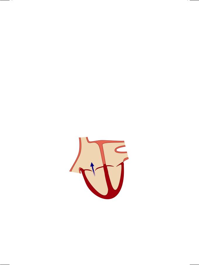
Ebstein’s anomaly is an uncommon congenital acyanotic heart disease characterized by abnormal tricuspid valve architecture, tricuspid regurgitation and association with paroxysmal supraventricular tachyarrhythmias. The physical examination of the patient and interpretation of simple cardiac investigations is a good exercise in bed-side clinical cardiology. A raised JVP with large v waves and prominent y descent are characteristic of tricuspid regurgitation into the right atrium (Fig. 4.1). So is an enlarged and pulsatile liver on abdominal examination. A sustained left parasternal heave is indicative of right ventricular volume overload.
Figure 4.1: Tricuspid regurgitation
The S1 is split because of delayed tricuspid valve closure (T1) due to right bundle branch block (RBBB) as well as the wide excursion of the septal tricuspid leaflet. The S2 is widely split due to delayed pulmonary closure (P2) because of the RBBB. Sometimes, the S2 is single because of soft P2 due to low pulmonary ejection volume. Rarely, the S2 is paradoxically split because of pre-excitation of the right ventricle caused by WPW syndrome Type B. The pansystolic murmur of tricuspid regurgitation is best audible over the lower left parasternal area and does not radiate towards the axilla or the base of the heart. Like all right-sided murmurs, it

Case 4 Ebstein’s Anomaly |
|
17 |
|
|
|
Figure 4.2: X-ray showing cardiomegaly due to enlargement of right atrium
increases in intensity during inspiration, provided the right ventricular function is normal.
In sinus rhythm, the ECG shows tall P waves (P. pulmonale) due to right atrial enlargement and wide QRS complexes due to right bundle branch block (RBBB). Sometimes, wide QRS complexes are due to WPW syndrome, in which case there is a short P-R interval (pre-excitation). At times, the rhythm is atrial fibrillation. On X-ray chest, the cardiac silhouette is enlarged towards the right of the midline, due to the large right atrium (Fig. 4.2). Superficially, this resembles a pericardial effusion. The differentiating feature is that the right lower portion of the cardiac silhouette curves inwards towards the center of the chest and not outwards, as it would in case of pericardial effusion.
On ECHO apical view, there is downward displacement of the tricuspid valve into the body of the right ventricle, towards the apex. The septal tricuspid
Figure 4.3: ECHO showing enlarged right atrium with displaced tricuspid valve

18 |
|
Section 1 Congenital Heart Diseases |
|
|
|
leaflet is attached to the IV septum, 10 mm or more distal to the anterior mitral leaflet. The tricuspid leaflet is large and shows wide excursion, often with a whip-like motion. The right ventricle is dilated and hyperkinetic due to volume overload. The right atrium is enlarged because of tricuspid regurgitation as well as due to “atrialization” of the upper portion of the right ventricle (Fig. 4.3). On long-axis view, because of downward displacement of the tricuspid valve, there is simultaneous recording of the mitral and tricuspid valves (MV and TV). On short-axis view, the tricuspid valve is shifted clockwise, from the normal 9 0’clock position to the 11 0’clock position.
The commonest reason for tricuspid regurgitation is dilatation of the tricuspid valve annulus secondary to right ventricular dilatation. Reason for annular dilatation is usually pulmonary hypertension due to congenital left-to-right shunt, rheumatic mitral valve disease or chronic cor pulmonale. Sometimes, dilated cardiomyopathy causes annular dilatation (Table 4.1). Primary tricuspid valvular regurgitation has several causes except coronary artery disease and systemic hypertension, the commonest forms of heart disease. Usual causes of tricuspid valve regurgitation are tricuspid leaflet prolapse, Ebstein’s anomaly, rheumatic heart disease, carcinoid syndrome and right-sided endocarditis. Uncommon causes include endocardial cushion defects, endomyocardial fibrosis and connective tissue disorders.
Table 4.1: Causes of tricuspid regurgitation
Valvular regurgitation
|
• |
Tricuspid valve prolapse |
|
• |
Right-sided endocarditis |
|
• |
Ebstein’s anomaly |
|
• |
Carcinoid syndrome |
|
• |
Endocardial cushion defect |
|
• |
Endomyocardial fibrosis |
|
• |
Connective tissue disease |
|
||
Annular dilatation |
||
|
• |
Pulmonary hypertension |
|
• |
Pulmonary regurgitation |
|
• |
Dilated cardiomyopathy |
MANAGEMENT ISSUES
The management of Ebstein’s anomaly includes the treatment of supraventricular tachyarrhythmias and the control of tricuspid regurgitation. Drugs that block the atrioventricular (AV) node to reduce the heart rate, such as betablockers and verapamil,aretheagentsofchoice.InthepresenceofWPWsyndrome,amiodarone is preferable. Digoxin may be used to treat atrial fibrillation in which case, an anticoagulant is also prescribed to reduce the risk of thrombo-embolism. If the tachyarrhythmias are refractory to drug treatment or an accessory bypass tract is present, radiofrequency ablation can be offered. In the presence of moderate to severe tricuspid regurgitation, valve repair with restrictive annuloplasty or even valve replacement may be considered.

|
|
C A S E |
|
|
|
|
|
|
|
|
|
||
|
|
|
|
|
||
|
|
5 |
|
Patent Ductus |
|
|
|
|
|
|
|
|
|
|
|
|
|
Arteriosus |
|
|
|
|
|
|
|
|
|
|
|
|
|
|
|
|
CASE PRESENTATION
A 6-year old boy was taken to the pediatrician by his mother, for treatment of fever with coryza and cough. While auscultating the chest of the child, the pediatrician incidentally heard a loud murmur over the upper precordium. Although the child had been taken to several doctors in the past for consultation and vaccination, nobody had noticed the murmur. The boy was born after normal vaginal delivery, without any intervention and was not cyanosed at birth. His mother had experienced no difficulty in nursing him. The boy’s growth milestones of early childhood were not delayed.
On examination, the child was irritable because of his respiratory catarrhe but not tachypneic. He was febrile but not anemic or icteric and there was no cyanosis or clubbing of the fingers or toes. The extremities were warm but not sweaty and his radial pulse was bounding in nature at a rate of 110 beats/min. The thyroid gland was not enlarged and there was no sign of congestive heart failure. The BP over the right arm in the supine position was 160/60 mm Hg and similar in the left arm. The child’s mother was quite sure that his blood pressure had never been checked earlier.
On examination of the precordium, the apex beat was hyperkinetic but there was no palpable parasternal heave. The loud murmur over the precordium was wide-spread but maximally audible in the 2nd left intercostal space, just below the middle of the left clavicle. On careful auscultation, the murmur was pansystolic but extended upto and through S2, well into diastole. No S3 or S4 sound could be appreciated because of the long murmur. The lung fields were clear on auscultation without any rhonchi or crepitations.
CLINICAL DISCUSSION
From the history and physical examination, this boy had a bounding pulse and an incidentally detected systolo-diastolic murmur. A good volume radial pulse, which can be appreciated even with the arm elevated above the head, is known as collapsing pulse. A collapsing pulse is indicative of a wide pulse pressure. Causes of a wide pulse pressure are:
•Severe anemia
•Thyrotoxicosis
•Paget’s disease
•Beri-beri disease
•Aortic regurgitation

20Section 1 Congenital Heart Diseases
•Arterio-venous fistula
•Patent ductus arteriosus.
A long murmur that extends throughout systole and crosses S2 to spill over into diastole, is known as a continuous murmur. Causes of a continuous murmur are as follows:
•Venous hum
•Mammary souffle
•Coarctation of aorta
•Patent ductus arteriosus
•Aortopulmonary window
•Coronary arterio-venous fistula
•Ruptured aneurysm sinus of Valsalva
Figure 5.1: X-ray showing cardiomegaly with prominent pulmonary artery
ECG of the patient showed tall R waves in left precordial leads and deep S waves in right precordial leads. X-ray chest findings were cardiomegaly with left ventricular contour and a prominent main pulmonary artery with pulmonary plethora ( Fig. 5.1). On ECHO, the left ventricle was dilated with normal systolic function. The left atrium was also dilated and all cardiac valves were structurally normal. On colour flow mapping, there was a retrograde mosaic jet extending from the left pulmonary artery to the dilated main pulmonary artery (Fig. 5.2).
Figure 5.2: ECHO showing retrograde jet from left branch to main pulmonary artery
Case 5 Patent Ductus Arteriosus |
|
21 |
|
|
|
On pulsed-wave Doppler, with the sample volume moving distally from the right ventricular outflow tract and across the pulmonary valve, increased velocity was detected in the pulmonary artery. The estimated pulmonary artery pressure was normal. Therefore, the definite diagnosis in this case is patent ductus arteriosus.
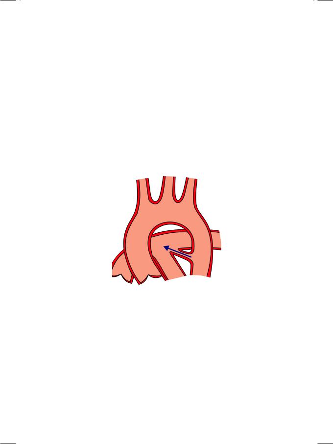
The ductus arteriosus is a channel that connects the descending aorta distal to the origin of left subclavian artery, to the left pulmonary artery just distal to the bifurcation of main pulmonary artery. The ductus remains open during intrauterinelifeandclosessoonafterbirthwhenitspurposeisfulfilled.Whenthe ductus fails to close physiologically within 24 hours after birth and anatomically within a week, it provides a communication between the aortic and pulmonary circulations. Flow from the aorta (at higher pressure) to the pulmonary artery (at lowerpressure)createsaleft-to-rightshuntacrossthePDA(Fig.5.3).Persistence of the ductus is sometimes associated with maternal rubella syndrome and premature delivery.
Figure 5.3: Patent ductus arteriosus
The continuous murmur of PDA is classically described as a “machinery” murmur and is referred to as Gibson’s murmur. It is maximally audible below the middle of the left clavicle, just before and just after the S2. The continuous murmur may be accompanied by a mid-diastolic murmur over the mitral area, due to torrential flow across the mitral valve. There may also be reverse splitting of S2, due to delayed closure of the aortic valve. When pulmonary hypertension develops, the murmur of PDA is confined to systole.
As already mentioned, there are several causes of a continuous murmur. In aorto-pulmonary window, there is a proximal communication between the aorta and the pulmonary artery. Although the murmur of aortic coarctation is typically systolic, in tight stenosis the murmur may extend into diastole. Venous hum is a low-pitched continuous murmur which is loudest over the supraclavicular fossa, but sometimes also heard over the precordium. It is accentuated by looking over the shoulder while sitting and abolished by compression of the jugular vein.

22 |
|
Section 1 Congenital Heart Diseases |
|
|
|
A mammary soufflé is heard widely over the precordium in pregnant women. It is better appreciated while lying down and during systole. Coronary fistula and ruptured aneurysm of sinus of Valsalva are rare arterio-venous communications that can also produce a continuous murmur.
Ventricular septal defect (VSD) produces a pansystolic murmur while aortic regurgitation causes an early diastolic murmur. When the two are associated as in perimembranous VSD, the murmur is systolo-diastolic. A similar systolodiastolic murmur occurs when an atrial septal defect (ASD) is associated with mitralstenosis,theLutembachersyndrome.Incontrasttoacontinuousmurmur, the two components of a systolo-diastolic murmur have a different character.
PDAisthecommonestcauseofcardiomegalyandheartfailureininfancyand childhood. Conversely, heart failure is the commonest cause of morbidity and mortality in PDA, at any age. Causes of heart failure in childhood are:
•Coarctation of aorta
•Patent ductus arteriosus
•Congenital cardiomyopathy
•Anomalous left coronary artery arising from pulmonary artery (ALCAPA).
When pulmonary hypertension develops in PDA, reversal of the shunt from pulmonary artery to aorta may occur. In that case, the continuous murmur gets shorter and quieter. The toes get more cyanosed and clubbed than the fingers. The reason for this differential cyanosis is that the ductus is distal to the left subclavian artery and predominantly the lower limbs get deoxygenated blood.
Other complication of PDA are endarteritis, aneurysm formation and rarely rupture of the ductus.
MANAGEMENT ISSUES
Percutaneous device closure of patent ductus arteriosus was one of the earliest non-surgical interventions in cardiology. All PDAs of significant size and shunt should be ligated, except in ductus dependent complex congenital cyanotic heart diseases of infancy.

S E C T I O N
2
Mitral Valve
Diseases