

новая папка / [libribook.com] 50 Cases in Clinical Cardiology_ A Problem Solving Approach 1st Edition
.PdfC A S E
43 Supraventricular
Tachycardia
. |
CASE PRESENTATION |
A 36-year old woman had been experiencing episodes of“fluttering”sensation in the chest, since two years. The episodes were described as a “machine-like” feeling over the precordium, that was not accompanied by chest pain or shortness of breath. During these episodes she did feel dizzy, but she never lost her consciousness. Once the episode was over, she would pass urine frequently. Her episodes were unrelated to physical exertion, food intake or to mental stress. At times she was able to abort the attack on her own, either by splashing cold water on her face or by applying firm pressure over her eyes. At other times, she had to visit a cardiologist who either performed carotid sinus massage or administered intravenous diltiazem. Initially, episodes would occur only once in a month or two but recently, their frequency had increased significantly. Therefore, although she was prescribed verapamil 120 mg twice a day which she took regularly, the patient was advised to undergo electrophysiological studies (EPS).
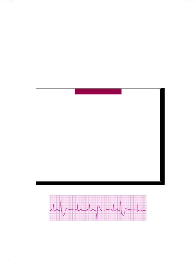
On examination, the patient was apprehensive but not tachypneic or in any distress. The heart rate exceeded 150 beats/min., with a BP of 96/70 mm Hg and she was afebrile. The JVP was not raised and there was no edema over the ankles. There was no tremor over fingers, thyromegaly or eye prominence to suggest thyrotoxicosis. The precordium was hyperkinetic in nature, with a normally located apex beat. On auscultation, S3 or S4 sound could not be appreciated because of extreme tachycardia, but no murmur or friction rub was audible. Breath sounds were vesicular without any rhonchi or crepts. A long-strip ECG recording of lead LII was obtained (Fig. 43.1).
Figure 43.1: ECG showing abrupt onset of narrow QRS tachycardia
196 |
|
Section 13 Cardiac Arrhythmias |
|
|
|
ECG INTERPRETATION
The ECG showed an abrupt onset of regular tachycardia at a rate exceeding 150 beats/min., with an RR interval of less than 10mm. The QRS complexes were narrow and the P or T waves were not visible. These findings are consistent with the diagnosis of paroxysmal supraventricular tachycardia (PSVT).
Figure 43.2: Diagram to illustrate circus movement in a closed re-entrant circuit
Supraventricular tachycardia is most often (in 90% cases) based on repeti tive circus movement of impulses in a closed reentrant circuit (reentrant tachycardia). In 50% cases, the circuit is composed of two pathways within the atrioventricular node (AV nodal reentrant tachycardiaAVNRT). In 40% cases, the circuit consists of an AV nodal pathway and an accessory bypass tract along its side (AV reentrant tachycardiaAVRT). An atrial impulse first passes anterogradely down one of the two pathways, the other pathway being in the refractory period. The impulses then returns retrogradely through the other pathway, which has by now recovered its conductivity. In this way, repetitive circulation of impulses occurs, to produce a sustained atrial tachycardia (Fig. 43.2). Least often (in about 10% cases), supraventricular tachycardia is due to rapid discharge of impulses from an ectopic atrial focus (ectopic atrial tachycardia).
The heart rate in paroxysmal atrial tachycardia is 150 to 200 beats per minute, if a reentrant circuit is involved. It tends to be slower in ectopic atrial tachycardia (120 to 150 beats/min), as the AV node cannot conduct more than 150 atrial impulses per minute. The heart rate can exceed a rate of 200 beats/min, if an accessory bypass tract is involved, as in case of WPW syndrome. This is because in WPW syndrome, the impulses can bypass the decremental influence of the AV node, by travelling down the accessory pathway. An atrial tachycardia arising
Table 43.1: Differences between ectopic and re-entrant atrial tachycardia
|
Ectopic tachycardia |
Re-entrant tachycardia |
|
|
|
Heart rate |
120-150/min |
More than 150/min |
|
|
|
Onset and offset |
Gradual |
Sudden |
|
|
|
P wave |
Ectopic, visible |
Inverted, Rarely visible |
|
|
|
A-V block |
Can coexist |
Never, 1:1 conduction |
|
|
|
Effect of vagal manoeuvre |
Slowing |
Termination |
|
|
|
Past history |
Not significant |
Previous episodes |
|
|
|
Organic heart disease |
May be present |
Generally absent |
|
|
|

Case 43 Supraventricular Tachycardia |
|
197 |
|
|
|
from an ectopic focus can be differentiated from a tachycardia due to a reentrant mechanism, by certain subtle features (Table 43.1).
Most often, a supraventricular tachycardia is characterized by narrow QRS complexes, due to synchronized ventricular activation through the specialized His bundle conduction system. Occasionally, the supraventricular impulses find one
Table 43.2: Differences between ventricular tachycardia and aberrant ventricular conduction
|
Ventricular tachycardia |
SVT with aberrancy |
|
|
|
Regularity of rhythm |
Slightly irregular |
Clock-like regularity |
|
|
|
P waves |
Not seen |
May be seen |
|
|
|
P-QRS relationship |
Unrelated |
Related |
|
|
|
QRS width |
> 0.14 sec |
0.12-0.14 sec |
|
|
|
QRS morphology |
Bizarre |
Triphasic |
|
|
|
QRS in V1 to V6 |
rS in V1 to V6 |
RsR’ in V1; Rs in V6 |
R-R’ height |
R > R’ |
R’ > R |
|
|
|
QRS axis |
Leftward |
Normal |
|
|
|
Capture/fusion beats |
May be seen |
Not seen |
|
|
|
Hemodynamics |
Compromised |
Stable |
|
|
|
Organic heart disease |
Often present |
Often absent |
|
|
|
Response to carotid massage |
No response |
Termination |
|
|
|
of the two bundle branches refractory to conduction. In that case, the impulses are conducted only through the other bundle branch, producing a situation of aberrant ventricular conduction. This needs to be differentiated from a wide QRS ventricular tachycardia. The differences between ventricular tachycardia and supraventricular tachycardia with aberrant ventricular conduction are given in Table 43.2.
CLINICAL DISCUSSION
Paroxysmal reentrant atrial tachycardia is most often based on a reciprocal mechanism involving a bypass tract or a dual intranodal pathway. Episodes of atrial tachycardia are one of the manifestations of preexcitation (WPW synd rome). In the absence of WPW syndrome, paroxysmal atrial tachycardia (PAT) is generally not associated with organic heart disease. If properly managed, PAT does not alter lifeexpectancy and carries an excellent prognosis. PAT coexisting with the WPW syndrome carries a poorer prognosis, because of the risk of degeneration into ventricular tachycardia. A paroxysm of atrial tachycardia in the presence of an underlying WPW syndrome is suggested, if it meets one of the following criteria:
•ECG during sinus rhythm shows short P-R interval and wide QRS complex
• The ventricular rate exceeds 200 beats/minute, indicating the absence of physiological AV block
•Inverted P waves are observed indicating retrograde atrial activation.

198 |
|
Section 13 Cardiac Arrhythmias |
|
|
|
Symptomsduetoatrialtachycardiadependupontheatrialrate,thedurationof the tachycardia and the presence of heart disease. A fast atrial tachycardia causes palpitation and neck pulsations. Angina pectoris may occur due to increased myocardial oxygen demand and reduced coronary filling time. Prolonged atrial tachycardia can cause dizziness or syncope due to decline in cardiac output (shortened ventricular filling time) and loss of atrial contribution to ventricular filling. Termination of the tachycardia is often followed by polyuria due to the release of atrial natriuretic peptide (ANP) by the stretching of atrial myocardium.
MANAGEMENT ISSUES
The first step in the management of supraventricular tachycardia is to attempt vagal stimulation, to block the atrioventricular (AV) node. Vagal manoeuvres include carotid sinus massage, supraorbital pressure, Valsalva manoeuvre and splashing icecold water on the face (Table 43.3). Before commencing carotid sinus massage, the carotid artery should be auscultated for a bruit. If a bruit is present, carotid massage should not be performed on that side, or else an embolus may dislodge from the plaque. Massage should be done on one side at a time and not simultaneously on both sides. If vagal manoeuvres fail to abort the tachycardia, a drug to block the AV node is administered intravenously. Drugs that are effective include adenosine, diltiazem and amiodarone. After restoration of sinus rhythm, oral diltiazem, verapamil, amiodarone or a betablocker such as metoprolol is prescribed to prevent recurrence.
Table 43.3: Management of supraventricular tachycardia
PSVT with nodal re-entry
• Vagal manoeuvres • Adenosine, diltiazem
PSVT with bypass tract
• Amiodarone therapy
• Radiofrequency ablation
The management of paroxysmal atrial tachycardia in the presence of WPW syndrome, is somewhat different. Vagal manoeuvres are useful, only if anterograde conduction proceeds through the AV node. Digitalis is contraindicated as it enhances conduction down the accessory pathway and may precipitate ventricular fibrillation. Diltiazem and metoprolol reduce the tolerance to the high ventricular rate and can precipitate congestive heart failure. Amiodarone is the antiarrhythmic agent of choice for the longterm treatment and prevention of arrhythmias associated with the WPW syndrome (Table 43.3).
The availability of sophisticated electrophysiological studies (EPS) to identify and locate bypass tracts and the development of latest ablative techniques, have revolutionalized the management of WPW syndrome. For ablation of the bypass tract, highfrequency AC current is delivered through a thermister tipped catheter, which leads to localized heat coagulation. Radiofrequency ablation (RFA) of the bypass tract can be offered to patients who report recurrent and frequent symptomatic episodes of PAT which are refractory to drug therapy or those that cause hemodynamic compromise.

|
|
C A S E |
|
|
|
|
|
|
|
|
|
||
|
|
|
|
|
||
|
|
44 |
Atrial |
|
|
|
|
|
|
|
|
|
|
|
|
|
|
Fibrillation |
|
|
|
|
|
|
|
|
|
|
|
|
|
|
|
|
. |
CASE PRESENTATION |
A 42-year old woman presented with the complaints of palpitation and extreme fatigue. She denied history of chest pain or dizziness, but she felt tired and breathless after routine activities. During the preceding 6 months, she had unintentionally lost 4 to 5 kg of weight, despite having a good appetite. There was no history of excessive thirst or frequent urination, but she passed 3 to 4 semiformed stools everyday. The patient felt particularly uncomfortable during the summer months when her restlessness, fatigue and palpitations increased considerably. There was no history of prolonged febrile illness with joint pains during her childhood and she had never received monthly shots of penicillin. The patient was married since 16 years and had two sons who were 13 and 9 years of age. Both of them were born after normal delivery and were in good health.
On examination, the patient was restless, anxious and tachypneic. Her extremities were warm and her palms were dry. There was no anemia, cyanosis, jaundice or ankle edema. The pulse was fast, irregular and of good volume, with a pulse rate of 90 to 100 beats/min. The heart rate by auscultation was 110 to 120 beats/min, with a BP of 144/92 mm Hg over the right arm. Her temperature was 99.20F and the respiratory rate was 24/min. There was tremor over her outstretched hand and the eye-balls were prominent with lid-retraction. The JVP was not raised and there were no palpable lymph-nodes, but the thyroid gland was diffusely enlarged. The goiter was not tender, but a bruit was audible. The precordium was hyperkinetic with a forceful apical impulse. The S1 was variable in intensity with a loud S2. No murmur or gallop sound was audible. A long-strip ECG recording of lead LII was recorded (Fig. 44.1).
Figure 44.1: ECG showing irregular rhythm with fine fibrillatory waves
200 |
|
Section 13 Cardiac Arrhythmias |
|
|
|
ECG INTERPRETATION
The ECG showed a fast irregular rhythm, with a variable R-R interval. The QRS complexes were narrow, but no P waves were visible. Instead, there were fine fibrillatory waves between the QRS complexes. These findings are consistent with the diagnosis of atrial fibrillation (AF). Atrial fibrillation is a grossly irregular fast rhythm produced by functional fractionation of the atria into numerous tissue islets, in various stages of excitation and recovery. Consequently, atrial activation is chaotic and ineffectual in causing atrial contraction (Fig. 44.2). Although 400 to 500 fibrillatory impulses reach the A-V node per minute, only 100 to 150 of them succeed in eliciting a ventricular response, while others are blocked in the A-V node. The random activation of the ventricles produces a grossly irregular ventricular rhythm.
Figure 44.2: Diagram to illustrate atrial islets with chaotic atrial activation
The hallmark of atrial fibrillation is the absence of discrete P waves. Instead, there are numerous, small, irregular fibrillatory waves (f waves) that are difficult to identify individually but produce a ragged baseline. In long-standing atrial fibrillation, these undulations are minimal and produce a nearly flat baseline. As mentioned, the ventricular rate is grossly irregular and varies from 100 to 150 beats per minute. Atrial fibrillation can be differentiated from atrial flutter by the absence of P waves and an irregular ventricular rate (Table 44.1). At times, precise differentiation between the two may be difficult and the rhythm is then known as “flutter-fibrillation”, “coarse fibrillation” or “impure flutter”.
Table 44.1: Differences between atrial flutter and atrial fibrillation
|
Atrial flutter |
Atrial fibrillation |
|
|
|
Atrial rate |
220-350 beats/min |
Over 350 beats/min |
|
|
|
Ventricular rate |
Regular. Half to one fourth |
Variable. No relation |
|
of atrial rate |
to atrial rate |
|
|
|
Atrial activity |
Flutter (F) waves |
Fibrillatory (f) waves |
|
Saw-toothed baseline |
Ragged baseline |
|
|
|
Ventricular activity |
Constant R-R interval |
Variable R-R interval |
|
|
|
In atrial fibrillation, the ventricular rate generally varies from 100 to 150 beats per minute. Faster rates are observed in children, patients of thyrotoxicosis and in the presence of WPW syndrome. Slower rates are observed during drug treatment

Case 44 Atrial Fibrillation |
|
201 |
|
|
|
with beta-blockers (propranolol, atenolol) or calcium-blockers (verapamil, diltiazem), as these drugs block the A-V node. Elderly patients with A-V nodal disease may also manifest slow atrial fibrillation. Regularization of the ventricular rate in a patient on digitalis for atrial fibrillation, indicates the onset of junctional tachycardia and is a manifestation of digitalis toxicity.
CLINICAL DISCUSSION
Atrial fibrillation (AF) can be classified into three groups. In paroxysmal AF, discrete episodes are self-terminating and last less than 48 hours. In persistent AF, fibrillation continues unabated for atleast 7 days, but can be converted to sinus rhythm by electrical or chemical cardioversion. In permanent AF, fibrillation continues indefinitely and conversion to sinus rhythm is not possible. Atrial fibrillation can occur in virtually any form of heart disease. Common causes of atrial fibrillation are given in Table 44.2.
Table 44.2: Causes of atrial fibrillation
Persistent atrial fibrillation
• Dilated cardiomyopathy • Constrictive pericarditis • Cardiac trauma/surgery
• Hypertensive heart disease • Valvular heart disease (MS) • Coronary artery disease (MI)
• Congenital heart disease (ASD).
Paroxysmal atrial fibrillation
• Thyrotoxicosis • WPW syndrome
• Sick sinus syndrome • Lone atrial fibrillation
• Acute alcoholic intoxication
• Pulmonary thrombo-embolism.
The symptoms of AF depend upon the ventricular rate, the nature and severity of underlying heart disease and the effectiveness of treatment. Palpitation is due to fast heart rate while syncope is caused by reduced cerebral perfusion. Angina may occur because of increased myocardial oxygen demand as well as shortened coronary filling time due to tachycardia. Dyspnea is due to pulmonary congestion, secondary to loss of atrial contribution to ventricular filling. Sometimes, regional ischemia in the form of limb gangrene, hemiparesis or blindness may occur because of systemic embolization from a left atrial thrombus.
Atrial fibrillation can be recognized clinically by several subtle signs. The pulse is irregularly irregular, with the pulse rate on palpation being less than the heart rate on auscultation (pulse deficit). The a waves are not observed in the neck veins. There is a beat-to-beat variability in the pulse pressure as well as the intensity of first heart sound, because of a variable ventricular diastolic filling period.

202 |
|
Section 13 Cardiac Arrhythmias |
|
|
|
MANAGEMENT ISSUES
The treatment of atrial fibrillation (AF) is governed by the patient’s symptoms and hemodynamic status. In the majority of patients, long-term rate control along with oral anticoagulation, is a reasonable therapeutic strategy. Drugs that prolong the refractory period of the atrio-ventricular (AV) node such as digoxin, diltiazem and metoprolol are effective for rate control in persistent AF. In patients of paroxysmal AF with infrequent episodes, a “pill in the pocket” (taken when symptomatic) strategy is preferable. Drugs used for this purpose are amiodarone, sotalol, flecainide and propafenone.
Long-standing atrial fibrillation produces stasis of blood in the left atrium and promotes the development of thrombi in the atrial cavity and the atrial appendage. Dislodged fragments of these thrombi can enter the systemic circulation as emboli and settle down in any arterial territory. Anti-coagulants such as caumarin and warfarin are required for long-term use in chronic atrial fibrillation, to reduce the likelihood of systemic embolization. This particularly applies to patients with rheumatic heart disease and to prosthetic valve recipients. Those patients with a previous history of thrombo-embolism (stroke or TIA) and those who have a documented atrial thrombus, are also candidates for long-term anticoagulant therapy.
Table 44.3: Management of atrial fibrillation
• Heart-rate control • Anticoagulation
• Electrical cardioversion • Radiofrequency ablation
Ifthepatient’sclinicalstatusispoorandhemodynamicsareunstable,electrical cardioversion with 100 to 200 Joules of energy is the treatment of choice, in an attempt to restore sinus rhythm. There are two requisites before cardioversion is attempted. Firstly, the patient should not have received digitalis in the previous 48 hours. Digitalis not only decreases the threshold for defibrillation, but also predisposes to the risk of life-threatening arrhythmias. Secondly, an oral anticoagulant should be initiated before cardioversion and continued for atleast four weeks. This is because atrial thrombi are more likely to dislodge as emboli, once sinus rhythm is restored. Cardioversion should not be attempted if there is a documented left atrial clot.
In patients who are symptomatic but either refractory or intolerant to several antiarrhythmic agents, the final option is of radiofrequency ablation (RFA). Advantages of RFA are not only freedom from symptoms but also avoidance of the toxic effects of antiarrhythmic drugs and the need to monitor anticoagulant therapy.
|
|
C A S E |
|
|
|
|
|
|
|
|
|
||
|
|
|
|
|
||
|
|
45 |
Ventricular |
|
|
|
|
|
|
|
|
|
|
|
|
|
|
Premature Beats |
|
|
|
|
|
|
|
|
|
|
|
|
|
|
|
|
. |
CASE PRESENTATION |
A 72-year old man arrived at the emergency room with shortness of breath, palpitation, anorexia and vomiting. He had been repeatedly hospitalized over the past 6 months, because of congestive heart failure due to ischemic cardiomyopathy. The patient had sustained myocardial infarction twice and his left ventricular ejection fraction was 25%. During his previous admission a month back, frequent multifocal ventricular premature complexes (VPCs) were detected, for which amiodarone 200 mg daily was added to his ongoing therapy. His dose of frusemide was escalated from 40 mg to 60 mg per day because of worsening heart failure. He was also receiving digoxin 0.25 mg once a day, 6 days in a week.
On examination, the patient was restless, orthopneic and in respiratory distress. There was mild pallor and the extremities were cold and clammy. The pulse was rapid, irregular and low in volume, with a BP of 104/66 mm Hg. The JVP was raised 5 cm above the angle of Louis and there was edema over the ankles and lower legs. The lower border of the liver was felt 4 cm below the costal margin and ascites was present. Fine inspiratory crackles were heard over the bases of lung fields bilaterally. The cardiac apical impulse was diffuse and displaced towards the axilla. On auscultation, both S3 and S4 were audible, producing a summation gallop rhythm. A soft pansystolic murmur was also heard at the cardiac apex, that radiated towards the left axilla and scapula. A 12-lead ECG was obtained, with a long-strip recording of lead LII (Fig. 45.1)
Figure 45.1: ECG showing multifocal ventricular premature beats

204 |
|
Section 13 Cardiac Arrhythmias |
|
|
|
ECG INTERPRETATION
The ECG showed a fast irregular rhythm with frequent premature beats. The premature beats had a wide and bizarre QRS morphology, were not preceded by P waves, but were followed by a compensatory pause. The morphology of the premature beats, as well as the interval between the premature beat and the precedingsinusbeat(couplinginterval),werevariable.Attimes,averypremature beat was superimposed upon the T wave of the preceding sinus beat. These findings are consistent with the diagnosis of multifocal ventricular premature complexes (VPCs) exhibiting the R-on-T phenomenon.
VPCs can be qualified on the basis of their pattern of occurrence and their location in the cardiac cycle. VPCs with an identical morphology and constant coupling interval are called unifocal VPCs. Those VPCs that have a variable morphology and changing coupling interval are called multifocal VPCs. That
VPC which occurs late in the diastolic period of the preceding sinus beat (long coupling interval), just about when the next sinus beat is expected, is called an end-diastolic VPC. A VPC that is so premature (very short coupling interval) that it is superimposed upon the T wave of the preceding sinus impulse, is said to exhibit the R-on-T phenomenon. A VPC during a slow rhythm, that does not allow any sinus beat to be missed and is not followed by a compensatory pause, is called an interpolated VPC. VPCs alternating with sinus beats constitute a bigeminalrhythm(extrasystolicventricularbigeminy).VPCaftereverytwosinus beats represents trigeminy and a VPC after every third sinus beat constitutes a quadrigeminy. The degree of ventricular ectopy has been categorized as per the Lown’s classification given in Table 45.1.
Table 45.1: Lown’s classification of ventricular ectopy
Category |
Degree of ectopy |
|
|
Class 0 |
No ectopy |
|
|
Class 1 |
Less than 30/hour |
|
|
Class 2 |
More than 30/hour |
|
|
Class 3 |
Multiform VPCs |
|
|
Class 4A |
Couplets |
|
|
Class 4B |
Runs of 3 or more |
|
|
Class 5 |
R-on-T phenomenon |
|
|
CLINICAL DISCUSSION
Ventricular premature complexes (VPCs) can occur even in normal individuals, although they are more often due to organic heart disease. Causes of VPCs in normal persons are:
•Drugs e.g. beta-agonists, theophylline
•Emotional stress and physical exercise
•Smoking and high intake of tea/coffee
•Anxiety neurosis and thyrotoxicosis.