

новая папка / [libribook.com] 50 Cases in Clinical Cardiology_ A Problem Solving Approach 1st Edition
.PdfCase 16 Aneurysm of Aorta |
|
75 |
|
|
|
abnormalities of the skeletal system are observed. The typical body habitus is a tall and slender figure with long tapering fingers and increased joint mobility. The pubis-to-heel measurement exceeds the crown-to-pubis length. Associated abnormalities include a high-arched palate, pectus carinatum, dislocated ocular lenses (ectopia lentis) and presence of inguinal hernias (Table 16.2).
Table 16.2: Clinical features of Marfan’s syndrome
Skeletal |
Cardiac |
Others |
|
|
|
Tall stature |
Aortic dilatation |
Dislocation of lens |
|
|
|
Joint hypermobility |
Aortic aneurysm |
High-arched palate |
|
|
|
Arm-span > height |
Aortic dissection |
Crowded dentition |
|
|
|
Long digits (arachnodactyly) |
Aortic regurgitation |
Pectus carinatum |
|
|
|
Scoliosis and sternal deformity |
Mitral valve prolapse |
Inguinal hernia |
|
|
|
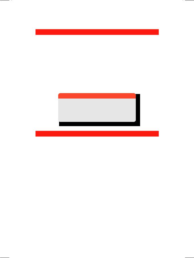
Cardiovascular complications are initiated by cystic medial necrosis of the ascending aorta. There is dilatation of the aortic root with aortic valve insufficiency and left ventricular volume overload (Fig. 16.2). Catastrophic dissection involving the proximal aorta is a serious and potentially lethal complication. Mitral valve prolapse is sometimes associated.
Figure 16.2: ECHO showing an aneurysmal aorta with jet of aortic regurgitation
Although syphilitic aortitis is now distinctly rare, Takayasu’s aorto-arteritis still afflicts young women in south Asia. It causes aneurysmal aortic dilatation and stenosis of the major branches emerging from the aorta. Blood pressure in the two arms may be different due to narrowing of subclavian artery on one side. On the other hand, hypertension may be present in the lower limbs due to reno-vascular stenosis. Therefore, Takayasu’s aorto-arteritis is also designated as “reverse coarctation”.
76 |
|
Section 5 Aortic Diseases |
|
|
|
MANAGEMENT ISSUES
The medical management of an aortic aneurysm depends upon its size, the blood pressure and the presence or absence of aortic dissection. If the aneurysm is small, stable in size and there is no dissection, it suffices to adequately control the blood pressure. A drug with negative ionotropic effect such as a beta-blocker is preferable, since it reduces the shearing force on the aorta. The patient is counselled to avoid strenuous and burst activity like weight-lifting. Follow-up echo is recommended every 6 to 12 months, to assess the size of aneurysm and degree of aortic regurgitation.
Surgical intervention is considered if the aneurysm is growing in size on serial imaging, there is evidence of impending rupture or dissection, or if the aortic regurgitation is severe (Table 16.3). The affected segment of the ascending aorta is resected and replaced with a prosthetic graft.
Table 16.3: Indications for aortic aneurysm surgery
• Severe dilatation more than 50 mm
• Rapid aneurysm growth > 10 mm/year • Dissection of the proximal aorta
• Severe aortic regurgitation
RECENT ADVANCES
Newer imaging techniques such as computed tomography (CT) and magnetic resonance imaging (MRI) have obviated the need for aortography, in the evaluation of aortic aneurysms. Serial imaging can identify an expanding size, impending rupture and an associated dissection.
|
|
|
C A S E |
|
|
|
|
|
|
|
|
|
|
|
|
||
|
|
|
|
|
|
|
||
|
|
|
17 |
Dissection |
|
|
|
|
|
|
|
|
|
|
|
|
|
|
|
|
|
|
of Aorta |
|
|
|
|
|
|
|
|
|
|
|
|
|
|
|
|
|
|
|
|
|
CASE PRESENTATION
A 64-year old woman presented to the emergency department with sudden onset of severe chest pain, one hour back. The chest pain was central in location and it peaked instantly. The pain was also felt over the back between her scapulae, but did not radiate to the arms or neck. There was no history of suffocation, choking, palpitation or syncope and she had never experienced such chest pain before. She had a history of hypertension for which she had been prescribed multiple drugs. However, she was irregular with her medication and often skipped her doses. As a result, her BP hovered around 150/100 mm Hg on most occasions. She was not a diabetic but was advised to take atorvastatin for her elevated LDL-cholesterol. She did not consume alcohol, but smoked a pack of cigarettes per day for the last 40 years. Although distressed by severe chest pain, she managed to tell the attending doctor that the grip of her left hand felt weak since one hour.
On examination, she was clearly distressed but not dyspneic or diaphoretic. Pulse rate was 104 beats/min., with a BP of 174/106 mm Hg in her right arm. All peripheral pulses in the lower extremities were palpable, but a bruit was audible over the right carotid artery. The JVP was not raised and there was no pitting edema around the ankles. The apex beat was heaving in nature and slightly displaced outwards towards the axilla. The S1 was normal and A2 was loud, but there was no gallop sound. A faint early-diastolic murmur was audible along the left sternal border.
CLINICAL DISCUSSION
In an elderly patient, the most frequent but by no means the sole cause of excruciating chest pain is myocardial infarction. Other causes of severe chest pain are pulmonary embolism and spontaneous pneumothorax. The pain of pleuro-pericarditis, oesophagitis or costo-chondritis is seldom excruciating. Acute aortic dissection is an uncommon but potentially fatal cause of severe chest pain that merits a high index of clinical suspicion.
TheECGshowedsinustachycardiabutnoS-Tsegmentshiftandthetroponin-T test was negative on admission. X-ray chest findings were mild cardiomegaly with a left ventricular contour and slight mediastinal widening due to aortic root dilatation. There was no basal atelactasis, pleural effusion or pneumothorax. ECHO revealed dilatation of the aortic root, with a false lumen cleaving the

78 |
|
Section 5 Aortic Diseases |
|
|
|
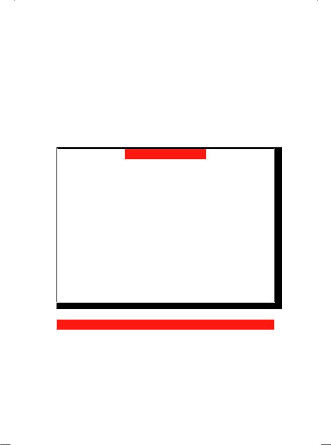
anterior aortic wall and having a blind end. An intimal flap separated the false lumen from the true aortic lumen (Fig. 17.1). On colour Doppler, a mosaic jet was observed in the left ventricular outflow tract, along the septum. There was no pericardial effusion. These findings were consistent with the diagnosis of acute dissection of the aorta.
Figure 17.1: ECHO showing cleavage of the anterior aortic wall
with intimal flap between true and false lumens
Acute myocardial infarction is an unlikely possibility in this case. The pain of myocardial infarction does not reach its maximum intensity instantly, but builds up gradually. Moreover, the radiation and accompaniments that are typical of the pain of myocardial infarction, were absent in this case. The ECG did not show S-T segment elevation and the troponin test was negative. On ECHO, cleavage of aortic wall and presence of aortic regurgitation are not typical features of acute myocardial infarction.
Table 17.1: Causes of aortic dissection
• Marfan’s syndrome • Coarctation of aorta
• Accelerated hypertension • Hypertension in pregnancy
• Trauma; accidental or surgical
Dissection of aorta is often due to accelerated hypertension and may complicate hypertension in pregnancy. Other causes are aortic coarctation and Marfan’s syndrome (Table 17.1). Aortic dissection cleaves the media of the aortic wall, with the adventitia and outer media forming the outer wall and the inner media and intima forming the inner wall. The false lumen produced by cleavage has a blind distal end while the proximal end communicates with the true lumen at the site of aortic tear. The intimal flap oscillates between the true and false lumens (Table 17.2). Other reasons for deformity of the anterior aortic wall are aortic root abscess in infective endocarditis and an aneurysm of the sinus of Valsalva.

Case 17 Dissection of Aorta |
|
79 |
|
|
|
Table 17.2: Typical features of aortic dissection
• Dilatation of the proximal aortic root > 42 mm • Anterior or posterior wall thickness >15 mm • Double echo-line of the involved aortic wall • Space between outer and inner wall >5 mm • False lumen within aortic wall with blind end • Intimal flap between true and false lumens.
Therearevariousclassificationsofaorticdissection.Thesimplestclassification is of the Stanford group, which categorizes dissection as Type A (proximal), where the ascending aorta is involved and Type B (distal), where the ascending aorta is spared. This classification has prognostic as well as therapeutic implications. DeBakey Type I dissection extends from the ascending to descending aorta while Type II is confined to the ascending aorta. Type III is confined to the descending aorta and akin to Stanford Type B (Table 17.3).
Table 17.3: Classification of aortic dissection according to location
DeBakey type |
Stanford group |
Location of dissection |
Incidence |
|
|
|
|
III } |
A |
Ascending to descending aorta |
10% |
|
Confined to ascending aorta |
70% |
|
|
|
||
|
|
|
|
III |
B |
Confined to descending aorta |
20% |
|
|
|
|
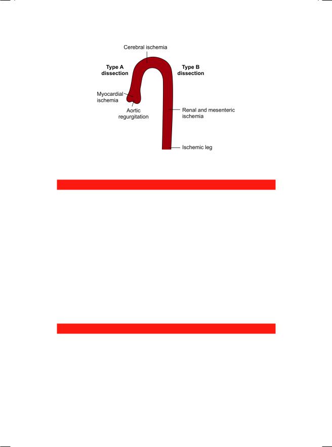
The complications of aortic dissection are related to the involvement of the aortic valve, pericardial sac and the arteries emerging from the aorta. Proximal involvement of the aortic valve can cause acute aortic regurgitation and heart failure. Hemorrhagic pericardial effusion can cause cardiac tamponade. Involvement of the coronary artery ostium can actually lead to myocardial infarction (Table17.4).Involvementofthesubclavianarteriescancauseupperlimbischemia and discrepancy between the radial pulse volume on both sides. Involvement of carotid arteries can cause cerebral ischemia as in our case, who had weakness of the left hand and a right-sided carotid bruit. In distal aortic dissection, there may be mesenteric, renal, spinal cord or lower limb ischemia (Fig. 17.2).
Table 17.4: Atypical features of aortic dissection
• Occlusion of neck vessels • Aortic valve regurgitation • Left ventricular dysfunction • Myocardial infarction
• Pericardial effusion
80 |
|
Section 5 Aortic Diseases |
|
|
|
Figure 17.2: Complications of aortic dissection
MANAGEMENT ISSUES
The management of aortic dissection depends upon its location and extent, the presence or absence of complications and the hemodynamics of the patient. In distal (Type B) dissection, it generally suffices to control the blood pressure adequately. Agents with negative ionotropic effect such as a beta-blocker or verapamil are preferable as they reduce the shearing force on the aorta and the progression of dissection. A vasodilator such as sodium nitroprusside or an ACEinhibitor may be added if the blood pressure remains significantly elevated. In the presence of abdominal, spinal or lower limb severe ischemia, an endovascular stent graft may be deployed to cover the dissection.
In proximal (Type A) dissection, surgical intervention is preferable unless the hemodynamics are compromised, in which case a conservative approach is adopted. The affected segment of the ascending aorta is resected and replaced with a prosthetic graft. The aortic valve and coronary ostia may need to be reconstructed. In extensive aortic dissection (DeBakey Type I), a combined approach that includes proximal resection with graft and distal endovascular stenting may be used.
RECENT ADVANCES
Newer imaging techniques such as computed tomographic (CT) angiography and magnetic resonance imaging (MRI) have obviated the need for aortography, in the evaluation of aortic dissection. The sensitivity and specificity of these tests, for a confirmatory diagnosis of aortic dissection, are in excess of 95%. The technique of percutaneous endovascular stenting to manage DeBakey Type I and III dissection, has also undergone considerable refinement.
|
|
C A S E |
|
|
|
|
|
|
|
|
|
||
|
|
|
|
|
||
|
|
18 |
Coarctation |
|
|
|
|
|
|
|
|
|
|
|
|
|
|
of Aorta |
|
|
|
|
|
|
|
|
|
|
|
|
|
|
|
|
CASE PRESENTATION
A young man 27 years of age, presented to the out-patient clinic with general fatigue and difficulty in climbing even two flights of stairs. He was diagnosed to have systemic hypertension five years back and had been on antihypertensive drugs ever since. Besides his fatigue, he also complained of episodic headache with dizziness. These episodes were often related to undue physical exercise, emotional upset or to missing of his medication. There was no history of cyanotic spells or fleeting joint pains during childhood. He denied abusing tobacco, alcohol or illicit drugs.
On examination, there were no facial features of an endocrine disorder such as thyrotoxicosis, acromegaly or Cushing’s syndrome, but his lower limbs were unusually thin. The radial pulse was 84 beats/min. and bounding in nature. The BP in his right arm was 170/100 mm Hg. Pulse amplitude over the femoral arteries was reduced in volume and delayed compared to the brachial pulsation. Also, there were visible pulsations in the suprasternal notch. The cardiac apex beat was heaving in nature and slightly displaced towards the left axilla. On auscultation, S1 was normal, A2 was loud and a presystolic S4 was audible. An ejection systolic murmur was heard at the upper left parasternal area. Incidentally, the same murmur was also audible over the interscapular region. Soft continuous murmurs were also heard over the scapulae. On abdominal auscultation, there was no bruit heard over the renal arteries.
CLINICAL DISCUSSION
When confronted with a young patient with moderate to severe systemic hypertension, the clinician should be alert to the possibility of secondary hypertension. Incidentally, secondary hypertension constitutes less than 5% of all hypertensive patients, while most have primary or essential hypertension. Usual causes of secondary hypertension are:
•Endocrine disorders
•Renovascular disease
•Coarctation of aorta.
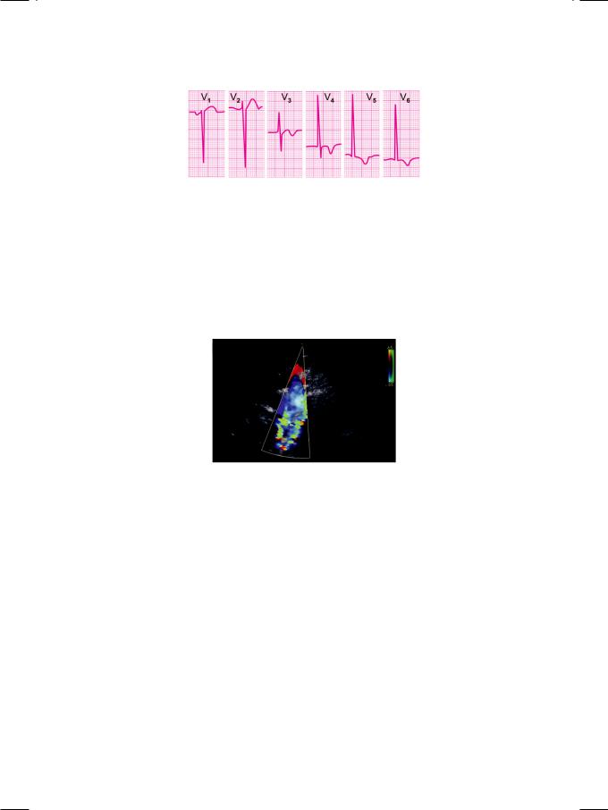
In this patient, ECG showed tall R waves in leads V5 and V6 with deep S waves in leads V1 and V2, indicating the presence of left ventricular hypertrophy (Fig. 18.1). X-ray showed mild cardiomegaly with a rounded contour of the left heart border. There was “notching” of the undersurface of the 4th to 8th ribs and
82 |
|
Section 5 Aortic Diseases |
|
|
|
Figure 18.1: ECG showing tall R waves in V5,V6 with deep S waves in V1,V2
a “figure-of-3” appearance of the aortic arch. ECHO revealed concentric left ventricular hypertrophy with normal ejection fraction. Aortic valve was bicuspid with thickened leaflets and an insignificant gradient across the valve. The mitral valve was normal and there was no ventricular septal defect. Doppler signal from the suprasternal notch showed a pulsatile aortic arch with a distal aortic gradient that tapered into diastole. A mosaic jet was seen in the descending aorta, directed away from the transducer (Fig. 18.2).
Figure 18.2: ECHO showing a mosaic jet in the descending aorta
Coarctation of aorta is the most likely cause of hypertension in this case.
There was clinical, radiological and cardiographic evidence of left ventricular hypertrophy. There were brachio-femoral delay, suprasternal pulsations, a parasternal and interscapular systolic murmur (coarctation itself) and continuous intercostal murmurs (collateral vessels). All these auscultatory findings are pathognomic of aortic coarctation. The murmur of coarctation itself may be continuous if the narrowing is severe, while presence of an ejection click indicates an associated bicuspid aortic valve. On X-ray chest, notching of the lower border of ribs indicates arterial collateralization, to by-pass the aortic obstruction. The “figure-of-3” appearance is produced by indentation of the aorta at the site of coarctation, with dilatation on either side of the narrowing. The suprasternal high velocity Doppler signal is characteristic of aortic coarctation.
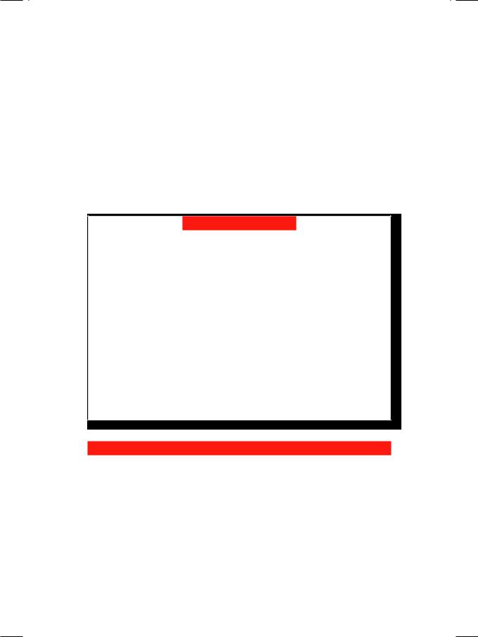
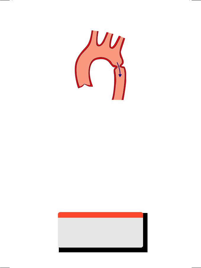
Coarctation of the aorta is a localized narrowing of the aortic arch (Fig. 18.3), due to a fibrous ring encircling the wall or a shelf projecting into the lumen. The narrowing is in the region of the ligamentum arteriosum (juxta-ductal) and may be pre-ductal or post-ductal. The aorta distal to the narrowing is often dilated and aneurysmal. Coarctation may present in the neonatal period (infantile type)
Case 18 Coarctation of Aorta |
|
83 |
|
|
|
Figure 18.3: Coarctation of aorta
with heart failure, in which case the narrowing is diffuse, tubular and severe (hypoplastic aorta). In adult type coarctation, the narrowing is discrete, ring-like, less severe and detected during evaluation of systemic hypertension.
Some patients may complain of pain and/or weakness in the legs (claudication), due to reduced femoral blood flow. Brachio-femoral delay pertains to the difference in amplitude and timing between the brachial and femoral pulsations. Therefore, the blood pressure is higher in the upper limbs than in the lower limbs. If the left subclavian artery is also narrowed by coarctation, the blood pressure is only elevated in the right arm. In Takayasu’s aorto-arteritis (pulseless disease), which is also called “reverse coarctation”, the blood pressure is reduced in the upper limbs compared to the lower limbs, because there is stenosis at the origin of aortic arch branches.
Hypertension in coarctation of aorta is multifactorial. The aorta proximal to the narrowing is stiff and the carotid sinus baroreceptors are set at a higher level. Additionally there is a renal component, due to narrowing proximal to the renal arteries. Coarctation of aorta is associated with a bicuspid aortic valve in half the patients. Other cardiac anomalies are ventricular septal defect and mitral valve prolapse (Table 18.1). In 10 to 20% patients, there are intracranial aneurysms around the circle of Willis. Aortic coarctation may be associated with Turner’s syndrome, characterized by a short-statured female with broad chest and a
Table 18.1: Abnormalities associated with coarctation
• Bicuspid aortic valve • Mitral valve prolapse
• Ventricular septal defect • Aneurysm sinus of Valsalva
• Intracranial Berry aneurysm
84 |
|
Section 5 Aortic Diseases |
|
|
|
webbed neck. Long-term complications of coarctation are heart failure and stroke due to hypertension, dissection of proximal aorta and ruptured aneurysm of the sinus of Valsalva. The bicuspid aortic valve can develop stenosis or regurgitation and be the site of endocarditis. Finally, sub-arachnoid hemorrhage can occur due to ruptured intracranial aneurysm (Table 18.2).
Table 18.2: Complications associated with coarctation
• Congestive heart failure • Endocarditis of aortic valve
• Dissection of proximal aorta • Sub-arachnoid hemorrhage • Stroke due to hypertension
• Aortic valve stenosis or regurgitation • Ruptured sinus of Valsalva aneurysm
MANAGEMENT ISSUES
Management of secondary hypertension due to aortic coarctation, is governed by the same principles that apply to the treatment of essential hypertension. This involves the judicious use of a combination of antihypertensive agents. Several drugs are used including diuretics, beta-blockers,calcium-antagonists andACEinhibitors (or ARBs), since the hypertension is generally moderate to severe in grade and severity. Definitive treatment of aortic coarctation (systolic gradient >
30mmHg)issurgicalexcisionoftheareaofaorticnarrowingwithanend-to-end anastomosis. A prosthetic tubular Dacron graft may also be interposed. Another technique is patch aortoplasty at the site of coarctation.
RECENT ADVANCES
Recently,thetechniqueofpercutaneousballoonaortoplastyhasbeendeveloped.
It is most suitable for discrete stenosis or if narrowing has recurred following surgical correction of coarctation. The incidence of restenosis after aortoplasty is reduced by stenting.