

новая папка / [libribook.com] 50 Cases in Clinical Cardiology_ A Problem Solving Approach 1st Edition
.PdfS E C T I O N
11
Typical ECG
Abnormalities


|
|
C A S E |
|
|
|
|
|
|
|
|
|
||
|
|
|
|
|
||
|
|
34 |
Left Ventricular |
|
|
|
|
|
|
|
|
|
|
|
|
|
|
Hypertrophy |
|
|
|
|
|
|
|
|
|
|
|
|
|
|
|
|
CASE PRESENTATION
A 56-year old obese gentleman visited his physician with the complaints of frequent headaches and spells of dizziness. He also found it difficult to carry out routine tasks and felt fatigued and breathless even on mild physical exertion. The patient was diagnosed to have systemic hypertension about 30 years back. At that time, he was extensively investigated for secondary hypertension, but no renal or endocrine disorder was detected. He was prescribed antihypertensive medications, but did not take them regularly and was not on periodic medical follow-up. The patient had always been overweight even at a young age and was diagnosed to have diabetes mellitus about 10 years back. He had not got his lipid profile checked recently. The man was sedentary and did not follow any particular diet plan. He smoked 8 to 10 cigarettes per day and consumed 2 to 3 large pegs of whiskey, on most days of the week.
On examination, the patient was grossly overweight, with a body mass index (BMI) of 34 kg/m2. He was tachypneic and afebrile. The pulse was regular, good in volume and the heart rate was 84 beats/min. All peripheral pulses were palpable and there was no carotid bruit or brachio-femoral delay. The BP was 164/106 mm Hg over the right arm in the sitting position. The JVP was not raised but mild edema was present over the ankles. The cardiac apex beat was sustained and heaving in nature and systolic pulsations were observed in the suprasternal notch. The S1 was normal but the A2 sound was accentuated; a S4 sound was heard in presystole. A soft systolic murmur was audible over the upper left parasternal area. Few basilar rales were audible over the lung fields. An ECG was obtained (Fig. 34.1).
Figure 34.1: ECG showing tall R waves in left chest leads with deep S waves in right chest leads
158 |
|
Section 11 Typical ECG Abnormalities |
|
|
|
ECG INTERPRETATION
The ECG showed tall R waves exceeding 25 mm in the left precordial leads and deep S waves in the right precordial leads. There also was ST segment depression and Twave inversion in the lateral leads. These findings are consistent with the diagnosis of left ventricular hypertrophy. The tall R waves in the lateral precordial leads, reflect the increased electrical forces generated by the thickened left ventricular myocardium. ST segment depression and T wave inversion in these leads indicate left ventricular strain. Additional findings can include notched bifid P waves (P. mitrale) due to left atrial enlargement and left axis deviation of the QRS vector.
Voltage criteria are often employed for the electrical diagnosis of left ventricular hypertrophy (Table 34.1). The Sokolow and Lyon criteria of S in V1 plus R in V5 or V6 greater than 35 mm is popularly used. Another criteria used is of Framingham wherein, R in V5 or V6 >25 mm or R in aVL >11 mm is taken as criteria for left ventricular hypertrophy. The left ventricular (LV) strain pattern of ST segment depression and Twave inversion is observed in LV pressure overload. In case of LV volume overload, the ST segment is isoelectric and the T wave is upright and tall.
Table 34.1: Voltage criteria for left ventricular hypertrophy
• Sokolow and Lyon Criteria
S. in V1 + R in V5 or V6 >35 mm
• Framingham Criteria
R in V5 or V6 >25 mm; R in aVL >11 mm
CLINICAL DISCUSSION
Causes of left ventricular hypertrophy are classified into those of systolic overload and the causes of diastolic overload. Systolic overload is due to systemic hypertension, aortic stenosis, coarctation of aorta and hypertrophic cardiomyopathy. Causes of diastolic overload are mitral and aortic valve regurgi tation and an intracardiac lefttoright shunt such as ventricular septal defect or patent ductus arteriosus (Table 34.2).
Echocardiography is 5 to 10 times more sensitive than an ECG, in detecting left ventricular hypertrophy (LVH). Presence of LVH is the most common ECHO abnormality in a hypertensive patient. Conversely, systemic hypertension is the most common cause of LVH. Besides detecting LVH, ECHO is used to evaluate left ventricular systolic and diastolic function and for the diagnosis of associated coronary artery disease. Other abnormalities seen on ECHO are mitral and aortic valve degeneration, dilatation of the aortic root and rarely aortic coarctation (Table 34.3). ECHO typically shows thickening of the interventricular septum (IVS) and LV posterior wall (LVPW) greater than 11 mm with virtual obliteration of the left ventricular cavity during systole (Fig. 34.2). Left ventricular hypertrophy is an

Case 34 Left Ventricular Hypertrophy |
|
159 |
|
|
|
Table 34.2: Causes of left ventricular hypertrophy
Systolic overload
• Aortic valve stenosis • Systemic hypertension
• Coarctation of the aorta
• Hypertrophic cardiomyopathy
Diastolic overload
• Mitral regurgitation • Aortic incompetence
• Ventricular septal defect • Patent ductus arteriosus.
Figure 34.2: ECHO showing thick septum and posterior wall with obliteration of left ventricular cavity
independent predictor of cardiovascular morbidity and mortality, as a risk factor for myocardial infarction, stroke, heart failure and sudden arrhythmic death. Serial echoes are performed annually to monitor the progress of hypertensive heart disease, particularly to assess the regression of LVH with antihypertensive drug therapy.
Table 34.3: ECHO findings in systemic hypertension
• Left ventricular hypertrophy (LVH) • Diastolic and systolic LV dysfunction • Regional wall motion abnormality • Mitral and aortic valve calcification • Dilatation of aortic root
• Coarctation of aorta.

160 |
|
Section 11 Typical ECG Abnormalities |
|
|
|
MANAGEMENT ISSUES
Current practice guidelines recommend that all hypertensive patients with left ventricular hypertrophy, target organ damage and associated cardiovascular risk factors particularly diabetes, should be offered antihypertensive drug treatment. The choice of firstline therapy has been the subject of debate, as well as the study design of several clinical trials. It is being believed that newer agents like calcium channel blockers (e.g. amlodipine) or an ACE inhibitor (e.g. ramipril) are more effective for regression of hypertrophy than older agents like diuretics (e.g. thiazide) or beta blockers (e.g. atenolol). Moreover, newer agents are less likely to increase the risk of diabetes than the older agents. The increased risk of diabetes with older drugs is particularly relevant in patients of South Asian origin, who are already at higher risk of developing diabetes and the metabolic syndrome.
|
|
C A S E |
|
|
|
|
|
|
|
|
|
||
|
|
|
|
|
||
|
|
35 |
Left Bundle |
|
|
|
|
|
|
|
|
|
|
|
|
|
|
Branch Block |
|
|
|
|
|
|
|
|
|
|
|
|
|
|
|
|
CASE PRESENTATION
A 63-year old man was brought to the emergency room past midnight, with the complaint of shortness of breath and difficulty in lying flat on the bed. The patient had sustained an anterior wall myocardial infarction 5 months back and ever since, he had complained of easy fatiguability and dyspnea on exertion. For the past 1 week, he required 2 or 3 pillows in bed to catch sleep and yet woke up often because of air-hunger. There was no recent history of chest pain, palpitation or syncope. His current daily medication was ramipril 5 mg, torsemide 20 mg, metoprolol 25 mm, aspirin 150 mg, clopidogrel 75 mg and atorvastatin 10 mg.
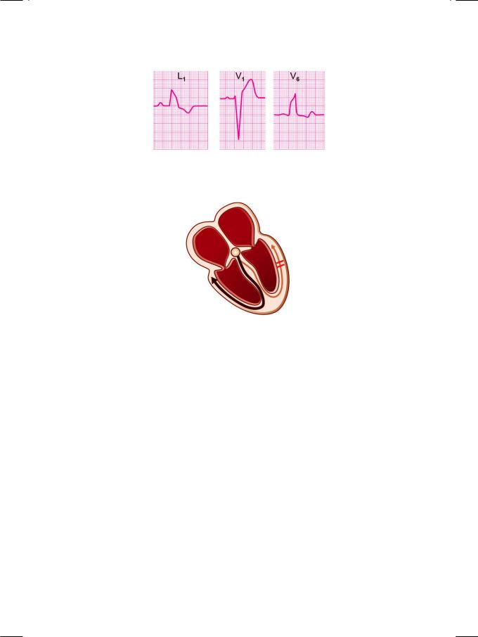
On examination, the patient was obviously tachypneic and looked anxious. He was pale and diaphoretic. The extremities were cold but there was no cyanosis. The JVP was not raised and there was no pitting edema around the ankles. The pulse was fast and low in volume with variable volume in successive beats. The BP was 104/74 mm Hg over the right arm in the supine position. The cardiac apex beat was diffuse and displaced towards the left axilla. The S1 was normal and the A2 was loud. There was paradoxical splitting of S2, that shortened during the phase of inspiration. A S3 was also appreciated in early diastole. A pansystolic murmur was audible over the cardiac apex, that radiated towards the axilla. Auscultation over the lung fields revealed bilateral crepts over the bases posteriorly. An ECG was obtained (Fig. 35.1).
ECG INTERPRETATION
The ECG showed broad ventricular complexes, that measured 0.14 sec. in width. In leads L1 and V6, each ventricular complex had two peaks, producing a M-shaped RsR’ pattern. The broad complex was followed by S-T segment depression and T-wave inversion. These findings are consistent with the diagnosis of left bundle branch block. Bundle branch block denotes delayed or interrupted conduction down the right or left branch of the bundle of His (Fig. 35.2). It leads to widening of the ventricular complex, due to delayed depolarization of the ventricle whose bundle branch is blocked. The QRS complexes of both the ventricles are “out of sync” with each other and produce two R waves in sequential order. Incomplete bundle branch block (BBB) results in QRS width of 0.11 to 0.12 sec while in complete BBB, the QRS width exceeds 0.12 sec. The S-T segment depression and
162 |
|
Section 11 Typical ECG Abnormalities |
|
|
|
Figure 35.1: ECG showing wide QRS complexes due to left bundle branch block
Figure 35.2: Diagram to represent left bundle branch block (LBBB)
T-wave inversion are repolarization abnormalities, secondary to the abnormal pattern of ventricular depolarization.
In left bundle branch block (LBBB), the M-shaped RsR’ pattern is observed in lead V6. The notched R wave represents abnormal sequence of septal and left ventricular free wall depolarization. There is total distortion of the normal QRS complex. In right bundle branch block (RBBB), the M-shaped rsR pattern is observed in lead V1. The septal (denoted by r) and free wall (denoted by S) depolarization through the left bundle branch are normal. Right ventricular depolarization (denoted by R’) is delayed. Therefore RBBB does not distort the normal QRS complex, but only adds a terminal R’ deflection.
Block in the conduction of impulses down one of the two fascicles of the left bundle branch constitutes a hemiblock (Fig. 35.3). Left anterior hemiblock (LAHB) is far more common than left posterior hemiblock (LPHB). Anterior hemiblock is more common because the anterior fascicle is compact, has a single blood-supply and lies close to the disease prone aortic valve. On the other hand, the posterior fascicle is fan-like, has a dual blood-supply and is far less likely to be involved in septal pathology. Hemiblock (LAHB) causes an abnormal pattern of left ventricular activation, resulting in left axis deviation of the QRS vector (Fig. 35.4).

Case 35 Left Bundle Branch Block |
|
163 |
|
|
|
Figure 35.3: Diagram to represent left anterior hemiblock (LAHB)
Figure 35.4: ECG showing left axis deviation due to left anterior hemiblock
CLINICAL DISCUSSION
Left bundle branch block (LBBB) often indicates the presence of organic heart disease. A variety of cardiac conditions can cause LBBB including myocardial infarction, systemic hypertension, aortic valve disease, cardiomyopathy and fibrocalcerous degeneration (Table 35.1). On the other hand, right bundle branch block (RBBB) is sometimes observed in normal individuals. Typical causes of RBBB are atrial septal defect, acute pulmonary embolism and chronic obstructive pulmonary disease.
RBBB does not distort the QRS complex, but only adds a terminal deflection due to delayed right ventricular depolarization. Therefore, changes of myocardial infarction such as appearance of Q waves and loss of R wave height, can be readily diagnosed in the presence of RBBB. However, it is difficult to diagnose myocardial infarction in the presence of LBBB, since the QRS complex is completely distorted.
Table 35.1: Causes of left bundle branch block
• Myocardial infarction • Aortic valve stenosis • Systemic hypertension
• Dilated cardiomyopathy
• Fibrocalcerous degeneration.

164 |
|
Section 11 Typical ECG Abnormalities |
||||
|
|
|
|
|
|
|
|
|
|
|
|
|
|
|
|
|
|
Table 35.2: Criteria for diagnosing MI in presence of LBBB |
|
|
|
|
|
• |
Presence of q wave in L1, V5 and V6 |
|
|
|
|
|
• |
Terminal S wave in leads V5 and V6 |
|
|
|
|
|
• |
S-T segment drift greater than 5mm |
|
|
|
|
|
• |
Upright T wave concordant with QRS |
|
|
|
|
|
|
|
|
|
The criteria for the diagnosis of myocardial infarction in the presence of LBBB are given in Table 35.2.
In left bundle branch block (LBBB), there is paradoxical splitting of the second heart sound (S2). The A2 component of S2 is delayed due to late depolarization of the left ventricle and follows the P2. During inspiration, when P2 gets delayed due to increased venous return, the splitting of S2 becomes narrow instead of physiological widening. Besides LBBB, another cause of paradoxical splitting of S2 is aortic valve stenosis in which left ventricular ejection time is prolonged. Other reasons for paradoxical splitting of A2 are pre-excitation of the right ventricle through an accessory bypass tract or premature activation of the right ventricle by an external pacemaker. LBBB causes a jerky motion of the interventricular septum on echocardiography. This should not be diagnosed as septal infarction, unless the septum fails to thicken during systole.
MANAGEMENT ISSUES
There is no specific treatment of left bundle branch block. The underlying heart disease is to be managed on its own merit. There is a word of caution regarding the use of drugs which block the A-V node such as verapamil, diltiazem and beta blockers. They may cause complete heart block, particularly if the block is bifascicular or trifascicular to begin with. Bifasicular block includes right bundle branch block and left anterior hemiblock. Trifasicular block is bifasicular block with a prolonged P-R interval.