

новая папка / [libribook.com] 50 Cases in Clinical Cardiology_ A Problem Solving Approach 1st Edition
.PdfC A S E
21 Pulmonary
Hypertension
CASE PRESENTATION
A 23-year old unmarried girl was brought to a physician with the complaints of progressively increasing breathlessness and fatigue on exertion, for the past 1 year. There was no history of fever, cough with purulent sputum, chest pain or hemoptysis. There was also no past history of seasonal allergy, bronchial asthma, anoxic spells or squatting attacks during her childhood. The patient had normal appetite without any weight-loss or history of night-sweats. On two occasions in the preceding month, she had fainted for less than five minutes. The fainting episodes were not preceded by palpitation, accompanied by tonic-clonic movements or followed by any abnormal behaviour. The parents of the girl denied any history of prolonged fever with painful joints, during her childhood or adolescence.
On examination, the pulse was feeble in volume at a rate of 92 beats/min. The BP was 106/68 mm Hg in the right arm and the patient was afebrile. A tinge of cyanosis was noticed over the tongue and lips but there was no clubbing of the finger-nails. Prominent a waves were observed in the venous pulsations at the neck, but there was no pedal edema. The apex beat of the heart was normal in position and had no special character. A parasternal heave was palpable along the left sternal border, with systolic pulsations felt over the 2nd left intercostal space. The S1 was normal but the P2 was accentuated. Splitting of S2 could be appreciated upto the cardiac apex. No S3 or S4 gallop sound was heard. A high-pitched diastolic murmur was audible along the sternum. The breath sounds were vesicular in nature without any rhonchi or crepitations.
CLINICAL DISCUSSION
From the history and physical examination, this young girl probably had cardiac outflow tract obstruction, that was responsible for her dyspnea and fatigue and also led to syncope. The most likely cause of low cardiac output in this case is pulmonary arterial hypertension. The prominent a waves in the jugular veins indicate forceful right atrial contraction against a noncompliant right ventricle. A left parasternal heave indicates right ventricular enlargement while systolic pulsations over the pulmonary area are due to dilatation of the main pulmonary artery.
Accentuation of the P2 is characteristic of pulmonary arterial hypertension. The fact that the P2 is audible even at the cardiac apex is itself indicative of the
96 |
|
Section 6 Pulmonary Diseases |
|
|
|
fact that the P2 is loud and the pulmonary hypertension is severe. Pulmonary hypertension causes pulmonary regurgitation even with a structurally normal pulmonary valve. This produces a high-pitched diastolic murmur known as the Grahm-Steel murmur. This murmur is short and does not increase during inspiration because the right ventricular end-diastolic pressure (RVEDP) is already high.
ECG showed tall R waves in right precordial leads and deep S waves in left precordial leads, indicative of right ventricular hypertrophy (Table 21.1).
Table 21.1: Criteria for right ventricular hypertrophy
• Clinical: Sustained left parasternal heave • ECG: Tall R waves in right precordial leads
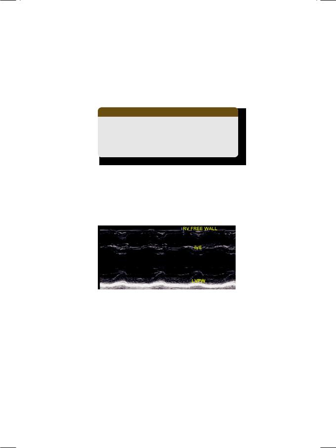
• X-ray: Cardiac silhouette to the right of midline • ECHO: Thickening of right ventricular free wall
Tall P waves (P. pulmonale) were due to right atrial enlargement. X-ray chest findings were an enlarged right ventricle to the right of midline, with a dilated main pulmonary artery and normal pulmonary vasculature. On ECHO, the left ventricular size and ejection fraction were normal and the left atrium was not dilated. The mitral and aortic valves were structurally normal without any pressure gradient or abnormal jet on color flow mapping. There was no shunt across the interventricular septum.
Figure 21.1: ECHO showing dilated right ventricle with paradoxical septal motion
The right ventricle was enlarged and globular in shape. There was paradoxical motion of the interventricular septum, that moved away from the left ventricular cavity in systole (Fig. 21.1). On short-axis view, the dilated main pulmonary artery with its right and left branches, gave a “pair of trousers” appearance (Fig. 21.2). On color flow mapping, a pulmonary regurgitant jet was observed in the right ventricular outflow tract (RVOT). The pulmonary artery pressure, as estimated from the peak trans-tricuspid velocity (Vmax) by using the Bernoulli equation (PG = 4 Vmax2), was markedly elevated (Fig. 21.3).
RVSP = PAP; |
|
RVSP – RAP = PG; |
RVSP = PG + RAP; |
RVSP = 4V2 + RAP |
PG |
: |
Pressure Gradient |
|
|
RAP |
: |
Right Atrial Pressure |
|
|
PAP |
: |
Pulmonary Artery Pressure |
|
|
RVSP |
: |
Right Ventricular Systolic Pressure |
|
|
Case 21 Pulmonary Hypertension |
|
97 |
|
|
|
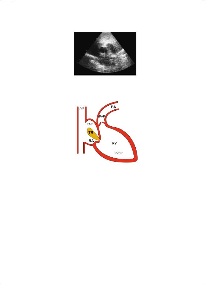
Figure 21.2: ECHO showing dilatation of the main pulmonary artery
Figure 21.3: Estimation of pulmonary artery pressure from tricuspid regurgitant velocity JVP: Jugular venous pressure; TR: Tricuspid regurgitation; RA: Right atrium; RV: Right ventricle; PA: Pulmonary artery
The clinical, electrographic and imaging evidence of right ventricular enlargement with pulmonary artery dilatation is also present in pulmonary valve stenosis but with certain differences. In pulmonary hypertension, the P2 is loud while in pulmonary stenosis, the P2 is muffled. On chest X-ray, in pulmonary hypertension there is normal pulmonary vasculature or pulmonary plethora while in pulmonary stenosis, the lung fields may be oligemic. On ECHO, thickening of pulmonary valve leaflets with systolic doming and restricted excursion is a feature of pulmonary valve stenosis. On color flow mapping, in pulmonary hypertension, a regurgitant jet is seen in the right ventricular outflow tract. In pulmonary stenosis, a systolic jet is observed in the proximal pulmonary artery.
There are several causes of pulmonary hypertension (Table 21.2). Elevated left atrial pressure due to mitral valve disease (stenosis or regurgitation) is a prominent cause. However, in our patient the mitral valve was structurally normal, without any increased flow velocity or a regurgitant jet. Increased pulmonary blood flow due to a left-to-right shunt is another important cause. But in our case there was

98 |
|
Section 6 Pulmonary Diseases |
|
|
|
Table 21.2: Causes of pulmonary hypertension
•Increased pulmonary flow
Lt. to Rt. shunt; ASD, VSD, PDA
•Raised left atrial pressure
Mitral valve disease; MS, MR
•Chronic pulmonary disease
Chronic bronchitis, fibrosis
•Obstruction to pulmonary flow
Thrombo-embolic, veno-occlusive
•Primary pulmonary hypertension
no ventricular septal defect or patent ductus arteriosus. Chronic obstructive or restrictive lung disease can also ultimately lead to pulmonary hypertension but there was no history of long-standing cough with expectoration or wheezing in our patient. Obstruction to pulmonary blood flow due to recurrent thromboembolism can also cause pulmonary hypertension but that is not the likely diagnosis in our case. Infrequent causes of pulmonary hypertension are intake of anorexigens, systemic sclerosis, sleep disordered breathing and HIV infection which are also unlikely possibilities in our patient. Therefore in all probability, our patient had idiopathic or primary pulmonary hypertension. The mild cyanosis could be due to minimal right-to-left shunting of blood across a patent foramen ovale.
MANAGEMENT ISSUES
In view of the pulmonary vasoconstriction and occlusion of the pulmonary vasculature associated with pulmonary hypertension, it is tempting to use vasodilators as therapeutic agents. Historically, a plethora of drugs have been used which includepapaverine,nicotinicacid,isoxsuprine,cyclandelateandpentoxyphylline. However, none of them has withstood the test of time or found to be beneficial during evaluation in clinical trials. Among the established cardiovascular drugs, isosorbide nitrate, hydralazine, prazosin and verapamil have also been tried in the treatment of pulmonary hypertension but their benefits have been modest. Antiplatelet drugs such as low dose aspirin, clopidogrel and the newer agent cilostazol have also been used with mixed results. Phosphodiesterase (PDE) inhibitors such as sildenafil and tadalafil are the only agents with proven clinical benefit.
RECENT ADVANCES
Given the success of phosphodiesterase inhibitors in the treatment of pulmonary hypertension, recently two new classes of vasodilators have been added to the therapeutic armamentarium. The prostaglandin (PG) analogues epoprostenol and iloprost have proven vasodilatory action. The endothelin (ET-1) antagonist bosentan is the most promising out of the newer agents. It has a vasorelaxant as well as antiproliferative action.
|
|
C A S E |
|
|
|
|
|
|
|
|
|
||
|
|
|
|
|
||
|
|
22 |
Pulmonary |
|
|
|
|
|
|
|
|
|
|
|
|
|
|
Embolism |
|
|
|
|
|
|
|
|
|
|
|
|
|
|
|
|
CASE PRESENTATION
A 72-year old elderly gentleman was rushed to the medical room of Toronto airport Canada, due to in-flight onset of severe chest pain, shortness of breath and profuse sweating. This happened minutes before the landing of his long-haul flight from New Delhi, India. He was a hypertensive on diuretic medication, but there was no past history of heart or lung disease. His son who received him at the airport, informed that the patient had undergone left knee replacement, six weeks prior to undertaking this journey.
On examination, the patient was restless, tachypneic, pale and diaphoretic. The pulse was rapid, irregular and feeble, at a rate of 110-120 beats/min. The BP in the right arm was 90/60 mm Hg. with a respiratory rate of 32/min and his temperature was 99.20 F. The JVP was elevated and there was edema, erythema and tenderness over the left calf. The apex beat was normal in location and a parasternal lift was felt. There was tachycardia, loud P2 and a right-sided S3. No murmur or pericardial friction rub was audible. On chest auscultation, breath sounds were diminished over the base of the left lung posteriorly.
CLINICAL DISCUSSION
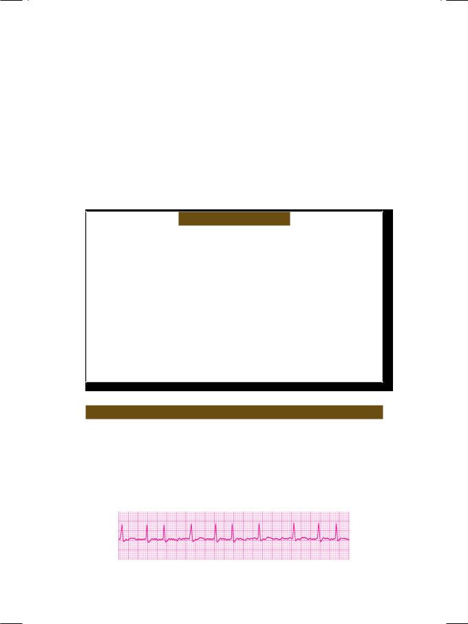
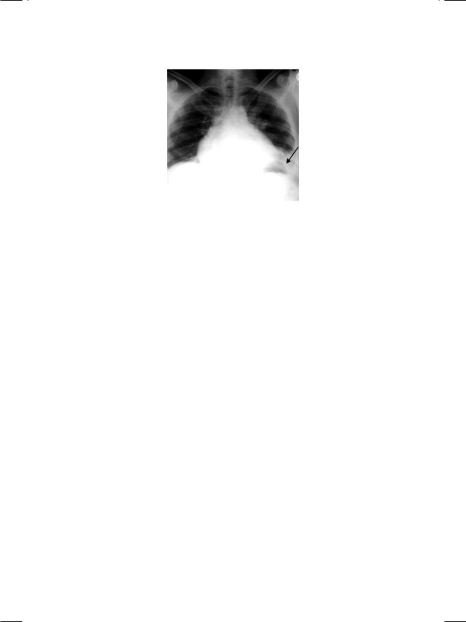
From the history and physical examination, the most probable diagnosis in this case is pulmonary embolism (PE) from deep vein thrombosis (DVT). ECG showed atrial fibrillation with fast ventricular response and T wave inversion in leads V1 to V3 (Fig. 22.1). There was no elevation of the S-T segment or presence of Q waves. X-ray chest findings were mild cardiomegaly, left basal atelectasis and a small left-sided pleural effusion (Fig. 22.2). There was no dilatation of the aortic root or widening of the mediastinum.
Figure 22.1: ECG showing atrial fibrillation with fast ventricular response
100 |
|
Section 6 Pulmonary Diseases |
|
|
|
Figure 22.2: X-RAY showing left basal atelectasis with small pleural effusion
ECHO revealed normal left ventricular size and ejection fraction, without any wall motion abnormality. There was a dilated and hypokinetic right ventricle with mild tricuspid regurgitation. The mitral and aortic valves showed some annular calcification, but there was no evidence of stenosis or regurgitation. There was no sign of aortic root dissection. There were also no vegetations on leaflets, ventricular thrombus or pericardial effusion.
There are several reasons for severe chest pain with dyspnea, sweating and hypotension. Acute myocardial infarction is the leading possibility, but the ECG did not show S-T segment elevation or presence of Q waves. Moreover, there was no regional wall motion abnormality of any left ventricular myocardial segment. Dissection of aorta may be considered, but there was no aortic root dilatation or cleavage of the aortic wall with a false lumen. Spontaneous pneumothorax generally occurs on a background of chronic lung disease with emphysema, but there was no such history in this patient. Moreover, there was no evidence of pneumothorax on the chest X-ray but only a small pleural effusion. Therefore, pulmonary embolism remains the strongest diagnostic possibility.
Pulmonary embolism is common and potentially life-threatening but often underdiagnosed clinical condition, associated with considerable morbidity and mortality. There is obstruction of pulmonary vasculature by a thrombus, fat, air or a tumour fragment. By and large, deep vein thrombosis (DVT) of the leg veins is the leading source. Predisposing factors are venous stasis, hypercoaguable state and injury to venous endothelium. Reasons for venous stasis are prolonged bed rest and immobility due to trauma, surgery, congestive heart failure or stroke. Long-distance air travel (economy-class syndrome) is also implicated. Hypercoaguability can be due to several reasons which may be inherited or acquired. Inherited defects are protein-C, protein-S or antithrombin-III deficiency and excess of homocysteine or fibrinogen. Acquired factors are estrogen therapy, malignancy, disseminated intravascular coagulation (DIVC) and antiphospholipid antibodies (APLA).
The clinical presentation of pulmonary embolism (PE) is extremely variable and is sometimes non-specific. Most patients present with pleuritic pain and dyspnea, with a variable degree of circulatory embarassment. In massive PE,

Case 22 Pulmonary Embolism |
|
101 |
|
|
|
there is hypotension and shock while in submassive PE, there is normotensive right ventricular dysfunction. Rarely, PE may present with insiduous onset of pulmonary hypertension. The clinical signs of acute PE are tachypnea and tachycardia, with or without hypotension. There is evidence of right ventricular strain in the form of elevated JVP, parasternal heave, loud P2 and right-sided S3. Signs of lower limb deep vein thrombosis (DVT) are edema, erythema and tenderness over the calf region.
PERTINENT INVESTIGATIONS
A large variety of investigations are performed for the diagnosis and assessment of pulmonary embolism. ECG invariably shows sinus tachycardia, sometimes with P. pulmonale, unless the patient is in atrial fibrillation. Right bundle branch block and T-wave inversion in leads V1 to V3 indicate right ventricular strain. The S1Q3T3 pattern, although specific for pulmonary embolism (PE), is seen in a minority of patients (Table 22.1). On chest X-ray, there may be basal atelactasis
Table 22.1: ECG features of pulmonary embolism
• Sinus tachycardia (invariable)
• Atrial fibrillation (sometimes)
• P. pulmonale (in sinus rhythm)
• Right bundle branch block
• Rightward QRS axis deviation
• Dominant R wave in lead V1
• T wave inversion in V1 to V3
• The S1 Q3 T3 Pattern prominent S in LI
significant Q in LIII inverted T in LIII
and a small pleural effusion. Classical finding of pulmonary infarction is a wedgeshaped homogenous opacity above the diaphragm known as Hampton’s hump or an oligemic lung segment which constitutes the Westermark’s sign (Table 22.2). ECHO may reveal a dilated and hypokinetic right ventricle with mild tricuspid regurgitation and sometimes a right ventricular thrombus. Echocardiography is more often used to exclude other entities that mimic PE such as myocardial infarction, aortic dissection and pericardial tamponade.
Table 22.2: X-ray findings in pulmonary embolism
• Basal atelectasis
• Small pleural effusion
• Oligemic lung segment (Westermark’s sign) • Wedge-shaped opacity (Hampton’s hump).

102 |
|
Section 6 Pulmonary Diseases |
|
|
|
On arterial blood gas (ABG) analysis, hypoxemia is invariably present if the PE is massive. Hypocapnia and respiratory alkalosis also occur unless there is ventilatory limitation, in which case there is hypercapnia. D-dimer is a degradation product of cross-linked fibrin and its quantitative assay is used for the diagnosis of PE. However, it is more useful as a “rule-out” test, with a high negative predictive value. Duplex ultrasound is a combination of Doppler venous flow detection and real-time B-mode imaging. It plays a pivotal role in the diagnosis of lower extremity deep vein thrombosis (DVT). The most reliable finding of DVT is non-compressibility of a venous segment.
Ventilation-perfusion (V-Q) scanning used to be popular for the diagnosis of PE, prior to the advent of high-resolution contrast-enhanced computed tomography (CT). Its diagnostic accuracy is greater when it is clubbed with a clinical probability score than when used alone. Presently, it is reserved for those who cannot tolerate intravenous contrast because of allergy or renal failure. Contrast enhanced computed tomography (CT) is now considered the ‘gold-standard’ imaging modality for the diagnosis of PE. New generation CT scanners can visualize sixth-order branches of the pulmonary vasculature with high resolution. They have virtually replaced the older technique of invasive pulmonary angiography. Classical findings on CT are filling defect and abrupt cut-off of a vessel. Magnetic resonance (MR) pulmonary angiography can only detect large proximal PE and is not reliable for segmental and sub-segmental emboli.
MANAGEMENT ISSUES
The choice of therapy in pulmonary embolism is largely individualized and depends upon the clinical severity. Needless to say, hemodynamic and respiratory support are of paramount importance in those patients who present with shock or hypotension. Anticoagulants are the mainstay of treatment and should be initiated instantly pending investigations, to prevent further extension of an already formed thrombus. Sub-cutaneous low molecular weight heparin (LMWH) such as Enoxaparin is preferable, because of predictable dose response and no need for laboratory monitoring. The new-drug Fondaparinux, a factor Xa inhibitor can also be used. When unfractionated heparin is used, the international normalized ratio (INR) should be 1.5 to 2.5 times the control, for full therapeutic effect. Importantly, certain conditions in the differential diagnosis of PE, such as aortic dissection and pericardial tamponade, are contraindications to anticoagulation.
Thrombolytic therapy accelerates the lysis of acute pulmonary embolism, but is associated with an increased risk of major hemorrhage. It is reserved for massive PE, with hypotension and severe right ventricular dysfunction. If thrombolysis fails or is contraindicated, percutaneous embolectomy can be performed. However, it can only be used in the main arteries and if attempted in smaller branches, can cause perforation. Finally, if there is an absolute contraindication to anticoagulation or failure of the same and there is a proximal venous thrombus, an inferior vena caval (IVC) filter can be placed. It provides a screen to prevent lower limb or pelvic emboli from travelling to the lungs.

|
|
C A S E |
|
|
|
|
|
|
|
|
|
||
|
|
|
|
|
||
|
|
23 |
Obstructive |
|
|
|
|
|
Pulmonary Disease |
|
|
||
|
|
|
|
|||
|
|
|
|
|
||
|
|
|
|
|
|
|
CASE PRESENTATION
A 63-year old man visited a chest physician with the complaints of fever, breathlessness, cough and wheeze, for the past 1 week. The fever was of moderate grade, without chills, rigors or night-sweats, but was associated with body-ache and fatigue. He did feel breathless for the past several years, but his dyspnea had worsened since the onset of fever. He was orthopneic in bed but denied paroxysms of nocturnal dyspnea. The cough was associated with copious purulent sputum especially during morning hours, but there was no hemoptysis. The patient also experienced tightness in the chest on walking and he was aware of wheezing. His appetite was moderate and there was no history of significant weight loss. On specific questioning, he admitted smoking about 20 cigarettes daily, for the last over 35 years.
On examination, he was obviously dyspneic and orthopneic in bed and his accessory muscles of respiration were working. The face was puffy and hyperemic and there was mild cyanosis over the tongue and lips. The extremities were warm and sweaty. Clubbing of the finger-nails was noticed and there was edema over the ankles. The JVP was raised, trachea was central and there were no palpable lymph nodes in the neck. The pulse rate was 104 beats/min with a good volume that decreased appreciably during inspiration. The BP was 136/84 mm Hg, temperature 101.60 F and the respiratory rate was 28 per minute.
The antero-posterior diameter of the chest was increased, giving it the shape of a barrel. The percussion note over both lung fields was hyper-resonant and the upper border of the liver was percussed in the 6th right intercostal space. Bilateral expiratory rhonchi with scattered crepts were audible over the entire lung fields, with normal air-entry. The cardiac apex beat could not be located but there was a palpable parasternal heave. The S1 was normal with a loud P2. A soft early-diastolic murmur followed the P2. A pansystolic murmur was also audible over the lower left parasternal area, that did not radiate towards the axilla. On abdominal examination, the liver edge was palpable 5 cm below the costal margin and was pulsatile. There was no other organomegaly or sign of free fluid in the abdominal cavity.
104 |
|
Section 6 Pulmonary Diseases |
|
|
|
CLINICAL DISCUSSION
From the history and physical examination, the most likely diagnosis in this case is chronic obstructive pulmonary disease (COPD). The fever with worsening dyspnea and purulence of sputum indicates acute exacerbation of COPD, which is not uncommon among smokers. There were signs of respiratory distress (working accessory muscles), polycythemia (facial hyperemia), deoxygenation (cyanosis) and chronic lung suppuration (finger-nail clubbing). The raised JVP, enlarged liver and ankle edema indicate right heart failure which, when it is due to COPD, is known as corpulmonale. An appreciable decline in pulse volume during inspiration, is termed as pulsus paradoxus. Besides COPD, other causes of pulsus paradoxus are status asthmaticus and constrictive pericarditis.
A barrel-shaped chest with hyper-resonance over the lung fields and a “pushed-down” liver are indicative of hyperinflation, which is termed as pulmonary emphysema. Because of hyperinflated lungs, the cardiac apex beat is difficult to locate. In acute exacerbation of COPD, the adventitious breath sounds (rhonchi and crepts) are audible over the entire lung fields and the airentry is equal. A parasternal heave with a loud P2 and an early-diastolic murmur of pulmonary regurgitation are indicative of pulmonary arterial hypertension. Pulmonary hypertension leads to secondary tricuspid regurgitation which produces a lower parasternal pansystolic murmur and an enlarged liver which is sometimes pulsatile. The neck veins are distended with a prominent y descent.
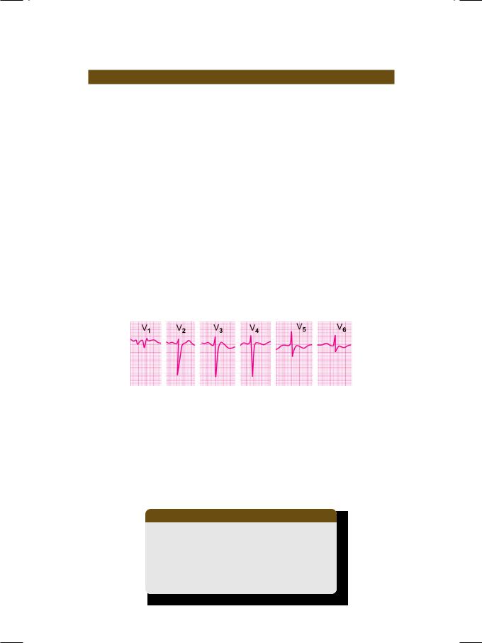
Figure 23.1: ECG showing non-progression of R wave
in the left precordial leads
The ECG shows low voltage of the QRS complexes, with non-progression of the R wave in precordial leads from V1 to V6 (Fig. 23.1). Due to clock-wise rotation of the heart, the right ventricle underlies most of the precordium, producing a rS pattern in most precordial leads. Other ECG features are tall R waves in right precordial leads (right ventricular hypertrophy), tall P waves (right atrial enlargement) and rightward deviation of the QRS vector (Table 23.1). The cardiac
Table 23.1: ECG abnormalities in COPD
• Low voltage QRS complexes (rS pattern) • Right ventricular hypertrophy (tall R in V1) • Right atrial enlargement (P. pulmonale)
• Right axis deviation of QRS vector (RAD) • Atrial tachyarrhythmias e.g.fibrillation (AF)