

новая папка / [libribook.com] 50 Cases in Clinical Cardiology_ A Problem Solving Approach 1st Edition
.Pdf66 |
|
Section 4 The Cardiomyopathies |
|
|
|
LVOT obstruction may be present at rest or become more pronounced after provocation. Provocation is provided by prolonged standing, Valsalva manoeuvre or giving sublingual nitrate. All these methods reduce left ventricular size and therefore increase the likelihood of LVOT obstruction. Conversely, squatting and isometric exercise such as hand-grip, increase left ventricular size and reduce the degree of LVOT obstruction.
MANAGEMENT ISSUES
The goals of treatment in hypertrophic cardiomyopathy are to control symptoms and to prevent sudden cardiac death (SCD). Symptoms include exertional dyspnea, typical or atypical angina, palpitations and syncope. Exertional symptoms are generally because of left ventricular hypertrophy with diastolic dysfunction and sometimes due to associated mitral regurgitation. Syncopal episodes are generally related to outflow tract obstruction and sometimes due to tachyarrhythmias. Patients should be advised to avoid strenuous exercise such as weight-lifting and not to participate in competitive sports.
Beta-blockers are the agents of first choice. They control heart rate, improve diastolic filling and reduce the outflow tract gradient. Calcium-channel blockers like verapamil are preferable, if beta-blockers are contraindicated. These agents are also used for rate-control if atrial fibrillation exists, although rhythm-control with amiodarone is preferable. This is because the loss of atrial contribution to ventricular filling, is often associated with clinical deterioration. All patients in atrial fibrillation should also receive an anticoagulant to reduce the risk of thromboembolism. Digitalis, diuretics and vasodilators should be avoided.
If the patient is symptomatic and refractory to medical treatment, surgical myomectomy is offered to relieve the outflow obstruction. In a patient who is at high-risk to undergo a surgical procedure, alcohol septal ablation can be performed by the percutaneous route. An injection of ethanol is given into one of the septal perforator branches to create an area of myocardial infarction and thereby widen the narrow outflow tract.
Sudden cardiac death (SCD) can be prevented by using an implantable cardioverter defibrillator (ICD), in selected high-risk candidates. These subjects are selected on the basis of identified risk factors for SCD. Risk factors include septal thickness >25 mm, outflow tract gradient >30 mm Hg, non-sustained ventricular tachycardia on 48 hour ambulatory Holter monitoring, history of recurrent syncope and atleast 1 SCD in a relative aged <45 years.
RECENT ADVANCES
Recently, cardiac magnetic resonance imaging (MRI) has been used to identify myocardial fibre disarray, as a risk factor for ventricular arrhythmias. Those subjectswiththisdemonstrableabnormalityshouldbecandidatesforimplantable cardioverter defibrillator (ICD), to prevent sudden cardiac death (SCD). Alcohol septal ablation as an interventional strategy, has gained popularity.
C A S E
15 Takotsubo
Cardiomyopathy
CASE PRESENTATION
A 33-year old woman came on her own to the emergency room at 2 am, with the complaint of severe central chest pain and suffocation sensation, of one hour duration. She was also short of breath and sweating, but her chest pain did not radiate to the arms or lower jaw. There was no past history of exertional angina or dyspnea and she did not have diabetes or hypertension. There was also no history of a “flu-like” illness in the preceding week. She was pre-menopausal and her blood lipid profile had never been checked. She did not smoke, consume alcohol or abuse illicit drugs, although she was on antidepressant medications off and on. There was no history of premature coronary artery disease is any of her family members. Upon close questioning, the lady disclosed that she lived alone ever since her acrimonious divorce 3 months back, five years after a traumatic marriage.
On examination, the patient was of average built, anxious, sweating and tachypneic. The extremities were warm and her palms were moist. The pulse was rapid, fair in volume, at a rate of 110 beats/min. with a BP of 140/90 mm Hg. There was no anemia, cyanosis or pedal edema and the JVP was not raised. The thyroid gland was not enlarged and there was no tremor or eye-sign of thyrotoxicosis. The apex beat was normal in location and character and the precordium was unremarkable. The S1 was loud and S2 normal in intensity with a soft S3 in early diastole. No murmur or friction rub was audible. There were few rales over the lower thirds of the lung fields.
CLINICAL DISCUSSION
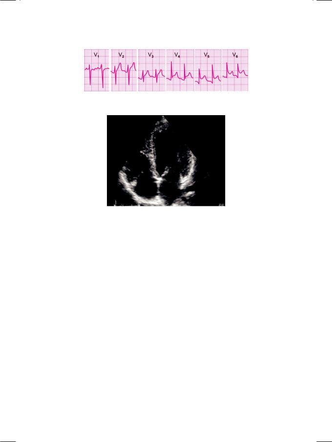
From the history and physical examination, this patient developed acute left ventricular dysfunction preceded by severe chest pain. ECG showed sinus tachycardia and S-T segment elevation in practically all the leads, with upright T waves (Fig. 15.1). X-ray chest findings were borderline cardiomegaly and pulmonary interstitial edema but there was no mediastinal widening or pleural effusion. ECHO revealed a mildly dilated left ventricle with an ejection fraction of 35%. The distal septum and the left ventricular apex were hypokinetic (Fig. 15.2). The mitral and aortic valves were structurally normal and no colour flow jet or abnormal velocity was detected. No intracardiac mass or pericardial effusion was seen. The proximal aorta did not show dilatation or any sign of aortic dissection.
68 |
|
Section 4 The Cardiomyopathies |
|
|
|
Figure 15.1: ECG showing sinus tachycardia with ST segment elevation
Figure 15.2: ECHO showing hypokinesia of distal septum and apex
Whenever a patient has severe chest pain with S-T segment elevation on ECG and wall motion abnormality on ECHO, the first possibility to cross the mind is an acute coronary syndrome. This patient had no major coronary risk factors barring psycho-social stress, but her troponin-T test was positive. Since she came to a tertiary-care hospital, primary coronary angioplasty was planned. To everyone’s surprise, the coronary angiogram turned out to be completely normal.
Another cause of severe chest pain with ECG findings is acute dissection of the aorta. These patients are usually hypertensive and develop acute aortic regurgitation with left ventricular volume overload. Our patient had no evidence of aortic root dilatation, cleavage of the aortic wall or aortic regurgitation. Rarely, dissection of the aorta can cause myocardial infarction, if the coronary ostia get occluded by the intimal flap.
Acute pericarditis also presents with chest pain and S-T segment elevation on the ECG. The pain is seldom excruciating and generally increases on deep inspiration. The troponin-T test is almost always negative. Presence of regional wall motion abnormality and reduced ejection fraction are unusual except if there is an additional component of myocarditis. Acute viral myocarditis is often preceded by a flu-like illness. It presents with acute left ventricular dysfunction but chest pain is unusual. Classical findings are global hypokinesia with depressed ejection fraction but regional wall motion abnormalities can occur if the myocardial inflammation is patchy in distribution.
Some other causes of acute left ventricular dysfunction also need to be considered. Low levels of serum electrolytes such as potassium, calcium and

Case 15 Takotsubo Cardiomyopathy |
|
69 |
|
|
|
magnesium can impair myocardial contractility. Similarly, nutritional deficiency of vitamins and trace elements like thiamine, selenium and carnitine can also cause ventricular dysfunction. At times, thyroid storm and pheochromocytoma can present with left heart failure and should be ruled out by appropriate assays including serum thyroid hormone levels and urinary catecholamines. In our case, the left ventricular dysfunction was clearly related to psycho-social stress. Therefore, the definite diagnosis is stress-induced cardiomyopathy which is also termed as Takotsubo cardiomyopathy.
Table 15.1: Synonyms of Takotsubo cardiomyopathy
• Broken heart syndrome
• Stress induced cardiomyopathy • Neurogenic myocardial stunning
• Transient ventricular apical ballooning
Takotsubo cardiomyopathy (Table 15.1) is a recently described entity also called the “broken heart syndrome”, neurogenic myocardial stunning or transient left ventricular apical ballooning. Takotsubo is a Japanese name given to a roundbottomed narrow-necked fishing pot, used to trap an octopus. The shape of the left ventricle after apical ballooning, resembles this fishing pot. It occurs more often in postmenopausal, middle-aged women. Acute intense emotional stress is the usual triggering factor and the surge of catecholamines either causes microvascular vasospasm or direct myocyte injury. Additionally, release of free fatty acids by adrenalin induced lipolysis and hypokalemia due to an excess of cortisol, impair myocardial contractility.
These patients usually present with severe chest pain and clinical signs of left ventricular dysfunction. The ECG shows S-T segment elevation in multiple coronary artery territories and the biomarker of myocardial injury, the troponin-T test, is generally positive. The ECHO shows wall motion abnormality of the left ventricular apex which does not conform to any particular coronary artery territory and typically spares the basal segments. Coronary angiography does not show any major occlusive lesion in most patients.
MANAGEMENT ISSUES
Before the clinical profile of this entity was clearly elucidated, many women with Takotsubo cardiomyopathy were probably managed as anxiety related panic attack or menopausal syndrome. Some were thought to have acute viral myocarditis while still others improved on their own while being investigated for thyroid hyperfunction and pheochromocytoma. When the patients present with chest pain, S-T segment elevation and positive troponin-T test, it is prudent to manage them as acute coronary syndrome. Aspirin, clopidogrel, beta-blocker and low molecular weight heparin (LMWH) can all be instituted. An angiotensin converting enzyme (ACE) inhibitor with a diuretic can be added for heart failure management. Most patients make a rapid and event-free recovery, without any long-term disability or recurrence.

70 |
|
Section 4 The Cardiomyopathies |
|
|
|
RECENT ADVANCES
Till a few years back, some stray case-reports of Takotsubo cardiomyopathy were published from Japan. With growing awareness about this entity, it is now being increasingly diagnosed across the globe. Recently carvedilol, a beta-blocker with additional alpha-blockade property, has been studied in a small cohort of cases and shown modest benefits.

S E C T I O N
5
Aortic Diseases


|
|
C A S E |
|
|
|
|
|
|
|
|
|
||
|
|
|
|
|
||
|
|
16 |
Aneurysm |
|
|
|
|
|
|
|
|
|
|
|
|
|
|
of Aorta |
|
|
|
|
|
|
|
|
|
|
|
|
|
|
|
|
CASE PRESENTATION
A young man 21 years of age, came to his family physician with the complaint of progressively increasing shortness of breath of 2 months duration. There was no history of fever, productive cough, chest pain, hemoptysis or wheezing. The severity of his breathlessness had compelled him to discontinue basketball, his favorite sport. In fact he had been a regular member of his school and college basketball teams. Interestingly, he was inducted into his senior school team in class 8, when he was only 13 years of age, because of his extraordinary height. There was no history of nasal allergy, recurrent sore-throat, bronchial asthma or prolonged fever with joint pains during his childhood. He denied abusing tobacco, alcohol or any illicit drug. During a routine medical check-up before joining college 3 years back, he had been told of a heart murmur.
On examination, pulse was 88 beats/ min. regular, good in volume, with a BP of 160/70 mm Hg in the right arm. The JVP was not raised and there was no ankle edema. The precordium was hyperdynamic and there were visible pulsations in the aortic area. The apex beat was heaving in nature and located in the sixth intercostal space, in the anterior axillary line. A soft early diastolic murmur was audible along the left sternal border, accompanied by a S3 sound in diastole.
The ECG was normal and X-ray chest showed mild cardiomegaly, with a hump over the upper right border of the heart. There was no sign of pulmonary congestion. ECHO revealed a dilated left ventricular cavity, with an end-systolic diameter of 52 mm and an ejection fraction of 45%. The aorta was enormously dilated, with a dimension of 68 mm between the leading edges of the anterior and posterior aortic walls. There was loss of the acute angle between the sinus of Valsalva and the ascending aorta. The aortic valve leaflets were not thickened, but appeared distant from the aortic walls. The mitral valve was structurally normal.
The patient was unusually tall, thin and lanky, with a height of 6 feet 1 inch and a body-weight of only 48kg. While examining the radial pulse, it was observed that his fingers were excessively long and tapering, with an extremely flexible wrist joint. Upon measurement, the distance between the finger-tips of the hands stretched sideways, was 6 feet 5 inches. In other words, the arm-span exceeded the height of the patient. Also, the pubis-to-heel length was more than the crown-to-pubis height. This physical appearance, coupled with the typical X-ray chest and ECHO findings of aortic root dilatation and aortic regurgitation, are highly suggestive of Marfan’s syndrome.

74 |
|
Section 5 Aortic Diseases |
|
|
|
CLINICAL DISCUSSION
The normal diameter of the aortic root in end-diastole, ranges from 20 to 37 mm. A dimension exceeding 40 mm constitutes aortic root dilatation, which has several causes (Table 16.1). If the dilatation is due to advanced age or systolic hypertension, the aortic annulus is normal in diameter. However, if the dilatation is caused by medial necrosis of the aortic wall or an inflammatory aortitis, the annulus is dilated with associated aortic regurgitation. In post-stenotic aortic dilatation, the aortic valve leaflets are thickened and calcific.
Table 16.1: Causes of aortic root dilatation
• |
Atherosclerosis |
Unfolded aorta |
|
|
Hypertension (elderly) |
|
|
|
• |
Medial necrosis |
Marfan syndrome |
|
|
Ehlers-Danlos syndrome |
|
|
|
• |
Aortoarteritis |
Syphilitic (rare) |
|
|
Takayasu disease |
|
|
|
• |
Collagen disease |
Reiter’s syndrome |
|
|
Ankylosing spondylitis |
|
|
|
• |
Post-stenotic |
Aortic stenosis |
|
|
|
• |
Post-traumatic |
Aortic aneurysm |
|
|
|
An aortic root diameter exceeding 60 mm, is a feature of aortic aneurysm which may be saccular or fusiform. The aneurysm appears as a balloon-like stretching of the aortic wall, which expands in systole and compresses the left atrium. The aortic valve cusps appear distant from the aortic walls, even when the valve is open in systole (Fig. 16.1).
Figure 16.1: Diagram showing aneurysmal dilatation of the ascending aorta. RV: Right ventricle; LV: Left ventricle; AO: Aorta; LA: Left atrium
Marfan’s syndrome is a connective tissue disorder, inherited as an autosomal dominant trait. The defect lies in the elastic fiber nature of the connective tissue. There is mutation in the gene encoding fibrillin-1, a connective tissue component and regulatory protein of transforming growth factor (TGF). Several striking