

новая папка / [libribook.com] 50 Cases in Clinical Cardiology_ A Problem Solving Approach 1st Edition
.Pdf
|
|
|
Case 49 Papillary Muscle Rupture |
|
225 |
||
|
|
|
|
|
|
|
|
|
|
|
|
|
|
|
|
|
Table 49.2: Differences between flail mitral leaflet and mitral valve prolapse |
|
|
|
|||
|
|
Flail leaflet |
|
Prolapsed leaflet |
|
|
|
|
Range of motion |
flaps freely |
|
just buckles |
|
|
|
|
Entry into left atrium |
|
|
|
|
|
|
|
extent |
deep into |
|
just enters |
|
|
|
|
duration |
for long time |
|
for short time |
|
|
|
|
Direction of tip |
towards LA |
|
towards LV |
|
|
|
|
|
|
|
|
|
|
|
|
|
|
|
|
|
|
|
MANAGEMENT ISSUES
Mitral regurgitation due to coronary heart disease is multifactorial and papillary muscledysfunctionorruptureisoneofthecauses.Otherreasonsaredysfunctional ventricular remodelling with increased sphericity and mitral annular dilatation due to ventricular enlargement. Medical management includes vasodilators along with a diuretic, provided the blood pressure allows their use. In surgical treatment, since the valvular anatomy is generally not distorted, mitral valve replacement is not the answer. Instead restrictive annuloplasty, which involves insertion of an undersized ring to improve leaflet apposition, is preferable. Moreover, annuloplasty preserves left ventricular geometry and spares the patient from the problems of anticoagulation (thrombosis, bleeding and monitoring).
|
|
C A S E |
|
|
|
|
|
|
|
|
|
||
|
|
|
|
|
||
|
|
50 |
Left Ventricular |
|
|
|
|
|
|
|
|
|
|
|
|
|
|
Aneurysm |
|
|
|
|
|
|
|
|
|
|
|
|
|
|
|
|
. |
CASE PRESENTATION |
A 68-year old man came with his son to his treating cardiologist, for a periodic heart check-up. Three months back, the patient has sustained an anterior wall myocardial infarction (MI), for which he was treated in this very hospital. At that time, he received oral aspirin, sub-cutaneous enoxaparin and an infusion of nitroglycerine. He was not given thrombolytic therapy because he presented over 24 hours after the onset of his chest pain and Q waves had already appeared on the ECG. The patient also declined coronary angiography, because he was not ready to undergo a revascularization procedure. Thereafter, he did not develop post-MI angina, but he did complain of some fatigue and exertional breathlessness.
On examination, the patient was tachypneic while lying on the couch in the doctor’s chamber. The pulse rate was 96 beats/min. with a BP of 110/74 mm Hg over the right arm. The JVP was not raised and there was no edema over the ankles. The precordium was remarkable because of a prominent bulge and a double apical impulse. The apex beat was diffuse and sustained and extended medially and upwards, upto the 3rd intercostal space. On auscultation, the S1 and S2 were normal but a soft S3 sound was audible in early diastole. No murmur or pericardial friction rub was appreciated. The breath sounds were vesicular with few crepitations audible over the lower lung fields.
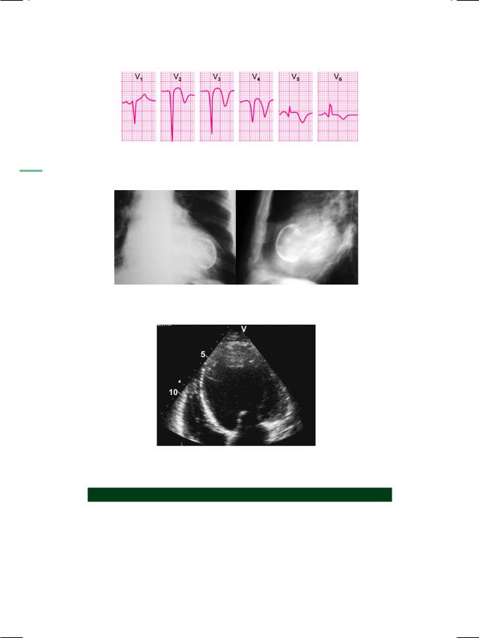
ECG showed attenuation of R waves in the antero-septal precordial leads. There was coving and elevation of the S-T segment with inversion of the T waves (Fig. 50.1). The cardiac Troponin -T test was negative. X-ray chest findings were increased cardio-thoracic ratio with a large bulge on the left border of the heart. An arc-like hemispherical calcification was seen within the bulge (Fig. 50.2).
ECHO revealed a dilated left ventricle with an ejection fraction of 35%. There was a large dyskinetic area involving the mid and distal interventricular septum as well as the left ventricular apex. The dyskinetic area underwent outward systolic expansion with a persistent deformity during diastole. The wall of the dyskinetic area was more echogenic than the adjacent myocardium. A laminated mass was observed contiguous with the dyskinetic area, with which it moved synchronously (Fig. 50.3). These findings are consistent with the diagnosis of left ventricular aneurysm with mural thrombus.
|
|
Case 50 Left Ventricular Aneurysm |
|
227 |
|
|
|
||
|
|
|
|
|
|
|
|
|
|
Figure 50.1: ECG showing R wave attenuation,
S-T coving and T wave inversion
Figure 50.2: X-ray showing an arc-like calcification within a ventricular bulge
Figure 50.3: ECHO showing a mural thrombus arising from the ventricular apex
CLINICAL DISCUSSION
A ventricular aneurysm usually develops after an anterior wall myocardial infarction, due to occlusion of the left anterior descending coronary artery. It rarely follows inferior wall infarction. On ECG, if the typical pattern of evolved myocardial infarction (MI) persists for three or more months after acute MI, ventricular aneurysm should be suspected. On precordial inspection, the ventricular aneurysm produces a double left ventricular apical impulse. If the aneurysm is particularly large, it causes a sustained and diffuse apical impulse that extends medially and upwards, by two or more intercostal spaces.

228 |
|
Section 14 Coronary Artery Disease |
|
|
|
|
|
|
|
|
|
|
|
|
|
|
|
|
|
Table 50.1: Differences between true LV aneurysm and Pseudo-aneurysm |
|
||
|
|
|
LV aneurysm |
Pseudo-aneurysm |
|
|
|
Shape |
Wide neck |
Narrow neck |
|
|
|
Location |
Apex of LV |
Posterior wall |
|
|
|
Motion |
Dyskinetic |
Expansile |
|
|
|
Wall |
Myocardium |
Pericardium |
|
|
|
Rupture |
Unlikely |
Liable |
|
|
|
Thrombus |
Laminar |
Fills cavity |
|
|
|
|
|
|
|
|
|
|
|
|
|
A ventricular aneurysm is a large bulge-like deformity with a wide neck, located at or near the apex of the left ventricle. It is more common after damage to the anterior wall than after inferior wall infarction. The aneurysm exhibits dyskinesia or outward systolic expansion and a persistent deformity during diastole. The wall of the aneurysm is made of myocardium and is more echogenic than adjacent areas because it is made of fibrous scar tissue. It does not rupture but is often associated with a pedunculated or laminated ventricular thrombus.
A false aneurysm (pseudo-aneurysm) follows rupture of the left ventricular free wall, where the resultant hemopericardium clots and seals the breach by pericardial adhesions. The neck of the pseudo-aneurysm that communicates with the left ventricle is narrower than the aneurysm itself. The pseudo-aneurysm appears as a globular extracardiac pouch. A false aneurysm is located on the posterolateral LV wall and is more common after inferior wall than after anterior wall infarction. It is not expansile and remains constant in size. The wall of the aneurysm is made of pericardium and it is less echogenic than adjacent areas. It is friable, more liable to rupture and it is often filled with a thrombus caused by hemopericardium. The differences between a true aneurysm and a pseudoaneurysm are given in Table 50.1.
The development of a left ventricular aneurysm after acute myocardial infarction is a serious complication. Most patients go on to develop intractable congestive heart failure requiring aggressive decongestive therapy. There is a higher likelihood of ventricular tachycardia originating from the myocardial scar. The incidence of post-infarction unstable angina is also increased. Moreover, fragments of the mural thrombus in the aneurysmal pouch may dislodge to produce distal embolism (Table 50.2).
Table 50.2: Complications of ventricular aneurysm
• Recurrent ventricular tachycardia • Post-MI unstable angina pectoris • Intractable congestive heart failure
• Mural thrombus with distal embolism

Case 50 Left Ventricular Aneurysm |
|
229 |
|
|
|
MANAGEMENT ISSUES
Presence of a thrombus within a ventricular aneurysm is an established indication for oral anticoagulant therapy. Other definite indications for anticoagulation are mechanical prosthetic valve and left atrial thrombus with mitral stenosis. While it takes 3 to 5 days for the therapeutic effect of an oral anticoagulant like warfarin to take over, heparin is given in this interim period. Unfractionated heparin requires monitoring of prothrombin time (PT) and aPTT, but it is cost-effective. Low molecular weight heparin (LMWH) like enoxaparin is more expensive but does not require monitoring.
It is established practice to prescribe aspirin, a statin, an ACE-inhibitor and a beta-blocker to every survivor of myocardial infarction, unless there is a specific contraindication. A diuretic is added to reduce cardiac workload, if there are symptoms of pulmonary congestion. An aldosterone antagonist like spironolactone or eplerenone can reduce cardiac workload and improve ventricular remodelling. A nitrate is prescribed for post-infarction angina while amiodarone is used if serious ventricular arrhythmias are documented.
It is not uncommon for patients who develop a ventricular aneurysm, to have left main or triple-vessel coronary artery disease (TVD). Resection of the aneurysm can be contemplated at the time of coronary artery bypass grafting (CABG) surgery which is performed for either unstable angina or for refractory heart failure.

Index
Page numbers followed by f refer to figure and t refer to table, respectively.
A
Acid-fast bacilli 106 Acute
alcoholic intoxication 201 AR 46
coronary syndrome 39,,217, 219, 221t, 220
mitral regurgitation 32, 223 myocardial infarction 32, 152,,184, 20 5, 218, 218f
myocarditis 127, 128, 129t pericarditis 109, 127, 185 pulmonary embolism 187 renal failure 171 rheumatic fever 125 valvular regurgitation 39 viral pericarditis 110
Addison’s disease 171 Adenosine 198 Amiodarone 18, 177
therapy 198 Amyloidosis 61 Aneurysm of
aorta 73
sinus of Valsalva 83, 86, 87 Aneurysmal
aorta with jet of aortic regurgitation 75f dilatation of ascending aorta 74f
Angiosarcoma 145
Angiotensin converting enzyme 129 inhibitor 69, 171
Ankylosing spondylitis 46,,50, 74 Annuloaortic ectasia 86
Anomalous left coronary artery arising from pulmonary artery 22
Anterior mitral leaflet 63 Antiarrhythmic drugs 177 Antistreptolysin O 111, 124
Anxiety neurosis and thyrotoxicosis 204 Aorta 13f
Aortic
aneurysm 74,,75, 76t dilatation 75 diseases 71 dissection 75, 79t
regurgitation 19, 26, 43, 44f, 45, 46t, 75, 125
root abscess 136 sclerosis 48
stenosis 39, 42, 50,,74, 125 valve 87f, 144f
calcification 50 endocarditis 133 leaflets 134f regurgitation 79 replacement 42,,46, 51 sclerosis 50
stenosis 39, 40f, 84, 215 Aortoarteritis 50, 74 Aortopulmonary window 20, 86 Arrhythmogenic right ventricular
dysplasia 178 Arterial blood gas 102, 106 Arteriovenous fistula 20, 44 Arthralgia 125
Assessment of severity of mitral regurgitation 31t
stenosis 27t
Asymmetrical septal hypertrophy 65 Atherosclerosis 74
Athlete’s heart 184 Atrial
fibrillation 101, 199-202 flutter 200t
myxoma 143, 145, 145t, 146, 149 natriuretic peptide 198
septal defect 7, 8, 8f, 9f, 13, 16, 22, 87 tachyarrhythmia 104
thrombus 145, 145t, 147, 149 Atriofascicular bypass tract 191

232 |
|
50 Cases in Clinical Cardiology: A Problem Solving Approach |
|
|
|
Atrioventricular block 181 connection 191 node 18, 198
Atypical causes of angina pectoris 215t Austin-Flint murmur 26
Automatic implantable cardioverter defibrillator 188
Autosomal dominant without deafness 177 AV nodal re-entrant tachycardia 192, 196
B
Bacterial pericarditis 119 Barlow’s syndrome 35 Basal atelectasis 101 Beri-beri disease 19, 44 Bicuspid aortic valve 46, 83 Billowing valve 35 Blalock-Taussig shunt 14
Blunt chest wall trauma 32, 86 Body mass index 186, 213, 217 Bradyarrhythmias 177
Brain hemorrhage 177 Broken heart syndrome 69
Brugada syndrome 185, 186, 187f, 188 Buckling of mitral leaflets 34f
Bundle of Kent 190f
C
Calcification of aortic valve leaflets 49f Calcium gluconate injection 172 Candida albicans 138
Carcinoid syndrome 12, 18, 94 Cardiac
arrhythmias 193 computed tomography 46
magnetic resonance imaging 66 resynchronization therapy 58 surgery 119, 205
trauma 201 Cardiomegaly with
prominent pulmonary artery 20f pulmonary congestion 45f
Carey-Coomb’s murmur 26, 125 Carotid sinus hypersensitivity 181
Catecholaminergic ventricular tachycardia 178, 188
Causes of acute
aortic regurgitation 86t mitral regurgitation 224t pericarditis 112t
aortic
dissection 78t regurgitation 46t root dilatation 50t, 74t
atrial fibrillation 201t constrictive pericarditis 119t continuous murmur 86t dilated cardiomyopathy 57t hyperkalemia 171t hypokalemia 167t
mitral regurgitation 32t pericardial effusion 116t prolonged Q-T interval 177t pulmonary
hypertension 98t regurgitation 12t stenosis 94t
restrictive cardiomyopathy 61t right ventricular overload 87t ST segment elevation 110t, 185f Stokes Adams attacks 181t tricuspid regurgitation 18f
Chaga’s disease 129 Chest
irradiation 119 wall trauma 46, 224
Chiari network remnant 139 Childhood asthma 11 Chronic
MR 32
obstructive pulmonary disease 104 pulmonary disease 98
stable angina 213 Circle of Willis 83 Cisapride 177 Classification of
aortic dissection 79t mitral valve prolapse 35t
Cleavage of anterior aortic wall 78f Coarctation of aorta 20, 22, 78, 81,
83f, 86, 159 Collagen disease 74 Complications of
aortic dissection 80f ventricular aneurysm 228t
Computed
tomographic angiography 80 tomography 14, 28, 61, 76, 87, 146
Confusion 181 Congenital
bicuspid aortic valve 41 cardiomyopathy 22 endocardial cushion defects 31 heart disease 201

|
Index |
|
233 |
|
|
|
|
long QT syndrome 178 |
E |
|
|
parachute valve 27 |
Ebstein’s anomaly 15, 16, 18 |
|
|
Congestive heart failure 14, 84, 205, 209 |
|
|
|
Eccentric jet of mitral regurgitation 224f |
|
|
|
Conn’s syndrome 167 |
|
|
|
Ectopia lentis 75 |
|
|
|
Connective tissue |
|
|
|
Ectopic and re-entrant atrial tachycardia |
|
|
|
disease 18 |
|
|
|
196t |
|
|
|
disorder 27, 32, 46, 143 |
|
|
|
Ehlers-Danlos syndrome 74 |
|
|
|
Constrictive pericarditis 61, 61t, 117, 119, |
|
|
|
Electrical cardioversion 202, 205 |
|
|
|
119t, 201 |
|
|
|
Electrolyte deficiency 177 |
|
|
|
Contrast enhanced computed tomography |
|
|
|
Endocardial |
|
|
|
102 |
|
|
|
cushion defect 18 |
|
|
|
Coronary |
|
|
|
infections 131 |
|
|
|
arteriovenous fistula 20, 86 |
|
|
|
Endocarditis of aortic valve 84 |
|
|
|
artery 220f |
|
|
|
Endocrine disorders 81 |
|
|
|
bypass graft 32, 46, 51, 221, 229 |
|
|
|
Endomyocardial fibrosis 18, 31, 32, 60, 61 |
|
|
|
disease 177, 185, 205, 201, 214, |
|
|
|
Enlarged globular left ventricle 128f |
|
|
|
216t, 229 |
|
|
|
Erythrocyte sedimentation rate 129 |
|
|
|
spasm 215 |
|
|
|
Ewart’s sign 114 |
|
|
|
vasospasm 110 |
|
|
|
Eye-signs of Grave’s disease 15, 16 |
|
|
|
Corrigan sign 44 |
|
|
|
|
|
|
|
C-reactive protein 111, 124, 129, 216 |
F |
|
|
Cushing’s |
|
|
|
disease 167 |
Fallot’s tetralogy 11, 13f, 14, 94 |
|
|
syndrome 81 |
|
|
|
Fatigue 181 |
|
|
|
Cyanotic spells 3 |
|
|
|
Fever 125 |
|
|
|
|
|
|
|
D |
Fibrinogen 216 |
|
|
Fibroelastoma 145 |
|
|
|
De Musset sign 44 |
Fibrosarcoma 145 |
|
|
Fistula formation 136 |
|
|
|
Deep vein thrombosis 99, 100, 101, 102 |
|
|
|
Flail mitral leaflet 225t |
|
|
|
Dementia 181 |
|
|
|
Flat domes of diaphragm 105 |
|
|
|
Diabetes mellitus 215 |
|
|
|
Floppy valve 35 |
|
|
|
Dilatation of |
|
|
|
Functional tricuspid regurgitation 11 |
|
|
|
aortic root 159 |
|
|
|
Fungal endocarditis 140 |
|
|
|
inferior vena cava 119f |
|
|
|
|
|
|
|
main pulmonary artery 106f |
G |
|
|
proximal aortic root 79 |
|
|
|
Dilated |
Gaucher’s disease 61 |
|
|
ascending aorta with prominent aortic |
|
|
|
Gibson’s murmur 21 |
|
|
|
knuckle 49f |
|
|
|
Glucose-insulin infusion 172 |
|
|
|
cardiomyopathy 18, 32, 55, 56, 201 |
|
|
|
Glyceryl trinitrate 215 |
|
|
|
pulmonary artery 105, 93f |
|
|
|
Glycogen storage disorder 61 |
|
|
|
Dislocation of lens 75 |
|
|
|
Granular-Sparkling appearance 120 |
|
|
|
Dissection of |
|
|
|
Grave’s disease 15 |
|
|
|
aorta 46, 77, 85, 86 |
|
|
|
|
|
|
|
proximal aorta 76, 84 |
H |
|
|
Disseminated intravascular coagulation |
|
|
|
|
|
|
|
100 |
Hammock appearance 34f |
|
|
Diuresis vomiting diarrhea 167 |
Hampton’s hump 101 |
|
|
Documented ventricular arrhythmias 209 |
Head injury 177 |
|
|
Dressler’s syndrome 110, 185 |
Heart |
|
|
Duroziez sign 44 |
block 136 |
|
|
Dyspnea 181 |
failure 22 |
|
|

234 |
|
50 Cases in Clinical Cardiology: A Problem Solving Approach |
|
|
|
Hemochromatosis 61 Hemodialysis 172 Hemoptysis 27 High-arched palate 75 Holter monitoring 209 Homocysteine 216 Hurler’s
mucopolysaccharidosis 27 syndrome 27
Hypercalcemia 171 Hypereosinophilic syndrome 61 Hyperinflated lungs 105 Hyperkalemia 169, 171 Hyperlipidemia 215 Hypertension 74, 205, 215
in pregnancy 78 Hypertensive heart disease 215 Hypertrophic
cardiomyopathy 63, 178, 215 obstructive cardiomyopathy 64, 65
Hypocalcemia 167 Hypokalemia 165, 167, 177
Hypokinesia of distal septum and apex 68f Hypoplastic aorta 83
Hypothyroidism 116
I
Idiopathic hypertrophic subaortic stenosis 65
Implantable cardioverter defibrillator 58, 66, 178, 210
Infarction 112, 205, 209
Infective endocarditis 32, 46, 86, 127, 134t, 139t, 143, 224
Inferior vena cava 9f Inguinal hernia 75
Interventricular septum 40, 63, 158 Intracardiac masses 141 Intracranial berry aneurysm 83
Intractable congestive heart failure 228 Intraventricular conduction defect 58 Ischemia 205
Ischemic cardiomyopathy 56 Isolated hypertrophy of left ventricular
apex 65f
J
James bundle 191 Jervell-Lange-Neilsen syndrome 177 Joint pains 3
Jone’s criteria 125t, 126
Jugular venous pressure 97f
Junctional escape rhythm 180
K
Kent bundle 191
Kerley B lines and pulmonary edema 114 Kussmaul’s sign 118
L
Lamb syndrome 146 Left
anterior descending artery 220f atrial
myxoma 26, 144f radiofrequency ablation 28 thrombus 28
bundle branch block 58 ventricular
aneurysm 226 diastolic dysfunction 60 dysfunction 79
end-diastolic pressure 39, 224 hypertrophy 65, 157-159, 206 outflow tract obstruction 39, 50,
63, 65 posterior wall 40
systolic dysfunction 56f Leptospirosis 129
Linear pericardial calcification 118f Lone atrial fibrillation 201
Long Q-T syndrome 178, 188 Low
molecular weight heparin 69, 150, 153, 229
voltage QRS complexes 104 Lower limb deep vein thrombosis 101 Lown’s classification 204
of ventricular ectopy 204t Lown-Ganong-Levine syndrome 191 Lyme’s disease 129
M
Magnetic resonance imaging 14, 46, 58, 61, 76, 80, 87, 146
Main pulmonary artery 20f Management of
atrial fibrillation 202t Fallot’s tetralogy 14 hyperkalemia 172t hypokalemia 168t