новая папка / [libribook.com] 50 Cases in Clinical Cardiology_ A Problem Solving Approach 1st Edition
.PdfCase 40 Early Repolarization Syndrome |
|
185 |
|
|
|
Table 40.2: Causes of S-T segment elevation
Coronary artery disease
• Myocardial infarction • Prinzmetal’s angina • Dressler’s syndrome • Ventricular aneurysm
Non-coronary disease
• Acute pericarditis
• Pulmonary embolism • Early repolarization • Brugada syndrome
Early repolarization is more frequently observed among young athletic males of Africo-Carribean descent. They are healthy subjects who are free of symptoms and their clinical examination is entirely normal. Acute viral pericarditis also presents with concave-upwards S-T segment elevation but sinus tachycardia is almost invariably present. Moreover, patients of acute pericarditis have a preceding flu-like illness, they present with chest pain and there is an audible pericardial rub.
MANAGEMENT ISSUES
Individuals who have features of early reploarization on the ECG, are healthy asymptomatic subjects without any objective evidence of organic heart disease. Therefore, no specific treatment apart from reassurance is advocated. However, lack of awareness about this entity may lead to unnecessary investigations includingstress-testing,myocardialperfusionimagingandcoronaryangiography.

|
|
C A S E |
|
|
|
|
|
|
|
|
|
||
|
|
|
|
|
||
|
|
41 |
Brugada |
|
|
|
|
|
|
|
|
|
|
|
|
|
|
Syndrome |
|
|
|
|
|
|
|
|
|
|
|
|
|
|
|
|
. |
CASE PRESENTATION |
A 28-year old man was asked to see a cardiologist by a general physician, for expert opinion on an abnormal ECG. The ECG had been performed by the physician empanelled with an insurance company, as part of the pre-insurance medical check-up. The man clearly denied any history of chest pain, dyspnea, palpitation or syncope. He was physically very active and undertook a brisk walk for 40 minutes, on most days of the week. He had also been a regular member of his school and college cricket teams. The man smoked about 5 cigarettes a day and consumed about 1 litre of beer on week-ends. His blood pressure and blood sugar levels were normal but his lipid profile showed an elevated LDL value. One of his cousin brothers had died of cardiac arrest, at the age of 32 years.
On examination, the man was well built and had a healthy appearance with a body mass index (BMI) of 26 kg/m2. He was conscious, comfortable and cooperative during examination. There was no anemia, cyanosis, jaundice or ankle edema. The pulse rate was 72 beats/min.; pulse was fair in volume and had no special character. The BP in the right arm was 120/80 mm Hg. The precordium was unremarkable and the cardiac apex beat was normally located. The S1 and S2 were normal and no S3 or S4 sound was heard. No murmur or pericardial friction rub was audible. The breath sounds were normal and no rhonchi or crepts were heard over the lung fields. A fresh ECG was obtained in the office of the cardiologist (Fig. 41.1).
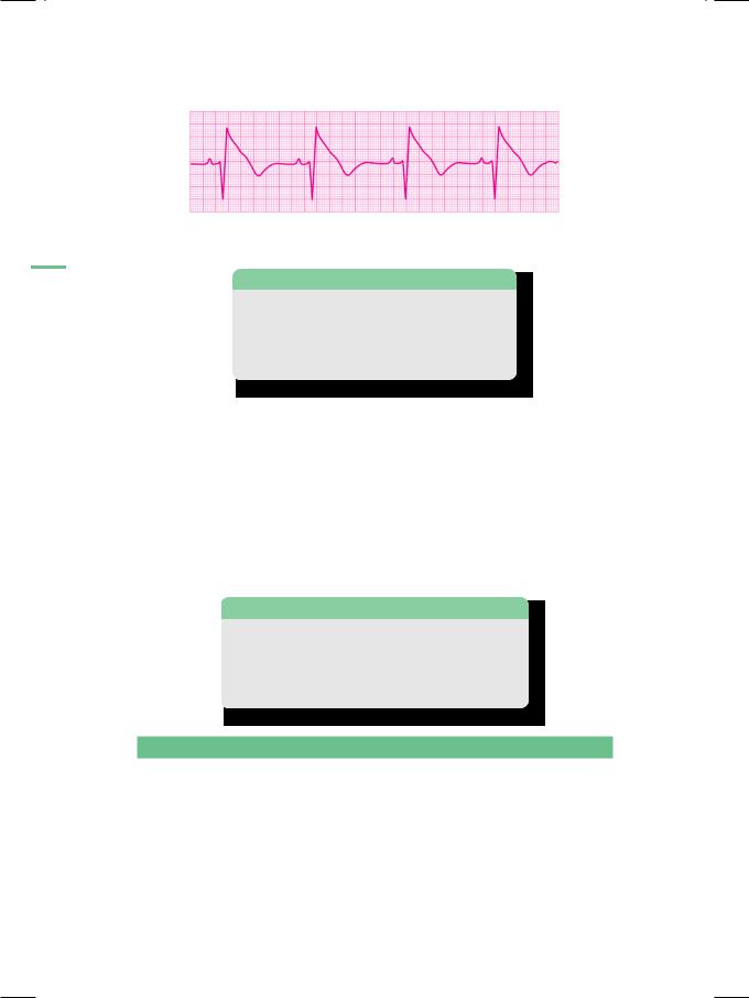
ECG INTERPRETATION
The ECG showed a triphasic rSR’ pattern in lead V1, which was 0.10 second in duration. Additionally, there was a “tent-like” coved S-T segment elevation of 0.2 mV with large inverted T waves (Table 41.1). These findings are consistent with the diagnosis of Brugada syndrome. Three types of S-T segment elevation are described in Brugada syndrome, depending upon the ventricular repolarization pattern.
•Type 1:“Tent-like” coved S-T segment, elevation >0.2 mV, negative T wave
•Type 2:“Saddle-back like” S-T segment, elevation >0.1 mV, positive T wave
•Type 3:“Saddle-back like” S-T segment, elevation <0.1 mV, positive T wave.
|
|
Case 41 Brugada Syndrome |
|
187 |
|
|
|
||
|
|
|
|
|
|
|
|
|
|
Figure 41.1: ECG showing features of the Brugada syndrome.
Table 41.1: ECG Features of Brugada Syndrome
• rSR’ pattern in lead V1 • rSR’ duration < 0.12 sec
• Large and inverted T wave • Coved S-T segment elevation
Besides Brugada syndrome, other causes of S-T segment elevation in lead V1 are right ventricular infarction, acute pulmonary embolism and arrhythmogenic right ventricular dysplasia (Table 41.2). The rSR pattern in lead V1 observed in case of Brugada syndrome, superficially resembles right branch block (RBBB). But unlike in RBBB, the ventricular complex is not more than 0.12 sec wide and there are no broad S waves in lead L1 and V6. The characteristic ECG abnormalities observed in Brugada syndrome may only be transient and not observed constantly. These may become exaggerated or unmasked after drug challenge with antiarrhythmic agents such as flecainide and procainamide.
Table 41.2 : Causes of S-T segment elevation in lead V1
• Brugada syndrome
• Right ventricular infarction • Acute pulmonary embolism
• Arrhythmogenic RV dysplasia.
CLINICAL DISCUSSION
The Brugada syndrome is a rare but striking electrocardiographic abnormality. It is believed to be a genetic disorder of sodium transport, across ion channels located in the right ventricle. This produces an abnormal pattern of right ventricular depolarization. Patients who have this abnormality are prone to develop sudden syncope because of malignant ventricular tachycardia or even cardiac arrest due to ventricular fibrillation. The genetic defect underlying Brugada syndrome may exist in more than one family member and form the basis of familial ventricular arrhythmias. The disorder is transmitted down subsequent generations by autosomal dominant inheritance.

188 |
|
Section 12 Electrocardiac Syndromes |
|
|
|
Brugada syndrome belongs to a class of congenital channelopathies which are responsible for nearly 5 to 10% cases of sudden cardiac death (SCD). Other members of this class are long Q-T syndrome (LQTS) and catecholaminergic ventricular tachycardia (CVT), as given in Table 41.3. Besides these channelo- pathies, congenital structural heart diseases responsible for SCD are hypertrophic cardiomyopathy and arrhythmogenic right ventricular dysplasia.
Table 41.3: Congenital channelopathies
• Brugada syndrome • Long Q-T syndrome
• Catecholaminergic VT* (*ventricular tachycardia)
MANAGEMENT ISSUES
There is no specific treatment of the underlying disorder in Brugada syndrome. However, insertion of an automatic implantable cardioverter defibrillator (AICD) may be considered in those patients with history of recurrent syncope, after cardiac resuscitation from ventricular fibrillation, or if there is history of sudden cardiac death in a family member.
C A S E |
|
42 |
WPW Syndrome |
|
. |
CASE PRESENTATION |
A 24-year old unmarried female arrived at the emergency-room, with the complaints of palpitation and light-headedness for the last 15 minutes. She drove herself to the hospital and admitted that she felt dizzy while driving. The patient also gave history of several such episodes in the past. At times, she was able to abort the attack by splashing cold water on her face, or by applying firm pressure over the eyes. At other times, she had to rush to the hospital, as on that day. During the “machinelike” sensation over the chest, she never experienced any chest pain or difficulty in breathing. However, after the palpitation stopped, she would pass a large volume of urine. The onset of these episodes was unrelated to physical exercise, emotional stress or to the intake of any particular food or beverage. There was no history of tremor, heat-intolerance, undue fatigue or significant weight-loss.
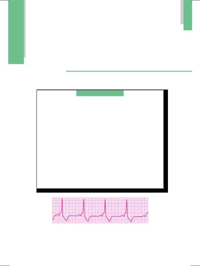
On examination, the patient was apprehensive but not tachypneic. The extremities were not cold but her palms were sweaty. The pulse rate was extremely rapid and exceeded 150 beats/min, although it could not be counted accurately. The BP was 91/64 mm Hg over the right arm and she was apyrexial. There were no signs of congestive heart failure. There was no goitre, bruit over the thyroid gland or any clinical sign of Grave’s disease. The precordium was normal and the apex beat was normally located. There was extreme tachycardia and the character of S1 and S2 could not be appreciated. The attending doctor performed right carotid sinus massage which resulted in sudden termination of the patient’s symptoms and a remarkable change in her heart rate. An ECG was obtained afterwards (Fig. 42.1).
Figure 42.1: ECG showing features of the WPW Syndrome
190 |
|
Section 12 Electrocardiac Syndromes |
|
|
|
ECG INTERPRETATION
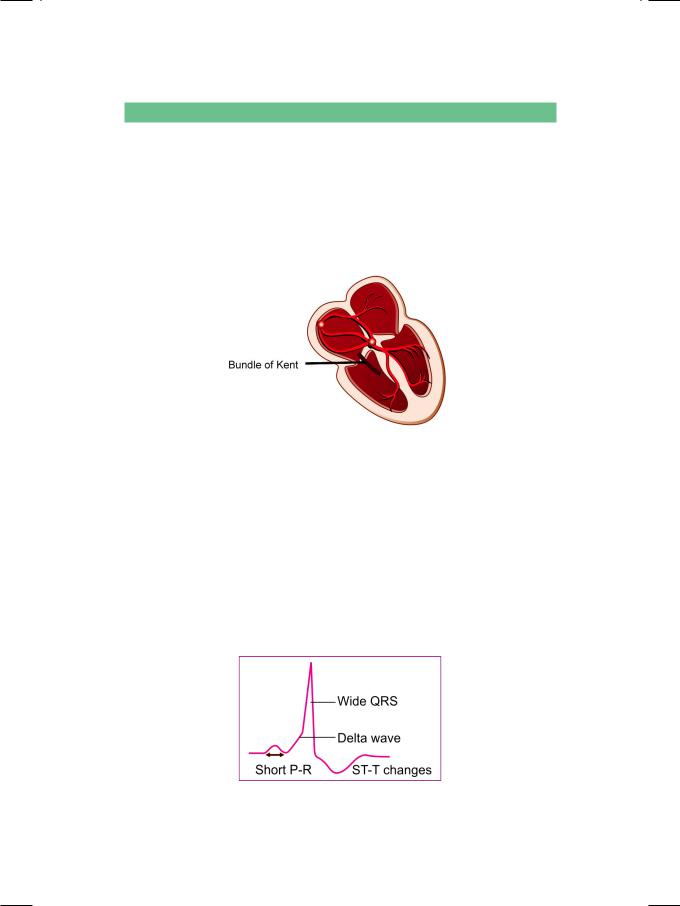
The ECG showed normal P waves with a P-R interval of 0.08 sec. The width of the QRS complex was 0.12 sec with a notch on the ascending limb of the R wave. There was depression of the S-T segment with inversion of the T wave. These findings are consistent with the diagnosis of WPW syndrome. The Wolff-Parkinson- White (WPW) Syndrome is a distinct electrocardiographic entity wherein an accessory pathway, the bundle of Kent, directly connects the atrial myocardium to the ventricular myocardium, bypassing the A-V node (Fig. 42.2). This produces abnormalities of the QRS complex, P-R interval, S-T segment and the T wave.
Figure 42.2: Diagram to illustrate the Bundle of Kent
The P-R interval is short because ventricular depolarization through the accessory pathway, bypasses the normal conduction delay at the A-V node. The notch on the ascending limb of R wave is the delta wave. It indicates preexcitation of the ventricle through the accessory pathway, before depolarization of the entire ventricle by the normal conduction system. The QRS complex is wide because it is a fusion beat, which is the sum of ventricular prexcitation and normal ventricular depolarization. The S-T segment and T wave inversion are repolarization abnormalities, secondary to the abnormal pattern of ventricular depolarization (Fig. 42.3).
Figure 42.3: ECG in case of WPW syndrome
Case 42 WPW Syndrome |
|
191 |
|
|
|
Three types of QRS configuration are described in the WPW syndrome, depending upon the direction of the accessory pathway.
•Type A (left septal connection) produces upright QRS complexes in all the precordial leads. It resembles right bundle branch block or true posterior wall myocardial infarction.
•Type B (right-sided connection) has negative QRS complexes in V1 and positive complexes in lead V6. It resembles left bundle branch block or left ventricular hypertrophy.
•Type C (left lateral connection) has positive QRS complexes in lead V1 and negative complexes in lead V6. It resembles right ventricular hypertrophy.
CLINICAL DISCUSSION
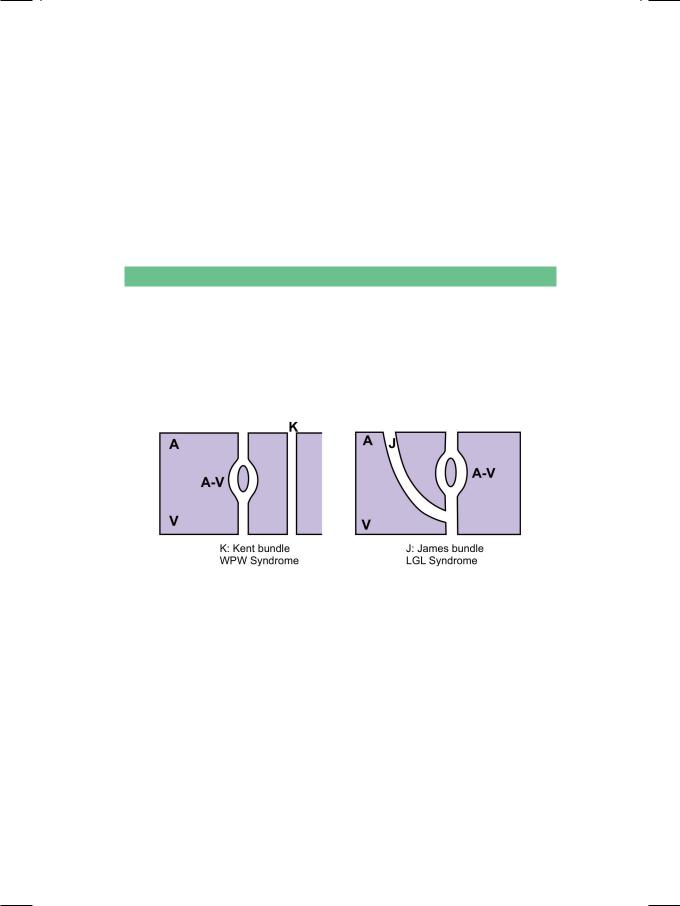
The standardized nomenclature of pre-excitation syndromes uses the term “tract” for pathways that insert into specialized conduction tissue and “connection” for pathways that enter the general myocardium. The Wolff-Parkinson-White (WPW) syndrome involves an atrio-ventricular connection (Kent bundle). Lown- Ganong-Levine (LGL) syndrome involves an atrio-fascicular bypass tract (James bundle) (Fig. 42.4). Mahaim fibers constitute a fasciculo-ventricular connection.
Figure 42.4: Diagram to illustrate the various pre-excitation syndromes
A: Atrium; V: Ventricle; A-V: Atrio-ventricular
The WPW syndrome is a masquerader of several other cardiac conditions:
•The delta wave as separate from the R wave can mimic bundle branch block.
•The dominant R wave in lead V1 may resemble right ventricular hypertrophy.
•Negative delta wave with S-T segment depression and T wave inversion, may appear as myocardial infarction.
•Antidromic A-V re-entrant tachycardia conducted anterogradely through the accessory pathway may be mistaken for ventricular tachycardia.
TheclinicalimportanceoftheWPWsyndromeliesinthefactthatitpredisposes to paroxysmal atrial tachycardia, since the bypass tract forms a re-entrant circuit with the regular conduction pathway. Paroxysmal atrial tachycardia (PAT) in the presence of WPW syndrome needs to be differentiated from a PAT without an accessory pathway, since the management is somewhat different.

192 |
|
Section 12 Electrocardiac Syndromes |
|
|
|
MANAGEMENT ISSUES
Presence of the WPW syndrome predisposes an individual to paroxysmal supraventricular tachycardia (PSVT), wherein the bypass tract constitutes a re-entrant circuit along with the normal conduction pathway. In almost 90% patients, conduction proceeds anterogradely down the A-V node and returns retrogradely through the accessory pathway to the atrium. This is known as orthodromic tachycardia and here the QRS complex is narrow. This arrhythmia responds to vagal manoeuvres and to drugs used in the treatment of A-V nodal re-entrant tachycardia (AVNRT). These drugs include verapamil, diltiazem, digitalis and adenosine.
In less than 10% patients, anterograde conduction proceeds down the accessory pathway and returns retrogradely through the normal A-V nodal pathway to the atrium. This is known as antidromic tachycardia and here the QRS complex is wide and demonstrates the WPW syndrome. This arrhythmia does not respond to vagal manoeuvres, but only to cardioversion with DC shock or to antiarrhythmic drugs that block the accessory pathway such as amiodarone.

S E C T I O N
13
Cardiac Arrhythmias