

новая папка / [libribook.com] 50 Cases in Clinical Cardiology_ A Problem Solving Approach 1st Edition
.PdfCase 8 Mitral Valve Prolapse |
|
35 |
|
|
|
According to the extent of motion, mitral valve prolapse (MVP) can be classified into 3 types (Table 8.1). In type 1, the anterior leaflet only moves upto the annulus while in type 2, it bows into the left atrium. In type 3, both the leaflets enter the left atrium. Strict echocardiographic criteria must be used to diagnose MVP because needless anxiety may be created by over-reporting this abnormality. Minor “technical” MVP may be observed in normal women due to high transducer position and caudal angulation. Conversely, true MVP may be missed due to low transducer position and cranial angulation.
Table 8.1: Classification of mitral valve prolapse
Type 1: AML and PML move upto the annulus
Type 2: Large AML bows into the left atrium
Type 3: Both AML and PML enter left atrium
Mitral valve prolapse is known as “floppy valve” or “myxomatous valve” or “billowing valve” and the condition is also designated as “Barlow’s syndrome”. MVP is far more commonly seen in females and occurs in 7% of middle-aged women. Often these women have a type A personality with history of panic attacks and migranous headaches. They may have a slender body habitus with thoracic skeletal deformities including pectus excavatum, straight back and scoliosis. The valvular abnormality is due to myxomatous degeneration leading to thickening, nodularity or redundancy of one or both mitral leaflets. Sometimes MVP is associated with other cardiac conditions such as ostium seundum ASD, Marfan syndrome and WPW syndrome (Table 8.2). Mitral regurgitation may be present due to faulty coaptation of leaflets and predisposes to endocarditis.
Table 8.2: Conditions associated with MVP
• Myxomatous degeneration • Rheumatic heart disease • Ostium secundum ASD
• WPW syndrome • Marfan syndrome • Turner syndrome
Atypical chest pain, palpitation, fatigue, orthostatic symptoms and neuropsychiatric complaints have all been well-described in patients with mitral valve prolapse. Whether these non-specific symptoms are directly attributable to MVP or due to autonomic dysfunction, continues to be widely debated and their causeeffect relationship remains unproven. Besides the classical symptoms, focal neurologic findings, such as transient ischemic attacks, amaurosis fugax, retinal artery occlusion and rarely hemiparesis have all been reported in patients with MVP. These neurologic findings probably occur as a result of thrombo-embolism from the prolapsing valve.

36 |
|
Section 2 Mitral Valve Diseases |
|
|
|
The ECG at rest frequently shows T-wave inversion in the inferolateral leads (LIII, aVF, V5, V6). False-positive ECG stress tests occur in up to 50% of patients with MVP. Premature beats are most common, although practically any arrhythmia can occur. The cause of the arrhythmia is not known but may be related to autonomic dysfunction or mechanical effects of the floppy valve. Incidence of syncope correlates poorly with the presence of arrhythmias.
PERTINENT INVESTIGATIONS
It is not unusual for patients of mitral valve prolapse to undergo a battery of sophisticated cardiac investigations, in the search for the diagnosis of a serious heart disease. Besides ECG and ECHO which do show some typical abnormalities, exercise stress test is done which is more often false-positive. Ambulatory 24-hour Holter monitoring frequently shows supraventricular and sometimes ventricular ectopic beats and rarely if ever reveals life-threatening arrhythmias. Myocardial perfusion imaging and coronary angiography expectedly do not show any significant abnormality. Hormonal assays are sometimes performed to rule out thyrotoxicosis and likewise, urinary catecholamines are rarely measured, to rule out the possibility of phaeochromocytoma.
MANAGEMENT ISSUES
Most women with mitral valve prolapse need reassurance that their cardiac condition is not serious or life-threatening. Anxiolytic drugs during the day with a mild tranquilizer at night are useful for those having overt anxiety and disturbed sleep. A beta-blocker such as propranolol has multiple benefits in these patients. It controls tachycardia and ectopic beats, reduces the degree of leaflet prolapse, treats the associated tremor and serves as a prophylactic drug against migraine. Low-dose aspirin is prescribed to prevent thrombo-embolism. Patients of MVP with mitral regurgitation (MR) and not those without demonstrable MR, require antibiotic prophylaxis against infective endocarditis, prior to a dental, endoscopic or surgical procedure.

S E C T I O N
3
Aortic Valve
Diseases


|
|
C A S E |
|
|
|
|
|
|
|
|
|
||
|
|
|
|
|
||
|
|
9 |
|
Aortic |
|
|
|
|
|
|
|
|
|
|
|
|
|
Stenosis |
|
|
|
|
|
|
|
|
|
|
|
|
|
|
|
|
CASE PRESENTATION
A 52-year old man presented to the out-patient cardiology clinic with easy fatiguability and breathlessness on exertion for the last 1 year. In the preceding month, he had experienced three distinct episodes of dizziness, followed by fainting. The syncopal episodes were unrelated to exercise and were not preceded by palpitation or chest pain. There was no history of prolonged febrile illness or joint pains during childhood.
On examination, the pulse was of low volume with a slow upstroke, at a rate of 84 beats/min. with a BP of 96/72 mm Hg. There was no anemia, cyanosis or icterus and the thyroid gland was not enlarged. The JVP was not raised and there was no ankle edema. The apex beat was normal in position and heaving in nature. The S1 was normal, S2 appeared single and S4 was audible in pre-systole. An ejection systolic murmur was audible over the aortic area, that was preceded by an ejection click and associated with a palpable thrill. The murmur and thrill typically radiated towards the carotid arteries.
CLINICAL DISCUSSION
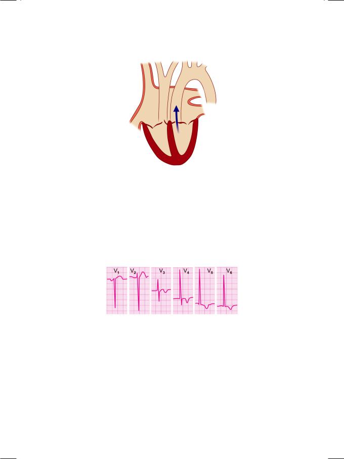
From the history and physical examination, this man had low cardiac output, possibly due to left ventricular outflow tract (LVOT) obstruction. The most likely diagnosis in this case is aortic valve stenosis (Fig. 9.1). A pulse of low volume (pulsus parvus) with slow upstroke (pulsus tardus) is a typical feature of aortic valve stenosis. A heaving apex beat indicates left ventricular hypertrophy and is also observed in uncontrolled systemic hypertension and in coarctation of aorta.
The low-pitched S4 sound in pre-systole, indicates forceful atrial contraction over a non-compliant left ventricle. It coincides with the a wave of the jugular vein. The S4 is always pathological in aortic stenosis, systemic hypertension and restrictive or hypertrophic cardiomyopathy. An acute rise in left ventricular enddiastolic pressure (LVEDP) as in acute coronary syndrome or acute valvular regurgitation causes acute onset of S4.
The S2 appears single because the A2 is muffled. If A2 is audible, the splitting of S2 is paradoxical or reverse, with the A2 following the P2 due to prolonged left ventricular ejection time. The ejection click heralds the onset of systole and the end of isovolumic relaxation. It indicates that the stenosis is valvular in nature
40 |
|
Section 3 Aortic Valve Diseases |
|
|
|
Figure 9.1: Aortic valve stenosis
and not subvalvular (infundibular) in location. The click may be absent if the valve is heavily calcified. The ejection systolic murmur of aortic stenosis indicates turbulent flow across the narrow aortic valve orifice. It is described as a diamondshaped murmur, since it builds up and declines gradually, with maximal intensity in mid-systole. This pattern coincides with the temporal profile of the pressure gradient across the valve. This murmur is also described as a “crescendodecrescendo murmur”. Length and loudness of the murmur does not correlate with the severity of aortic stenosis.
Figure 9.2: ECG showing tall R waves in lateral chest leads
ECG showed tall R waves in left precordial leads with T wave inversion, indicative of left ventricular hypertrophy with strain (Fig. 9.2). X-ray chest findings were a boot-shaped heart with a prominent ascending aorta, suggestive of poststenotic dilatation. On ECHO, the left ventricular cavity was small in size, with a good ejection fraction. There was concentric thickening of the interventricular septum (IVS) and the left ventricular posterior wall (LVPW). The aortic valve leaflets were thickened and calcific with restricted excursion and reduced opening of the valve. Due to fusion at the leaflet tips, there was systolic doming of leaflets. On colour flow mapping, a mosaic jet was observed in the proximal aorta (Fig. 9.3) with an increased systolic velocity across the valve on CW Doppler.

Case 9 Aortic Stenosis |
|
41 |
|
|
|
Figure 9.3: ECHO showing a mosaic colored jet in the proximal aorta
Table 9.1: Assessment of the severity of aortic stenosis
|
|
|
Mild |
Moderate |
Severe |
|
|
|
|
||
Vmax (m/sec) |
<2.5 |
2.5-4 |
>4 |
||
PG (mm Hg) |
<2.5 |
25-60 |
>60 |
||
|
|
|
|
|
|
AVa (cm2) |
|
>1.5 |
1.0-1.5 |
<1.0 |
|
Vmax |
: |
Peak velocity |
|
|
|
PG |
: |
Pressure gradient |
|
|
|
AVa |
: |
Aortic valve area |
|
|
|
Transthoracic echocardiography (TEE) generally suffices for the diagnosis of aortic stenosis. However, the severity of stenosis is determined from the peak velocity and pressure gradient across the valve on Doppler (Table 9.1). Measurements from various echo views are taken to obtain the peak aortic flow velocity. In rheumatic aortic stenosis, assessment of concomitant mitral valve abnormalities is crucial as the mitral valve is almost invariably involved. Finally, left ventricular wall thickness, end-diastolic diameter and ejection fraction are to be measured. There are some fallacies associated with the calculation of aortic stenosis severity. Reverberation artifacts in a heavily calcified valve may overestimate AS severity. The peak velocity and pressure gradient depend on the heart rate and stroke volume. They also depend upon the degree of parallelism obtained between the Doppler beam and aortic flow direction.
Table 9.2: Causes of aortic stenosis
• Congenital bicuspid valve • Rheumatic heart disease • Senile calcific degeneration
The main causes of AS are congenital bicuspid aortic valve, rheumatic heart disease and senile calcific degeneration (Table 9.2). Rheumatic aortic stenosis

42 |
|
Section 3 Aortic Valve Diseases |
|
|
|
usually presents in the 2nd or 3rd decade of life and is almost always associated with mitral valve disease. Congenital bicuspid aortic valve leads to aortic stenosis because of increased mechanical and shear stress on the valve and usually presents in the 4th of 5th decade. Calcific aortic stenosis is akin to atherosclerosis and accompanied by multiple cardiovascular risk factors. It typically presents in the 6th or 7th decade of life. Rarely, a discrete membrane or ring may cause subvalvular or supravalvular AS. Complications of AS are left ventricular hypertrophy with diastolic dysfunction earlier on and systolic heart failure in the later stages. Syncopal episodes are due to tachyarrhythmias or LVOT obstruction. Angina may occur because of coronary ostial stenosis or the increased oxygen demand of the hypertrophied myocardium.
PERTINENT INVESTIGATIONS
Exercise ECG testing and Dobutamine stress ECHO may be undertaken in asymptomatic patients of aortic stenosis, to assess their functional capacity. The characteristic response is a drop in blood pressure during the test due to vasodilatation and failure of the cardiac output to rise. Patients with a positive test may be taken up for valve replacement. Coronary angiography is indicated to assess the coronary circulation and need by bypass graft surgery (CABG), at the time of aortic valve replacement. Besides showing occlusive coronary disease, angiography may reveal coronary ostial stenosis due to calcification of the aortic valve.
MANAGEMENT ISSUES
There is no medical treatment for aortic stenosis barring perhaps low-does aspirin with a statin in those who have associated coronary risk factors. Aortic valve replacement (AVR) is the procedure of choice in symptomatic severe aortic stenosis or in asymptomatic stenosis with left ventricular dysfunction or a positive stress test. AVR may also be undertaken in moderate AS at the time of CABG surgery or in those engaged in high profile jobs such as aircraft pilots or military personnel (Table 9.3). Complications of aortic valve replacement are prosthetic valve malfunction, thrombo-embolism or bleeding and infective endocarditis.
Table 9.3: Indications for surgery in aortic stenosis
• Moderate to severe AS with symptoms • Moderate AS with a positive stress test
• Moderate to severe AS with LV dysfunction • Moderate AS with high profile job (e.g. pilot)
• Moderate AS with planned surgery (e.g. CABG)
RECENT ADVANCES
The technique of percutaneous aortic valve replacement (AVR) has been recently refined and its feasibility is now clearly established. It is particular suitable for candidates at high risk for a surgical procedure under general anesthesia. The role of percutaneous aortic balloon valvuloplasty is currently limited.

|
|
C A S E |
|
|
|
|
|
|
|
|
|
||
|
|
|
|
|
||
|
|
10 |
Aortic |
|
|
|
|
|
|
|
|
|
|
|
|
|
|
Regurgitation |
|
|
|
|
|
|
|
|
|
|
|
|
|
|
|
|
CASE PRESENTATION
A 54-year old man presented with breathlessness on exertion and throbbing headache. His exertional dyspnea started about 8 months back and progressed to the extent that he found it difficult to climb even one flight of stairs. His throbbing headache was a daily feature and he also felt some pulsations over the neck. There was no history of exertional chest pain, palpitation or syncopal episodes. The patient had visited an orthopedician for a still back with difficulty in bending forward. On X-ray of the lumbo-sacral spine, it was diagnosed as ankylosing spondylitis. He was taking oral steroids prescribed by an ophthalmologist for iridocyclitis, in addition to his antihypertensive medication.
On examination, the radial pulse was of good volume and bounding in nature at a rate of 84 beats/min. The BP was 160/64 mm Hg in the right arm. Pulsations were visible over the carotid arteries and the femoral pulses were also bounding in nature. There was no clinical sign of congestive heart failure. The apex beat was diffuse and hyperkinetic in nature and significantly displaced towards the axilla. The S1 was normal and the S2 was soft, with a S3 gallop sound in early diastole. A highpitched blowing murmur was heard in diastole soon after the S3, on either side of the upper sternum. A short diastolic rumble was also heard over the cardiac apex. Few scattered crepts were audible over the lower lung fields.
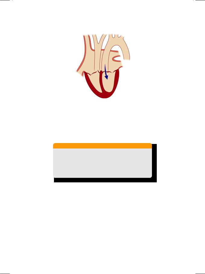
CLINICAL DISCUSSION
From the history and physical examination, this man had aortic regurgitation (Fig. 10.1) with possibly mitral stenosis and evidence of left ventricular dilatation. Aortic root dilatation with AR is a known complication of ankylosing spondylitis. The early-diastolic decrescendo murmur of AR is soft and blowing in character, best heard with the patient sitting up and leaning forward. Interestingly, when AR is due to aortic root dilatation, the murmur is louder along the right sternal border. When AR is due to aortic valve pathology, the murmur is louder along the left sternal border. In acute AR, for instance due to dissection of aorta, the murmur is very short and soft and associated with a S4 and not S3.
The diastolic rumble at the apex does not necessary indicate concomitant mitral stenosis. It is due to fluttering of the anterior mitral leaflet between the AR jet and mitral valve inflow and is known as Austin-Flint murmur. A bounding
44 |
|
Section 3 Aortic Valve Diseases |
|
|
|
Figure 10.1: Aortic regurgitation
(“water-hammer”) pulse that can be appreciated even with the arm elevated above the head is known as a collapsing pulse and is indicative of a wide pulse-pressure. Besides aortic regurgitation, causes of wide pulse pressure are severe anemia, thyrotoxicosis, beri-beri disease, patent ductus arteriosus and arteriovenous fistula. Certain classical clinical signs of wide pulse-pressure, have been described in aortic regurgitation (Table 10.1).
Table 10.1: Clinical signs of aortic regurgitation
• |
Corrigan sign: |
Vigorous pulsations in the carotid vessels |
|
|
|
• |
De Musset sign: |
Nodding of the head with each heart beat |
|
|
|
• |
Quincke sign: |
Visible capillary pulsations in the nail bed |
|
|
|
• |
Traube sign: |
Pistol-shot sounds over femoral arteries |
|
|
|
• |
Duroziez sign: |
Diastolic murmur over femoral arteries |
|
|
|
ECG of the patient showed tall R waves in left precordial leads with upright T waves indicating left ventricular diastolic overload. X-ray chest findings were cardiomegaly and pulmonary congestion, giving it a “bat-wing” appearance (Fig. 10.2). On ECHO, the left ventricle was dilated with an ejection fraction of 45%. There was also dilatation of the aortic root but the aortic valve leaflets were not thickened or calcific and had no vegetations. The mitral valve was structurally normal and the left atrium was normal in size. On colour flow mapping, a regurgitant jet was seen entering the left ventricular outflow tract (Fig. 10.3). The volume of AR (ml/beat), the width of the AR jet as percentage of LVOT diameter
(%) and the depth of the jet entry into the left ventricle are used to gauge the severity of AR (Table 10.2). However, there are some fallacies associated with these calculations. The narrow jet of mild AR may extend deep into the left ventricle while the broad jet of severe AR may not extend that far if it is “eccentric” or “off-centre”.