

новая папка / [libribook.com] 50 Cases in Clinical Cardiology_ A Problem Solving Approach 1st Edition
.PdfCase 25 Pericardial Effusion |
|
115 |
|
|
|
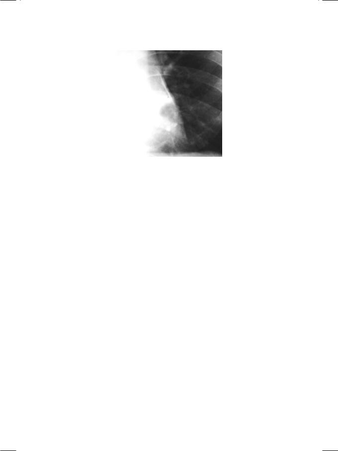
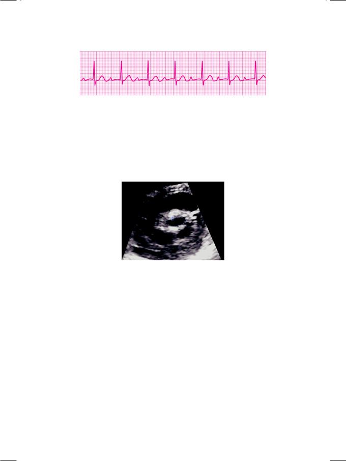
Figure 25.2: ECHO showing an echo-free space all around the heart
Table 25.1: Quantification of pericardial effusion
Amount |
Volume |
Posterior space |
Anterior space |
|
|
|
|
Small |
< 100 ml |
< 1 cm |
— |
|
|
|
|
Moderate |
100-500 ml |
1-2 cm |
< 1 cm |
|
|
|
|
Large |
> 500 ml |
> 2 cm |
> 1 cm |
|
|
|
|
fibrinous strands while malignant effusion may show echo-dense metastasis deforming the smooth pericardium.
A large pericardial effusion imposes constraint on filling of the cardiac chambers and hence impairs cardiac output. This serious clinical situation is known as cardiac tamponade. Tamponade results from a large volume of effusion, or rapid collection of a small effusion. A large effusion may accumulate gradually without causing tamponade, if the pericardial sac is compliant and gets adequate time to stretch itself. Therefore, rapidity of fluid accumulation is more important than absolute volume. The right-sided chambers are low-pressure structures and can easily get compressed by a large pericardial effusion. Right atrial collapse is a sensitive but less specific sign of cardiac tamponade. Right ventricular collapse, particularly if it lasts for more than one-third of diastole, is a less sensitive but more specific sign of cardiac tamponade.
Due to the constraint imposed by the effusion on the filling of the cardiac chambers, increased filling of the right ventricle (during inspiration) reduces filling of the left ventricle (hence cardiac output). Therefore during inspiration, tricuspid flow velocity increases while mitral flow velocity decreases. The converse occurs during expiration. A change in flow velocity exceeding 25% is considered to be diagnostic. This phenomenon of ventricular interdependence also forms the basis of pulsus paradoxus, which is observed clinically. This reciprocal relationship is also seen in constrictive pericarditis, where it is an important differentiating feature from restrictive cardiomyopathy.

116 |
|
Section 7 Pericardial Infections |
|
|
|
Table 25.2: Causes of pericardial effusion
• |
Infective |
: |
Tubercular pericarditis |
• |
Traumatic |
: |
Accidental, surgical |
• |
Malignant |
: |
Metastasis, irradiation |
• |
Metabolic |
: |
Uremia, myxedema |
• |
Autoimmune |
: |
Rheumatoid arthritis |
There are several causes of pericardial effusion but inflammatory causes lead the list (Table 25.2). Tuberculosis is the commonest infection that causes pericardial effusion in the developing countries. Auto-immune disorders like rheumatoid arthritis and systemic lupus are also sometimes responsible for pericardial effusion. Idiopathic or viral pericarditis is usually associated with only a small effusion. Hemorrhagic effusion may follow accidental or surgical trauma or may be due to metastatic deposits in malignant disease. Metabolic disorders that cause pericardial effusion are uremia and myxedema (hypothyroidism).
PERTINENT INVESTIGATIONS
Pertinent investigations in pericardial effusion are completed blood count, tuberculin test, antinuclear antibodies, renal function tests and thyroid profile. The pericardial fluid is tested by cytology, bacterial culture and for adenosine deaminase activity and tumour markers when tuberculosis or malignancy are suspected.
MANAGEMENT ISSUES
Drainage of a large pericardial effusion by means of echo-guided (for safe needle entry) pericardiocentesis is the treatment of choice, particularly if there is cardiac tamponade. Most often the hemodynamic response to drainage is dramatic and gratifying. Fluid resuscitation may cause only transient improvement, but ionotropes has no role as cardiac contractility is not compromised. If there is no evidence of tamponade and the hemodynamics are stable, immediate drainage is unnecessary and the patient is managed conservatively. However, careful periodic assessment is mandatory. In tamponade due to aortic dissection, pericardiocentesis worsens hemodynamic stability and surgical drainage is preferable.
Antitubercular drugs are sometimes prescribed empirically if tubercular etiology is likely, or definitely if it is proven upon pericardial fluid analysis. Antinflammatory agents including steroids are employed for the treatment of auto-immune and inflammatory disorders. Malignant disease is treated with suitable chemotherapy or even radiotherapy which however, carries the risk of pericardial constriction.
|
|
C A S E |
|
|
|
|
|
|
|
|
|
||
|
|
|
|
|
||
|
|
26 |
Constrictive |
|
|
|
|
|
|
|
|
|
|
|
|
|
|
Pericarditis |
|
|
|
|
|
|
|
|
|
|
|
|
|
|
|
|
CASE PRESENTATION
A 52-year old man visited the out-patient clinic of the department of internal medicine, with complaints of generalized fatigue, reduced appetite, increased abdominal girth and swelling over both ankles. He denied history of heavy alcohol intake, prolonged jaundice, hematemesis or altered bowel habits. About one year back, he was diagnosed to have pulmonary tuberculosis, for which he was prescribed anti-tubercular drugs for 6 months. But he was able to take his full medication only for 3 months, because he developed drug-induced hepatitis and therefore his rifampicin and isoniazid were stopped. However, he did take ethambutol and levofloxacin for the remaining 3 months. At present there were no complaints of fever, night-sweats, productive cough or hemoptysis. There was no history of diabetes, hypertension or heart disease in the patient or any of his family members.
On examination, the patient looked ill with a pinched face, thin emaciated arms, swelling around the ankles and a protuberant abdomen. He was not tachypneic or orthopneic and not in any form of distress. His conjunctiva and tongue were pale but there was no cyanosis or icterus. The pulse was fair in volume at a rate of 92 beats/min., with an appreciable fall in pulse volume during inspiration. The JVP was elevated 5 cm above the angle of Louis and it failed to fall appreciably during inspiration. The abdomen was distended, but there were no dilated veins or spider naevi on the skin surface. Shifting dullness was demonstrated, indicating free fluid in the abdominal cavity. The liver edge was palpable 8 cm below the right costal margin; it was slightly tender but not pulsatile. The precordium was silent and the apex beat was difficult to locate. There was no murmur or pericardial rub, but a highpitched sound was audible in mid-diastole. There was some retraction of the apex of right lung on inspection, but both the lung fields were clear on auscultation.
CLINICAL DISCUSSION
From the history and physical examination, this patient definitely was in right heart failure. ECG showed sinus rhythm with low QRS voltages and non-specific T wave inversion. X-ray chest did not show any cardiomegaly, but there was a striking area of linear calcification along the left heart border (Fig. 26.1). There was fibrosis over the apical segment of the right lung. ECHO revealed normal size of all cardiac chambers, with normal left ventricular ejection fraction. There
118 |
|
Section 7 Pericardial Infections |
|
|
|
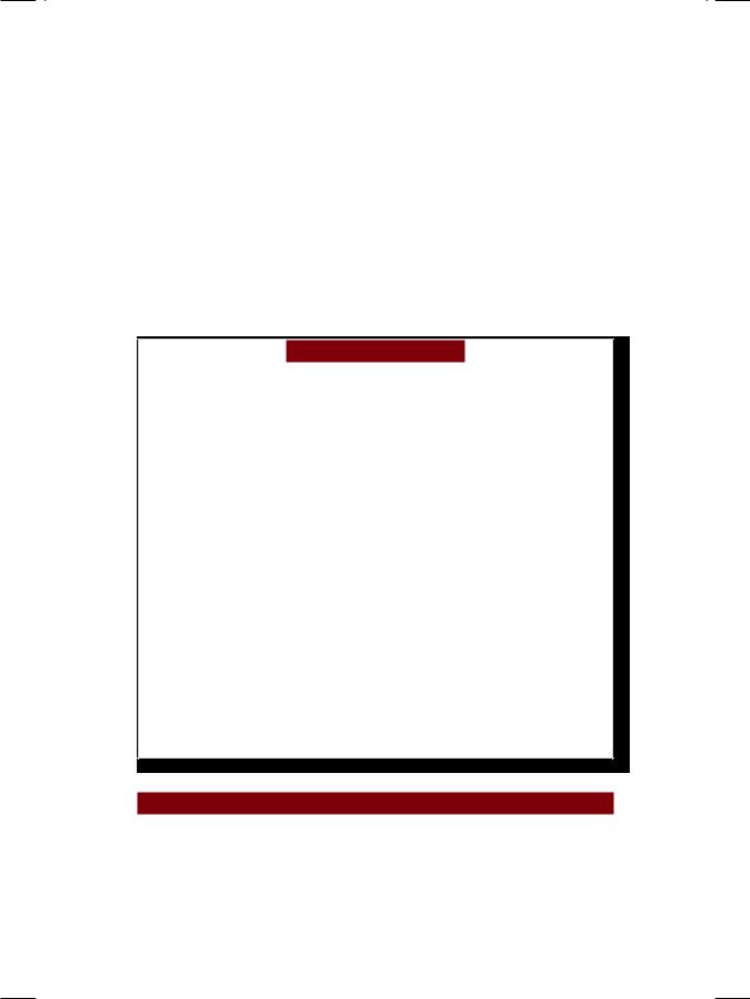
Figure 26.1: X-ray showing linear pericardial calcification
was no myocardial thickening or wall-motion abnormality and all cardiac valves were structurally normal. However, there was thickening of the pericardium, with multiple parallel echo-lines casting a bright reflection. Therefore, the most probable diagnosis in this case is constrictive pericarditis.
There were several typical clinical findings in this case. These were a variable pulse volume, raised JVP, silent precordium, no cardiac murmur and a sharp third heart sound in diastole. An appreciable decrease in pulse volume during quiet respiration is known as pulsus paradoxus. Besides constrictive pericarditis, pulsus paradoxus is observed in cardiac tamponade, chronic obstructive lung disease and status asthmaticus. A raised JVP that paradoxically rises even further during inspiration is known as Kussmaul’s sign. Besides constrictive pericarditis, the Kussmaul’s sign is observed in restrictive cardiomyopathy and after right ventricular infarction. The deep x descent on the JVP, represents the rapid phase of early diastolic ventricular filling. This is known as the “dip and plateau” pattern or the “square-root” sign of the ventricular pressure trace.
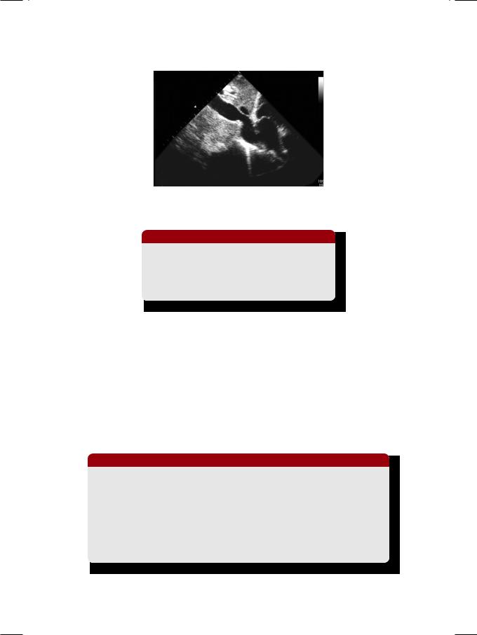
A silent precordium with a ‘lost’ apex-beat is a feature of pericardial effusion or constriction, morbid obesity and pulmonary emphysema. The third heart sound in constrictive pericarditis is the pericardial knock. It is a sharp and highpitched sound, when compared to the classical S3. The pericardial knock marks the termination of rapid early-diastolic phase of ventricular filling. Due to pericardial constriction, the inferior vena cava is dilated (Fig. 26.2) and there is congestive hepatomegaly. The spleen is also enlarged and ascites is present. These findings are picked up on clinical examination as well as by ultrasonography.
Constrictive pericarditis is a masquerader of several clinical conditions. Signs and symptoms are similar to those of congestive heart failure, but right-sided failure is more prominent and ascites is out of proportion to the degree of pedal edema. Hepatomegaly, ascites and edema may be misdiagnosed as cirrhosis of liver, if the neck veins are not observed carefully. Finally, if a patient is in atrial fibrillation, the diastolic knock may be misinterpreted as an opening snap and the diagnosis of mitral stenosis may be entertained.
Case 26 Constrictive Pericarditis |
|
119 |
|
|
|
Figure 26.2: Abdominal ultrasound showing dilatation of the inferior vena cava
Table 26.1: Causes of constrictive pericarditis
• Tubercular pericarditis • Bacterial pericarditis • Cardiac surgery
• Chest irradiation
Constrictive pericarditis is most often preceded by a tubercular infection. Sometimes it may follow bacterial pericarditis but never viral pericarditis. Rheumatic pancarditis rarely causes constriction because the effusion is serofibrinous in nature and usually gets absorbed. Constrictive pericarditis may also follow chest irradiation or cardiac surgery (Table 26.1).
It is often difficult if not impossible to differentiate constrictive pericarditis from restrictive cardiomyopathy and cardiac catheterization may be required for their distinction. Restrictive cardiomyopathy is suggested by the presence of certain subtle echo features which are not observed in pericardial constriction. These features are small ventricles and large atria, thick mitral and tricuspid valve leaflets and mild impairment of left ventricular systolic function (Table 26.2).
Table 26.2: Differences between constrictive pericarditis and restrictive cardiomyopathy
|
Constrictive pericarditis |
Restrictive cardiomyopathy |
|
|
|
Pericardium |
Thick |
Normal |
|
|
|
Myocardium |
Normal |
Thick |
|
|
|
Ventricles |
Normal |
Obliterated |
|
|
|
Atria |
Normal |
Dilated |
|
|
|
LV function |
Normal |
Mildly impaired |
|
|
|
MV and TV |
Normal |
Regurgitation |
|
|
|

120 |
|
Section 7 Pericardial Infections |
|
|
|
Moreover, the ventricular free walls may give a “granular-sparkling” appearance. Clinically speaking, restrictive cardiomyopathy generally presents with biventricular heart failure while constrictive pericarditis presents with sole or predominant right heart failure.
MANAGEMENT ISSUES
A two to three month period of conservative treatment can be tried in constrictive pericarditis, if the symptoms are mild. In the presence of signs indicating systemic venous congestion, it is naturally tempting to prescribe a diuretic to a patient of constrictive pericarditis. However, this approach is often counterproductive since ventricular filling declines further and cardiac output falls. Ionotropes like digoxin are unhelpful as there is no impairment of myocardial contractility. It is crucial to maintain sinus rhythm and to cardiovert atrial fibrillation by electrical or pharmacological means. This is because atrial fibrillation leads to loss of atrial contribution to ventricular filling and shortens the diastolic filling time. Pericardectomy by surgical means is the only effective treatment of constrictive pericarditis. It releases the ventricles from the restriction to diastolic filling, imposed by the rigid pericardial sac.
RECENT ADVANCES
The classical hemodynamic findings of constrictive pericarditis on cardiac catheterization not only helps to arrive at a definitive diagnosis but also help to differentiate between pericardial constriction and restrictive cardiomyopathy. These findings have been recently ellucidated. Due to the restriction imposed by the rigid pericardium on right ventricular filling, there is a sharp rise in early diastolic pressure followed by a flat pressure profile in mid and late diastole. This is reflected as a “dip and plateau” wave form on right ventricular pressure tracing and constitutes the “square-root sign”. On inspection of the JVP, this pattern is observed as a prominent x descent.
Another hemodynamic finding is the reciprocal variation in the mitral and tricuspid inflow velocities with the phases of respiration, due to pericardial constraint. This reflects ventricular interdependence wherein right ventricular filling increases during inspiration at the expense of left ventricular filling. The converse happens during expiration when right ventricular filling decreases and left ventricular filling increases. This is reflected as an exaggerated (>25%) rise or fall of mitral and tricuspid inflow velocities, during inspiration and expiration.

S E C T I O N
8
Myocardial
Infections

|
|
C A S E |
|
|
|
|
|
|
|
|
|
|
|
|
|
|
|
|
|
27 |
Rheumatic |
|
|
|
|
|
|
|
|
|
|
|
Fever |
|
|
|
|
|
|
|
|
CASE PRESENTATION
A 19-year old girl visited her family physician with the complaints of high-grade fever, pain in lower-limb joints and rash over the arms and legs, for the past 1 week. The fever started with pain in her throat and dry cough and was associated with chills but not rigors. There was no history of purulent sputum, chest pain, dyspnea, wheezing or hemoptysis. She also denied pain abdomen, vomiting, loose stools or burning sensation during micturition. The pain in her joints started in her right wrist and went on to affect both her ankles, which limited her walking ability. The skin rash was macular and erythematous, but there were no petechial spots or history of pruritus. The girl had not undergone dental extraction or any surgical procedure in the recent past. She did have sore-throat frequently during her childhood, but there was no history of anoxic spells or squatting attacks while playing.
On examination, the young girl looked ill, toxic and anxious. The pulse rate was 104 beats/min. with a BP of 104/66 mm Hg and her temperature was 100.6o F. Her extremities were warm and dry and there was no tremor or clubbing of the fingers. She was mildly anemic but not cyanosed or icteric. The erythematous rash over the extremities blanched on pressure. Her both ankles were mildly swollen and tender to touch, but there was no redness over the skin. On throat examination, there was mild pharyngeal congestion with enlarged tonsils that had few pustules. At the neck, the thyroid gland was not enlarged, JVP was not raised and there was no significant lymphadenopathy. The apex beat was normal in location and character and the precordium was quiet. The S1 was soft and S2 normally audible; no S3 gallop or pericardial friction rub was appreciated. A low-pitched, mid-diastolic murmur was heard at the cardiac apex. The murmur was not preceded by an opening snap and did not undergo pre-systolic accentuation. The lung fields were clear without any rhonchi or rales.
CLINICAL DISCUSSION
From the history and physical examination, this young girl had a febrile illness with arthralgias, erythematous rash and a diastolic murmur. The obvious diagnosis with this constellation of clinical findings is acute rheumatic fever. The ECG showed sinus rhythm with a heart rate of 86 beats per minute and a
124 |
|
Section 8 Myocardial Infections |
|
|
|
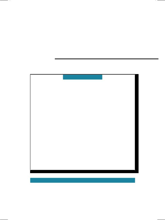
Figure 27.1: ECG showing prolongation of the P-R interval
prolonged P-R interval (Fig. 27.1). The QRS complexes were narrow and there was no S-T segment shift or T wave inversion. The X-ray chest did not show increased cardio-thoracic ratio or signs of pulmonary edema. ECHO revealed normal left ventricular size and systolic function, mild left atrial dilatation and a normal sized right ventricle. The mitral valve leaflets were mildly thickened (Fig. 27.2) with normal excursion and there was no diastolic doming of either leaflet.
Figure 27.2: ECHO showing thickened mitral valve leaflets
The hemoglobin was 10.4 g/dL with a leucocyte count of 11,800/cu.mm. of which 73% were neutrophils. The ESR was 55 mm in the 1st hour. Urine analysis showed traces of albumin but no WBCs or RBCs. The peripheral blood smear did not show any plasmodium parasite. The anti-streptolysin O (ASLO) titre was 366 IU with a C-reactive protein(CRP) value of 66 mg/L. Biochemical parameters including blood glucose, serum cholesterol and the renal and liver function tests were within normal limits. Urine and blood culture did not yield any bacterial growth. However, culture of the swab taken from the throat was positive for betahemolytic Streptococcus.
The clinical diagnosis of acute rheumatic fever is based on the presence of Jone’s criteria (Table 27.1). Two major criteria or one major and two minor criteria, along with evidence of recent Streptococcal infection, are required to clinch the diagnosis. Major Jone’s criteria are carditis, polyarthritis, chorea, erythema marginatum and subcutaneous nodules. Minor criteria are fever, arthralgia, history of rheumatic fever, elevated ESR, CRP and prolonged P-R interval. Evidence of a recent Streptococcal infection include positive throat swab culture and increased anti-Streptolysin O (ASLO) titer.