

новая папка / [libribook.com] 50 Cases in Clinical Cardiology_ A Problem Solving Approach 1st Edition
.Pdf
|
|
C A S E |
|
|
|
|
|
|
|
|
|
||
|
|
|
|
|
||
|
|
38 |
Prolonged Q-T |
|
|
|
|
|
|
|
|
|
|
|
|
|
|
Syndrome |
|
|
|
|
|
|
|
|
|
|
|
|
|
|
|
|
. |
CASE PRESENTATION |
A 62-year old woman visited her cardiologist along with her daughter, because of two episodes of syncope in the preceding week. The fainting episodes were preceded by palpitation and associated with blurring of vision. The patient was a known case of coronary heart disease and she had sustained an anterior wall myocardial infarction ten months back. At that time, she did not receive thrombolytic therapy because of late presentation to the hospital and she had also declined coronary angiography. The patient also felt breathless on exertion and her echocardiogram showed a large anterior wall motion abnormality with an ejection fraction of 30+5%. About a month back, she underwent 24-hour Holter monitoring which showed multifocal ventricular premature beats, with short runs of ventricular tachycardia. Therefore, she was prescribed amiodarone, in addition to her usual medication which included ramipril, frusemide, aspirin, atorvastatin and isosorbide mononitrate. An initial loading dose of 1200 mg per day of amiodarone, was tapered to a maintenance dose of 400 mg daily after one week.
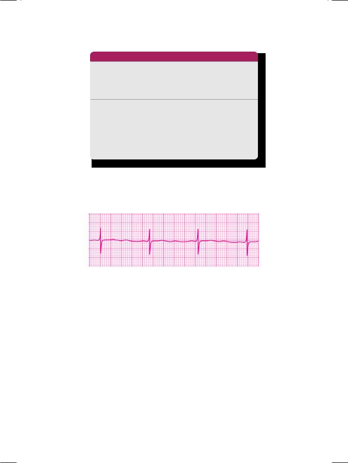
On examination, the patient was apprehensive and tachypneic. The pulse was fast and low in volume with a few “missed beats”. The heart rate was 104 beats/min. with a BP of 110/66 mm Hg. The cardiac apical impulse was diffuse and displaced towards the axilla. The S1 and S2 were normal with an S3 gallop sound in diastole. No murmur or friction rub was audible. There were bilateral basilar rales over the lung fields. An ECG was obtained (Fig. 38.1).
Figure 38.1: ECG showing broad T waves with prolonged Q-T interval
ECG INTERPRETATION
The ECG showed normal regular sinus rhythm. The P waves were normal in morphology and large Q waves were seen in the anterior precordial leads. The
176 |
|
Section 12 Electrocardiac Syndromes |
|
|
|
T wave was upright, tall and broad, which occupied most of the R-R interval. The measured Q-T interval was 0.60 sec. This finding is consistent with the diagnosis of long Q-T syndrome.
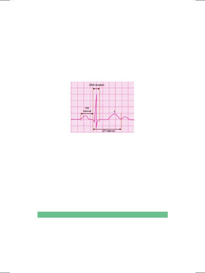
The duration of the QRS complex represents ventricular depolarization time and the width of the T wave represents ventricular repolarization time. Therefore, the Q-T interval is a measure of the total duration of ventricular electrical systole. The Q-T interval is measured on the horizontal axis, from the onset of the Q wave to the termination of the T wave (not the U wave). The duration of the QRS complex, the length of the S-T segment and the width of the T wave are included in the measurement of the Q-T interval (Fig. 38.2).
Figure 38.2 : Measurement of the Q-T interval
The upper limit of normal Q-T interval depends upon several variables, including the age, gender and the autonomic tone. It tends to be shorter in young individuals (0.44 sec) and longer in the elderly (0.45 sec). It is slightly shorter in males than in females, the upper limit being 0.43 sec in men. The Q-T interval shortens at fast heart rates and lengthens at slow heart rates. Since the Q-T interval varies with tachycardia and bradycardia, the measured Q-T interval needs to be corrected for heart rate.
The corrected Q-T interval is known as the Q-Tc interval. For heart rate correction, the Bazett’s formula is generally used, where Q-Tc interval is equal to the measured Q-T interval divided by the square-root of the R-R interval. When the heart rate is 60 beats/min. and the R-R interval is 1 sec (25 x 0.04sec), the Q-Tc interval and the Q-T interval are the same. As a general rule, a Q-T interval that exceeds half of the R-R interval, is taken as a prolonged Q-T interval.
CLINICAL DISCUSSION
There are several causes of Q-T interval prolongation. These have been enlisted in Table 38.1. Congenital long Q-T syndromes may present dramatically with syncope. Indeed, congenital long Q-T syndromes are characterized by prolongation of the Q-T interval, syncope, ‘seizures’ and sudden death due to ventricular arrhythmias (Torsade de pointes), in apparently healthy children and young adults. Some cases of sudden infant deaths have been attributed to congenital long Q-T syndrome. Therefore, an ECG should be performed in all
Case 38 Prolonged Q-T Syndrome |
|
177 |
|
|
|
Table 38.1: Causes of prolonged Q-T interval
Congenital causes
• Jervell -Lange-Neilsen syndrome
(autosomal recessive with deafness)
• Romano -Ward syndrome
(autosomal dominant without deafness)
Acquired causes
• Electrolyte deficiency e.g. calcium, potassium
• Antiarrhythmic drugs e.g. quinidine, amiodarone • Coronary artery disease e.g. myocardial infarction • Myocarditis e.g. viral myocarditis, rheumatic fever
• Intracranial event e.g. head injury, brain hemorrhage • Bradyarrhythmias e.g. A-V block, sinus bradycardia • Drug induced e.g. terfenadine, cisapride, olanzapine
infants on anticonvulsants for seizure prophylaxis. The prognosis and triggers of sudden cardiac death in patients with congenital long Q-T syndrome, are related to the Q-T interval and the genotype.
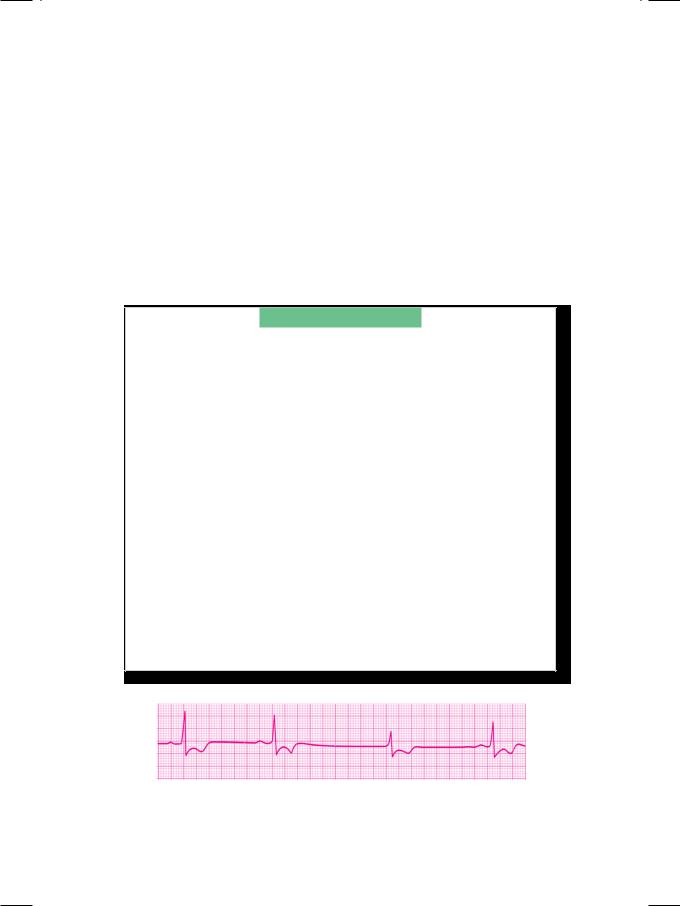
Figure 38.3 : ECG showing pseudo-prolongation of the Q-T interval
In hypokalemia, the T wave is flattened and the prominent U wave may be mistaken for the T wave. This may falsely suggest prolongation of the Q-T interval, whereas it is actually the Q-U interval. Hypokalemia therefore, cause pseudo-prolongation of the Q-T interval (Fig. 38.3). Antiarrhythmic drugs such as quinidine, procainamide and amiodarone can prolong the Q-T interval. They also cause widening of the QRS complex which, if it exceeds 25% of baseline, is an indication for withdrawing the culprit drug. Since Q-T interval prolongation predisposes to arrhythmias, this is the mechanism to explain the arrhythmia enhancing property or proarrhythmic effect of antiarrhythmic drugs. Besides antiarrhythmic agents, certain non-cardiovascular drugs can also prolong the Q-T interval. These drugs are listed in Table 38.2.
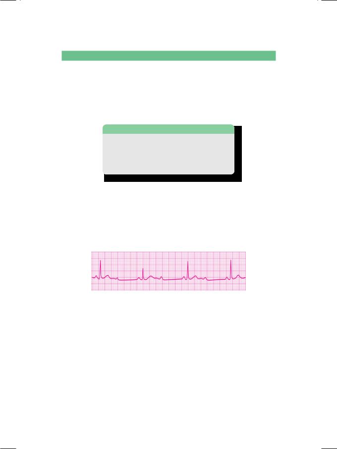
The clinical importance of Q-T interval prolongation lies in the fact that it predisposes to a typical type of polymorphic ventricular tachycardia.This tachycardia is known as “Torsade de pointes”, a ballet term which literally means “torsion around a point”. This term explains the morphology of the ventricular tachycardia, which consists of polymorphic QRS complexes that keep changing

178 |
|
Section 12 Electrocardiac Syndromes |
|
|
|
Table 38.2: Drugs causing Q-T interval prolongation
Antiarrhythmics |
Anti-infectives |
|
|
Quinidine |
Erythromycin |
Procainamide |
Gatifloxacin |
Amiodarone |
Ketoconazole |
|
|
Psychiatry drugs |
Miscellaneous |
|
|
Imipramine |
Cisapride |
Haloperidol |
Terfenadine |
Amitryptyline |
Ketanserin |
|
|
in amplitude and direction (Fig. 38.4). The polymorphic QRS complexes give the appearance of periodic torsion or twisting around the isoelectric line.
Long Q-T syndrome (LQTS) belongs to a class of congenital channelopathies, that are responsible for about 5 to 10% cases of sudden cardiac death (SCD). Other conditions belonging to this class are the Brugada syndrome and catecholaminergic ventricular tachycardia (CVT). Besides these above channelopathies, structural heart diseases responsible for SCD are hypertrophic cardiomyopathy and arrhythmogenic right ventricular dysplasia.
Figure 38.4: Polymorphic ventricular tachycardia ( ‘Torsades de pointes’)
MANAGEMENT ISSUES
The treatment of long Q-T syndrome depends upon the cause. When the cause is reversible, it suffices to correct the electrolyte abnormality or to withdraw the offending drug. The Q-T interval should be carefully assessed at peak plasma concentration, if multiple drugs with Q-T prolonging effect are used. In Q-T interval prolongation due to a cardiovascular or intracranial event, the underlying condition has to be managed. Since patients with congenital long Q-T syndrome are prone to syncope and sudden death due to ventricular arrhythmias, an implantable cardioverter defibrillator (ICD) is advocated.
The management of polymorphic ventricular tachycardia (“Torsades de pointes”) due to long Q-T interval, is different from the management of monomorphic ventricular tachycardia. In the acute setting, an infusion of magnesium sulphate or isoproterenol (a beta-blocker) is used. If the patient does not respond, overdrive ventricular pacing or electrical cardioversion is performed. An implantable cardioverter defibrillator (ICD) is offered to those who develop recurrent syncope, have family history of sudden cardiac death (SCD) and to survivors of cardiac arrest. To patients of congenital long QT syndrome (LQTS), cervical sympathetic ganglionectomy can be offered.
|
|
C A S E |
|
|
|
|
|
|
|
|
|
||
|
|
|
|
|
||
|
|
39 |
Sick Sinus |
|
|
|
|
|
|
|
|
|
|
|
|
|
|
Syndrome |
|
|
|
|
|
|
|
|
|
|
|
|
|
|
|
|
. |
CASE PRESENTATION |
A 78-year old elderly gentleman was paid a domiciliary visit by his physician, to evaluate frequent spells of dizziness over the past 2 weeks. The patient felt lightheaded when he stood up from the sitting or lying down position. Fortunately, he had never fallen down or fainted because he used a walking aid and was regularly looked after by a personal attendant. There was no history of breathlessness, chest pain or palpitation. The patient was hypertensive for over 40 years and took his medicines regularly. There was no past history of myocardial infarction or paralytic stroke. The only time he was hospitalized was for prostate surgery five years back. The patient’s family members had also noticed recent mental confusion and lapses in his memory.
On examination, the patient was conscious and cooperative but somewhat confused. The pulse was irregular, good in volume, at a rate of 48 to 52 beats/min. The dorsalis pedis pulsations were diminished on both sides and a bruit was audible over the left carotid artery. The BP was 160/82 mm Hg in the supine position and 150/74 mm Hg while standing. The JVP was not raised and there was no pitting edema over the ankles. The S1 was normal and A2 was loud; no S3 or S4 sound was audible. A short harsh systolic murmur was heard over the upper parasternal area. A different blowing pansystolic murmur was heard over the cardiac apex, that radiated towards the axilla. Breath sounds were vesicular, without any crepitations. An ECG was obtained (Fig. 39.1). At the time of prostate surgery, an ECHO had shown mild concentric left ventricular hypertrophy with an ejection fraction of 52% and no regional wall motion abnormality. CT scan of the head at the same time showed periventricular lacunar infarcts and changes of diffuse cerebral atrophy.
Figure 39.1: ECG showing sinus bradycardia, asystole and junctional escape
180 |
|
Section 12 Electrocardiac Syndromes |
|
|
|
ECG INTERPRETATION
The ECG showed sinus bradycardia at a rate of 50 beats/min., with short periods of asystole. At times the R-R interval was twice the usual R-R interval, suggesting 2:1 sino-atrial exit block (S-A block). At other times, the period of asystole was followed by a delayed beat without a preceding P wave, consistent with a junctional escape beat. This constellation of ECG findings which includes sinus bradycardia, S-A block and junctional escape, is consistent with the diagnosis of sinus node dysfunction, the so called sick sinus syndrome (Table 39.1).
Table 39.1: ECG findings in sick sinus syndrome
• Sinus bradycardia • Sino-atrial exit block
• Slow atrial fibrillation
• Junctional escape rhythm
The sick sinus syndrome usually presents with bradyarrhythmias such as those mentioned above. At times the indications of sinus node dysfunction are an inadequate tachycardia with sympathomimetic drugs, excessive sensitivity to beta-blocker drugs and atropine resistant bradycardia. At other times, there may be atrial fibrillation with slow ventricular response or a junctional rhythm. The coexistence of fast and slow cardiac rhythms constitutes the so called “tachy-brady” syndrome.
Figure 39.2: ECG showing 2:1 atrio-ventricular (A-V) block
In a pause due to 2:1 sino-atrial (S-A) exit block, the entire P-QRS-T complex is missing, since neither atrial nor ventricular activation occurs. By contrast in 2:1 atrio-ventricular (A-V) block, the P wave is inscribed normally but not followed by a QRS complex (Fig. 39.2). S-A block may coexist with A-V block and even left bundle branch block, if fibrocalcerous degeneration involves the entire conduction system. Quite often, sinus node dysfunction is suspected clinically but difficult to prove because the ECG is normal and Holter monitoring does not show up the arrhythmia, during the period of observation. A prolonged sinus node recovery time (SNRT) and sino-atrial conduction time (SACT) on electrophysiological studies (EPS) is then taken as a diagnostic criteria for sick sinus syndrome (SSS).

Case 39 Sick Sinus Syndrome |
|
181 |
|
|
|
CLINICAL DISCUSSION
The “sick sinus syndrome” (SSS) is a clinical condition caused by a diseased sinus node, which fails to produce or successfully conduct a sufficient number of cardiac impulses. It is observed in elderly patients and is believed to be caused by a degenerative condition like amyloidosis or infiltration of the atrium by a fibrocalcerous process. Certain cardiovascular drugs notably beta-blockers (atenolol, metoprolol), calcium-blockers (verapamil, diltiazem) and digoxin may also cause sinus node dysfunction, which is reversible after discontinuation of therapy.
Table 39.2: Symptoms of sick sinus syndrome
• Dizziness and syncope • Dyspnea and fatigue • Palpitation and angina
• Confusion and dementia
The most frequent symptoms of sick sinus syndrome are dizziness, mental confusion and fainting attacks (Table 39.2). Spells of dizziness and syncope in sick sinus syndrome are due to transient ventricular asystole, causing a precipitous decline in stroke volume and cerebral perfusion. Such episodes are known as Stokes-Adams attacks. Besides SSS, other causes of Stokes-Adam’s attacks are advanced atrio-ventricular block, malignant ventricular arrhythmias, carotid sinus hypersensitivity and subclavian steal syndrome (Table 39.3).
Table 39.3: Causes of Stokes Adams Attacks
• Advanced atrio-ventricular block • Serious ventricular arrhythmias • Carotid sinus hypersensitivity
• Subclavian steal syndrome • Sick sinus syndrome
Since patients are in the advanced age group, many of them have had a cerebrovascular accident or a prior myocardial infarction. They may also complain of dyspnea and fatigue due to heart failure. Palpitation and angina pectoris may occur due to tachyarrhythmias or associated coronary artery disease. Dizziness and syncope in an elderly patient may be multifactorial. Even those with documented sick sinus syndrome may additionally have volume depletion, electrolyte imbalance or hypoglycemia. They may have orthostatic hypotension due to autonomic failure or vertebro-basilar insufficiency. Still others may have cardiac outflow obstruction due to aortic sclerosis or ventricular arrhythmias due to an old myocardial scar or heart failure. Therefore, a detailed evaluation of these patients is warranted.

182 |
|
Section 12 Electrocardiac Syndromes |
|
|
|
MANAGEMENT ISSUES
Drugs that can increase the rate of discharge from the sinus node such as sympathomimetic (adrenaline) and vagolytic (atropine) drugs, can only temporarily increase the heart rate. Permanent pacemaker implantation (PPI) is the definitive form of treatment, particularly if the symptoms are severe and disabling. Dual-chamber pacemakers are more physiological and carry a lower risk of atrial fibrillation and stroke. They also have a distinct advantage over single-chamber pacing if there is coexistent A-V nodal disease or bundle branch block. Pacemaker implantation makes it possible to use antiarrhythmic drugs to treat tachyarrhythmias, which otherwise would have caused severe bradycardia.

|
|
C A S E |
|
|
|
|
|
|
|
|
|
||
|
|
|
|
|
||
|
|
40 |
Early Repolarization |
|
|
|
|
|
|
|
|
|
|
|
|
|
|
Syndrome |
|
|
|
|
|
|
|
|
|
|
|
|
|
|
|
|
. |
CASE PRESENTATION |
A 29-year old well-built man of African origin, sought an appointment with the cardiologist, for opinion on an abnormal ECG. The ECG was performed as part of routine pre-employment medical evaluation. The man vehemently denied complaints of fatigue, breathlessness, chest pain, palpitation or syncope. He had been actively involved in competitive sports during his college days and still played tennis on week-ends. The man did not smoke or take alcohol, but was fond of caloriedense food. He did not suffer from diabetes or hypertension and had never got a serum lipid analysis done. There was no family history of coronary heart disease or of cerebrovascular accident.
On examination, the man was of stocky built, with a muscular physique. His body mass index (BMI) was 28 kg/m2. He was fully conscious, comfortable and cheerful. There was no anemia, cyanosis, icterus or edema. The trachea was central, thyroid gland was not palpable and the JVP was not raised. The pulse rate was 58-62 beats/ min. with a normal pulse volume and no special character. The BP was 120/80 mm Hg over the right arm. The precordium was unremarkable, with a normally located apex beat. The S1 and S2 were normal without any gallop sound. No murmur or pericardial friction rub was audible. The breath sounds were vesicular without any audible rhonchi or crepitations. A fresh ECG was performed in the cardiologist’s office (Fig. 40.1).
Figure 40.1: ECG showing features of early repolarization syndrome

184 |
|
Section 12 Electrocardiac Syndromes |
|
|
|
ECG INTERPRETATION
The ECG showed tall R waves in the lateral precordial leads, preceded by narrow Q waves. The S-T segment was elevated concave upwards, with an initial slur on the S-T segment (J wave). The T waves were upright, tall and symmetrical in the lateral leads, with prominent U waves in the mid-precordial leads. These findings are consistent with the diagnosis of early repolarization syndrome (Table 40.1). Since early repolarization is frequently observed in healthy athletic persons, this entity is also known as the “athlete’s heart”.
There are several causes of S-T segment elevation (Table 40.2), of which acute myocardial infarction is the leading cause. The S-T segment elevation of early repolarization, can simulate the injury pattern of acute myocardial infarction. However, there are several classical differentiating features:
•S-T segment elevation is concave upwards in lead V6
•Ratio of S-T elevation : T wave height is below 0.25
•There is no reciprocal S-T depression in other leads
•ECG changes do not evolve as in case of infarction
•ECHO does not show abnormal regional wall motion
•Serial level of cardiac enzyme titers are not increased.
Table 40.1: ECG features of early repolarization syndrome
• Tall R wave in lead V6
• Narrow and deep Q wave
• Concave S-T segment elevation • Initial J wave on the S-T segment
• Upright and tall symmetrical T wave
CLINICAL DISCUSSION
The “early repolarization” variant is an alarming electrocardiographic entity, which presents with S-T segment elevation. It represents early repolarization of a portion of the ventricle, before the entire myocardium has been depolarized. There is an early uptake of the S-T segment, before the descending limb of the R wave has reached the baseline. This causes an initial slur on the S-T segment, known as the J wave. The S-T segment is elevated and concave upwards. There is an associated increased amplitude of the R wave. The T wave is also tall, but the ratio of S-T segment elevation to T wave height is less than 0.25. Interestingly, the degree of S-T elevation and T wave height may vary on a day-to-day basis and the S-T segment may normalize after exercise. Besides the features already mentioned, other characteristics of this syndrome are sinus bradycardia with sinus arrhythmia, voltage criteria of left ventricular hypertrophy and persistent juvenile pattern of T wave inversion in leads V1 to V3.