

новая папка / [libribook.com] 50 Cases in Clinical Cardiology_ A Problem Solving Approach 1st Edition
.PdfCase 47 Chronic Stable Angina |
|
215 |
|
|
|
coronary artery disease due to atherosclerosis. Stable angina is due to coronary occlusion by one or more atherosclerotic plaques that have a small lipid core, a thick fibrous cap and are less prone to rupture. Conventional risk factors for coronary disease are advanced age, male sex, being overweight, tobacco abuse, diabetes mellitus, hypertension, hyperlipdemia, sedentary lifestyle, psychosocial stress and family history of coronary artery disease (Table 47.1).
Table 47.1: Conventional risk factors for coronary artery disease
• |
Male Sex |
• |
Hypertension |
|
|
|
|
• |
Advanced age |
• |
Hyperlipidemia |
|
|
|
|
• |
Being overweight |
• |
Diabetes mellitus |
|
|
|
|
• |
Tobacco abuse |
• |
Sedentary lifestyle |
|
|
|
|
• |
Family history |
• |
Psychosocial stress |
|
|
|
|
Besides atherosclerotic coronary artery disease, there are some other atypical and uncommon causes of angina pectoris, which too cause an imbalance between myocardial oxygen supply and demand. These uncommon causes of angina are given in Table 47.2.
Table 47.2: Atypical causes of angina pectoris
Non-cardiac
• Severe anemia • Thyrotoxicosis
Vascular
• Coronary artery spasm • Microvascular disease
Cardiac
• Aortic valve stenosis
• Hypertensive heart disease • Hypertrophic cardiomyopathy
MANAGEMENT ISSUES
The first step in the management of stable angina pectoris is suitable life-style modification and control of cardiovascular risk factors. Patients should be advised to cut-down on calorie-dense foods, follow a regular exercise regimen, to quit smoking and manage psycho-social stress. Major risk factors such as hypertension, diabetes and hyperlipidemia should be treated with suitable drugs, in addition to correction of life-style.
Glyceryl trinitrate (GTN) is used sub-lingually for rapid relief from angina, while long-acting isosorbide mononitrate (ISMN) is given for long-term prophylaxis. Beta-blockers are the mainstay of therapy, as they reduce the rate and force of cardiac contraction and therefore the workload and oxygen demand.

216 |
|
Section 14 Coronary Artery Disease |
|
|
|
In the scenario of beta-blocker contraindication or intolerance, a rate-limiting calcium antagonist like verapamil or diltiazem is used. Metabolic agents like nikorandil and trimetazidine are sometimes used. Low-dose aspirin prevents thrombotic events. If the patient is allergic or intolerant to aspirin, clopidogrel is employed. All patients should receive a statin, irrespective of their cholesterol level. Statins have amply proven their worth in preventing cardiovascular events. The ACE-inhibitor ramipril has also been shown to reduce CV events and allcause mortality in patients with cardiovascular disease.
RECENT ADVANCES
Besides the conventional risk factors of coronary artery disease, certain new risk factors have been recently identified (Table 47.3). These include high levels of C-reactive protein (CRP), homocysteine (Hcy), lipoprotein (a) [Lp (a)], and fibrinogen (Fgn). However, the INTERHEART study clearly showed that majority of heart attacks across the globe can be conveniently explained on the basis of conventional risk factors. Modest intake of alcohol with high intake of fruits and vegetables were found to be important “anti-risk” factors.
Ranolazine is a new metabolically active agent used for the treatment of stable angina. Ivabradine is a recently introduced drug for heart-rate control, in patients who cannot tolerate uptitration of beta-blocker dose.
Table 47.3: Non-conventional risk factors for coronary artery disease
• C-reactive protein (CRP) • Lipoprotein (a) [Lp(a)] • Homocysteine (Hcy)
• Fibrinogen (Fgen)
|
|
C A S E |
|
|
|
|
|
|
|
|
|
||
|
|
|
|
|
||
|
|
48 |
Acute Coronary |
|
|
|
|
|
|
|
|
|
|
|
|
|
|
Syndrome |
|
|
|
|
|
|
|
|
|
|
|
|
|
|
|
|
. |
CASE PRESENTATION |
A 56-year old gentleman was brought to the emergency department by his wife, in the wee hours of the morning. The man had complained of severe central chest pain with profuse sweating, during sexual intercourse. He was a known case of diabetes mellitus and hypertension. However, he was irregular with his medication and did not follow-up with his doctor periodically. The patient had not undergone a blood lipid analysis despite advice from his doctor and persistent request by his wife. He had a sedentary life-style and was on no particular dietary restrictions. He smoked 8 to 10 cigarettes everyday and took 3 to 4 pegs of whiskey, on most days of the week. A treadmill test performed one year earlier had been inconclusive, because the patient had failed to achieve the target heart rate. Recently, he had been complaining of frequent episodes to restlessness and belching, which he attributed to acidity and indigestion.
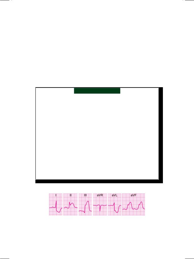
On examination, the patient was tachypneic and diaphoretic. He was markedly overweight with a body mass index (BMI) of about 32 kg/m2. The pulse was irregular and low in volume with “skipped” beats. The extremities were cold and clammy. His heart rate was 104 beats/min. with a BP of 104/74 mm Hg. Skin tags and acanthosis nigricans were seen at the nape of the neck and xanthelasma were observed over the upper eyelids. The precordium and apex beat were unremarkable. The S1 and S2 were loud with a S3 gallop; no murmur was audible. Few basilar rales were heard over the lung fields bilaterally. An ECG was obtained (Fig. 48.1).
Figure 48.1: ECG showing S-T segment elevation in inferior leads with S-T segment depression in lateral leads
218 |
|
Section 14 Coronary Artery Disease |
|
|
|
ECG INTERPRETATION
The ECG showed sinus rhythm with normal P waves and no prolongation of the
Q-T interval. There was a 5 mm elevation of the S-T segment in leads LII, LIII and avF. There was also S-T segment depression (reciprocal changes) in leads LI and
avL. There was no attenuation of the R wave or appearance of significant Q waves. These findings are consistent with the diagnosis of hyperacute phase of inferior wall myocardial infarction.
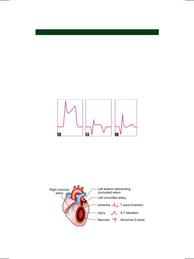
The phases of acute myocardial infarction are divided into hyperacute phase (0 hr to 6 hrs), recent phase (7 hrs to 7 days), evolved phase (8 days to 28 days) and stabilized phase (more than 29 days). In the hyperacute phase, the S-T segment is markedly elevated, which blends with the proximal limb of the tall T wave. In the evolved phase, the S-T segment elevation begins to settle down, T waves get inverted and Q waves appear. In the stabilized phase, there is normalization of the S-T segment and T waves but the Q waves persist (Fig. 48.2).
Figure 48.2: Diagram to illustrate the various phases of
S-T elevation myocardial infarction
Myocardial infarction due to coronary occlusion, consists of a central necrotic core surrounded by a zone of injury and skirted by a water-shed area of ischemia. These areas form the pathological basis of the ECG changes observed in myocardial infarction. Q wave represents necrosis, S-T elevation is due to injury and T wave inversion is indicative of myocardial ischemia (Fig. 48.3).
Figure 48.3: The pathological basis of ECG changes after acute myocardial infarction

Case 48 Acute Coronary Syndrome |
|
219 |
|
|
|
The location of an infarct is identified from the particular ECG leads that show the classical findings of myocardial infarction (Table 48.1).
Table 48.1: Location of infarction from leads with Q waves
Leads with Q waves |
Location of infarction |
|
|
V1V2 |
Septal |
V3V4 |
Anterior |
V5V6LIaVL |
Lateral |
V1-4 |
Anteroseptal |
V3-6LIaVL |
Anterolateral |
V1-6LIaVL |
Extensive anterior |
LIaVL |
High lateral |
LIILIIIaVF |
Inferior |
|
|
CLINICAL DISCUSSION
Acute coronary syndrome is classified into ST elevation myocardial infarction (STEMI), non-ST elevation myocardial infarction (NSTEMI) and unstable angina (UA). Since myocardial infarction (not angina) involves tissue necrosis, the levels of cardiac enzymes (creatine kinase and troponin), are elevated. The distinction between unstable angina and NSTEMI is therefore based on the cardiac enzyme levels. STEMI is also referred to as Q wave or transmural MI. NSTEMI is also designated as non-Q or subendocardial MI. This classification has therapeutic as well as prognostic implications. Acute coronary syndrome is due to coronary occlusion by an atherosclerotic plaque, which has a large lipid core, a thin fibrous cap and is more prone to rupture. A thrombus forms over the ruptured vulnerable plaque.
In variant angina, which is also known as Prinzmetal’s angina, the basis of myocardial ischemia is not coronary thrombosis but arterial spasm. In an ischemic episode of vasospastic angina, the ECG changes are similar to those of hyperacute phase of infarction with S-T segment elevation and tall T waves. The difference is that the ECG changes do not evolve serially but settle down rapidly. Q wave never appear and levels of cardiac enzymes are not raised as there is no myocardial necrosis.
Myocardial infarction is classified on the basis of the age of infarct (recent or stabilized), site of infarct (anterior or inferior wall) and type of infarct (transmural or subendocardial). There may paucity of ECG findings if the infarction is small, atrial, posterior in location, or right ventricular MI. ECG findings of infarction are difficult to interpret in the presence of left bundle branch block, left ventricular hypertrophy, WPW syndrome and digoxin therapy. Reasons for a disparity between ECG changes and the clinical findings are left circumflex disease, hibernating myocardium, attenuation phenomenon or the presence of a mechanical complication.
220 |
|
Section 14 Coronary Artery Disease |
|
|
|
Table 48.2: Types of regional wall motion
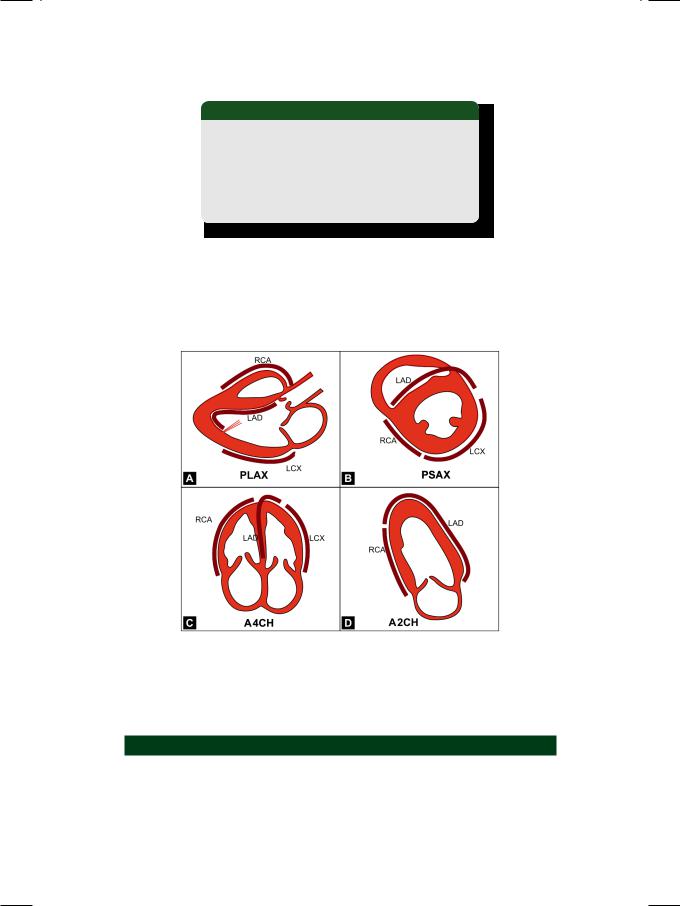
• Normal motion: full inward motion • Hypokinesia:<50% inward motion • Akinesia: no inward wall motion • Dyskinesia: outward movement • Aneurysmal: outpouching of wall.
On echocardiography, the systolic inward motion of the infarcted myocardial segment is reduced in extent, altogether absent or may even be paradoxically outward. The regional wall motion abnormality can be classified as given in Table 48.2. From the location of the regional wall motion abnormality (RWMA), it is possible to identify the coronary artery which is occluded (Fig. 48.4).
Figure 48.4: Identification of the occluded coronary artery from the location of wall motion abnormality
PLAX: Parasternal long axis; PSAX: Parasternal short axis; RCA:
Right coronary artery; LAD: Left anterior descending artery; LCX:
Left circumflex artery
MANAGEMENT ISSUES
All patients with an acute coronary syndrome (ACS), should ideally be managed in a coronary care unit (CCU), where the speed of instituting treatment improves myocardial salvage (time is muscle). The long-term survival after myocardial infarction heavily depends upon the extent of myocardial salvage (muscle is time). The essentials of treatment are bed-rest, oxygen, opiate analgesia, aspirin

Case 48 Acute Coronary Syndrome |
|
221 |
|
|
|
162.5 mg stat and clopidogrel 300 mg stat. Patient of STEMI may be taken up for percutaneous coronary intervention (PCI), if there is rapid access to a Cath-Lab facility. If not, the patient should receive thrombolytic therapy, provided there are no contraindications.
Patient of NSTEMI should receive heparin, nitrate and a beta-blocker. High risk patients are taken up for coronary angiography with revascularization. Coronary artery revascularization is undertaken by percutaneous transluminal coronary angioplasty (PTCA) or by coronary artery bypass graft (CABG) surgery. PTCA is suitable for significant (>70% stenosis) single-vessel disease or 2-vessel disease. CABG is preferable for 3-vessel disease, 2-vessel disease including proximal LAD and left main-stem occlusion.
RECENT ADVANCES
A number of scoring systems have been developed, to risk-stratify patients presenting with an acute coronary syndrome. The most commonly used is the TIMI (Thrombolysis In Myocardial Infarction) Risk score, that was derived from trials of low molecular weight heparin. The TIMI system assigns a binary score (0 or 1) to seven independent risk factors, which have a similar predictive value. Higher the score, greater is the risk of death or recurrent ischemia at 14 days. The seven risk factors of the TIMI score, are given in Table 48.3.
Table 48.3: TIMI risk score after acute coronary syndrome
• Sixty-five years of age or older • Three or more CV risk factors • Significant coronary stenoses • S-T segment deviation, > 2 mm
• Use of aspirin prior to admission
• Two or more anginal episodes within previous 24 hours
• Elevated cardiac enzyme levels
(creatine kinase or troponin)

|
|
C A S E |
|
|
|
|
|
|
|
|
|
||
|
|
|
|
|
||
|
|
49 |
Papillary Muscle |
|
|
|
|
|
|
|
|
|
|
|
|
|
|
Rupture |
|
|
|
|
|
|
|
|
|
|
|
|
|
|
|
|
. |
CASE PRESENTATION |
A 63-year old woman was wheeled into the emergency room, with the complaints of vague chest discomfort, profuse sweating and sinking sensation lasting about 5 hours. She also had retching and vomiting for which she took some antacid, thinking that it was “gas-trouble”. When there was no relief, the patient’s daughter deemed it necessary to get an ECG done. The ECG findings were alarming and therefore she was admitted to the coronary care unit (CCU). There she received thrombolytic therapy and remained stable for the next 3 days. On the 4th day of her admission, she felt breathless and was unable to lie flat in bed. There was no fresh chest pain and the cardiac monitor did not show any serious arrhythmia, except for an occasional ventricular premature complex.
On examination, the patient was tachypneic, diaphoretic and appeared to be in distress. She looked pale and her extremities were cold and clammy. The pulse was fast, irregular and of low volume, with a rate of 104 beats/min. Her BP was 106/74 mm Hg in the right arm and the respiratory rate was 28/min. The JVP was not raised and there was no edema over the ankles. The precordium was hyperkinetic and the cardiac apex beat was displaced towards the left axilla. The S1 and S2 were normal but an S3 gallop sound was appreciated in diastole. A soft systolic murmur was audible over the cardiac apex. The murmur radiated towards the axilla and could be heard upto the left scapula. Bilateral coarse crackles were heard over the lung fields.
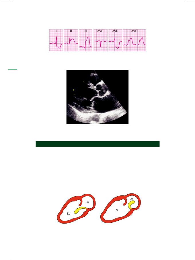
ECG showed sinus tachycardia with 5 mm elevation of the S-T segment in leads LII, LIII and aVF. There was reciprocal S-T segment depression in leads LI and aVL. These findings were consistent with the diagnosis of hyperacute inferior wall myocardial infarction (Fig. 49.1). Additionally, there was elevation of the S-T segment in the right-sided chest leads V3R and V4R. This was indicative of right ventricular infarction.
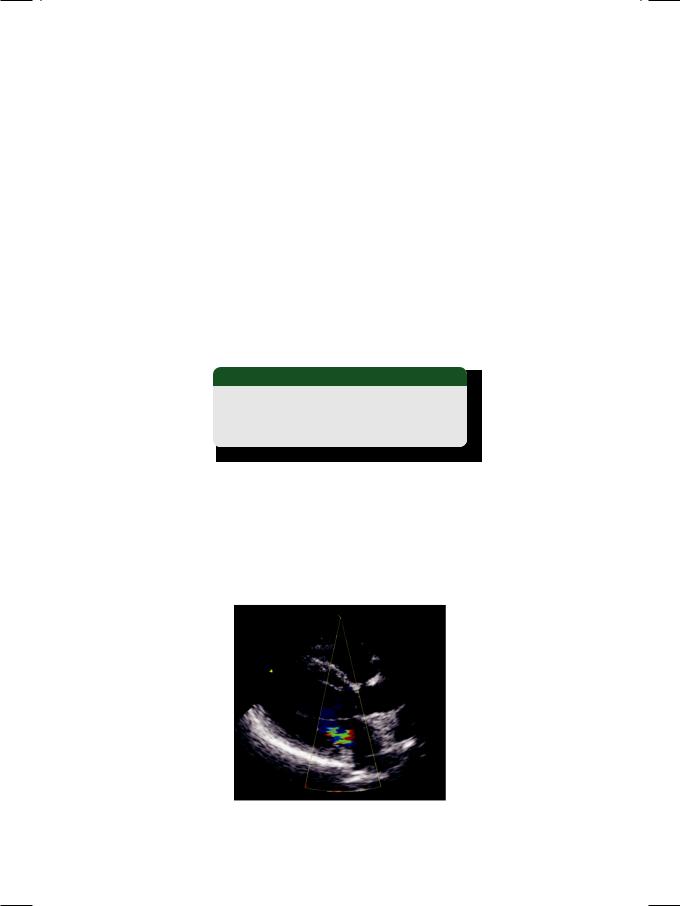
ECHO revealed a normal sized left ventricle with an ejection fraction of 45%. There was dilatation of the right ventricle and right atrium. The postero-basal segment of the left ventricle and free wall of the right ventricle were hypokinetic. There was no mass or thrombus in any chamber. The posterior leaflet of the mitral valve exhibited an exaggerated whip-like motion (Fig. 49.2). The tip moved past the mitral annular plane and deep into the left atrium. It failed to coapt with the anterior leaflet at the end of diastole. On colour flow mapping, an eccentric jet was seen in the left atrium, that was directed towards the posterior left atrial wall. These findings are consistent with the diagnosis of flail mitral leaflet due to papillary muscle rupture.
|
|
Case 49 Papillary Muscle Rupture |
|
223 |
|
|
|
||
|
|
|
|
|
|
|
|
|
|
Figure 49.1: ECG showing the hyperacute phase of inferior wall myocardial infarction
Figure 49.2: ECHO showing exaggerated motion of a flail posterior mitral leaflet
CLINICAL DISCUSSION
Acute mitral regurgitation (MR) in a setting of acute myocardial infarction, occurs either due to papillary muscle rupture or because of papillary muscle dysfunction. Rupture of a papillary muscle due to ischemic necrosis causes a flail mitral valve leaflet (Fig. 49.3). Since rupture of the postero-medial papillary muscle is more common than that of the antero-lateral muscle, often it is the posterior mitral leaflet (PML) that is flail. It generally follows inferior wall infarction due to occlusion of the posterior descending branch of the right coronary artery. Papillary muscle dysfunction is due to ischemic restriction of papillary function or akinesia of the infero-basal wall that does not adequately shorten in systole. As
Figure 49.3: Diagram to illustrate whip-like motion of a flail posterior mitral leaflet
LV: Left ventricle; LA: Left atrium
224 |
|
Section 14 Coronary Artery Disease |
|
|
|
a result, the posterior mitral leaflet fails to reach the plane of the valve annulus and the coaptation point of mitral leaflets is distally located into the left ventricle.
In acute MR due to myocardial infarction (MI), the left ventricular (LV) systolic function often remains unimpaired. In pump failure due to extensive MI, there is severe LV dysfunction. Although there is no time for left ventricular dilatation to develop, an acute rise in left ventricular end-diastolic pressure (LVEDP) rapidly produces frank pulmonary edema. The systolic murmur of acute MR is short and soft compared to the long and loud murmur of MR due to chronic valvular disease. This is because of two reasons. Firstly, the large mitral orifice created by acute MR, does not generate much turbulence across the valve. Secondly, the rapid rise in left atrial pressure and decline in pressure gradient impedes regurgitation, during the later half of systole. Besides papillary muscle rupture due to myocardial infarction, other causes of acute mitral regurgitation are blunt trauma to the chest wall and valve dehiscence in case of infective endocarditis (Table 49.1). Another cause of a systolic murmur after myocardial infarction is ventricular septal rupture.
Table 49.1: Causes of acute mitral regurgitation
• Myocardial infarction • Infective endocarditis • Chest wall trauma
On Doppler or on color flow mapping, the MR flow velocity or color jet is eccentric and directed towards the posterior left atrial wall (Fig. 49.4). The jet area may be much less than the actual amount of MR; hence there is a risk of underestimating the severity of acute MR. The flail leaflet of papillary muscle rupture resembles the floppy leaflet of mitral valve prolapse with subtle differences. The prolapsing leaflet just buckles but does not flap freely and it enters the left atrium only for a brief period (Table 49.2).
Figure 49.4: ECHO showing an eccentric jet of mitral regurgitation