

новая папка / [libribook.com] 50 Cases in Clinical Cardiology_ A Problem Solving Approach 1st Edition
.Pdf56 |
|
Section 4 The Cardiomyopathies |
|
|
|
Figure 12.1: X-ray showing the classical findings of left ventricular systolic dysfunction
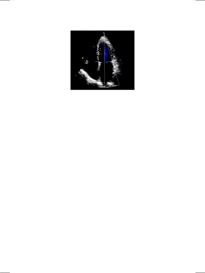
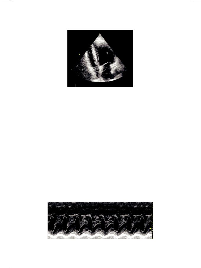
Figure 12.2: ECHO showing reduced excursion of the septum and mitral leaflets
•Organic mitral regurgitation
•Ischemic cardiomyopathy
•Dilated cardiomyopathy.
Organic mitral regurgitation with volume overload leading to severe left ventricular dysfunction is unlikely in this case, because the mitral valve architecture was normal and valvular regurgitation was only mild in severity.
Ischemiccardiomyopathyisadiagnosticpossibility,butthereareseveralreasons against it. The patient had no significant coronary risk factors or family history of heart disease. ECG did not show any S-T segment shift or the presence of Q-waves. On ECHO, there was global hypokinesia instead of regional wall motion abnormality. Moreover, there were no dyskinetic segments in the left ventricular

|
|
Case 12 Dilated Cardiomyopathy |
|
57 |
||
|
|
|
|
|
|
|
|
|
|
|
|
|
|
|
Table 12.1: Differences between dilated and ischemic cardiomyopathy |
|
|
|
||
|
|
Dilated CMP |
Ischemic CMP |
|
|
|
|
Hypokinesia |
Global |
Regional |
|
|
|
|
RWMA and the coronary territory |
Do not conform |
Conform |
|
|
|
|
Dyskinesia |
Not seen |
Seen |
|
|
|
|
RV involvement |
Often |
Rare |
|
|
|
|
|
|
|
|
|
|
|
|
|
|
|
|
|
wall while right ventricular dilatation was present (Table 12.1). Therefore, the most probable diagnosis in this case is dilated cardiomyopathy.
There are several causes of dilated cardiomyopathy (DCMP). The idiopathic varietyisbelievedtofollowanacuteviralmyocarditis.Prominentdefinitecauses of DCMP are heavy alcoholism, diabetes mellitus and the peri-partum period. Familial DCMP is seen in Duchenne’s muscular dystrophy and Friedrich’s ataxia. Deficiency of thiamine (vitamin B1), selenium and carnitine can cause a reversible form of DCMP. Drugs implicated in the causation of DCMP are doxorubicin, imatinib, cyclophosphamide and trastuzumab. Uncommonly, a persistent tachycardia due to any cause, can lead to permanent DCMP
(Table 12.2).
Table 12.2: Causes of dilated cardiomyopathy
• Idiopathic • Alcoholism • Diabetes • Peripartum • Nutritional
• Drug induced
• Tachycardia related
PERTINENT INVESTIGATIONS
While echocardiography is a dependable diagnostic modality for dilated cardiomyopathy, a battery of tests can be performed to ascertain the etiological diagnosis. This is because there are several causes of DCMP and a vigorous search is warranted to identify a treatable cause, with the optimism of preventing relentless progression of the disease.
Inflammatory markers (ESR and CRP) are elevated in the presence of myocarditis while levels of biomarkers (CPK, LDH) are increased in myocardial necrosis. Patients are screened for a chronic infection (HIV, Hepatitis C) or nutritional deficiency (thiamine, selenium, carnitine). Serum iron, ferritin and transferrin are ordered if anemia or hemochromatosis are suspected and T3, T4 and TSH if thyrotoxicosis is to be ruled out. Renal and liver function tests are ordered to assess additional causes of fluid overload.

58 |
|
Section 4 The Cardiomyopathies |
|
|
|
Cardiacmagneticresonanceimaging(MRI)isusefultoassessleftventricular size and geometry, LV remodelling and for tissue characterization with gadolinium enhancement. Coronary angiography is performed in the presence of atherosclerotic risk factors. Endomyocardial biopsy is rarely performed because of its low diagnostic yield.
MANAGEMENT ISSUES
The first step in the management of DCMP is to identify and treat reversible factors. Measures include withdrawal of the offending drug and correction of any nutritional deficiency. Abstinence from alcohol and control of diabetes are equally important. The heart rate has to be controlled in tachycardia related cardiomyopathy.
Treatment of most of these patients is on the lines of standard heart failure therapy.Diureticsareprescribedtotreatfluidoverloadandmodulatorsofrenin- angiotensin system (ACE-inhibitors or ARBs) to reduce cardiac afterload. Betablockers are used particularly if extreme tachycardia or atrial fibrillation are present. If the patient has persistent atrial fibrillation, rate control with digoxin is required. Additionally, the patients are initiated on an oral anticoagulant to reduce the risk of thrombo-embolic complications.
RECENT ADVANCES
A significant proportion of patients with dilated cardiomyopathy succumb to malignant ventricular arrhythmias and not to heart failure per se. Antiarrhythmic drugs reduce ejection fraction and have proarrhythmic potential, besides their systemic side-effects. The use of automatic implantable cardioverter defibrilla- tors (AICDs) is now advocated as a primary preventive strategy against sudden cardiac death (SCD) in such patients. Anticoagulant therapy iswidely prescribed to patients who are in atrial fibrillation and in those where a mural thrombus has been documented. Currently, trials are underway to evaluate the role of anticoagulants against thromboembolic complications in patients who are in sinus rhythm.
In patients with moderate to severe symptoms (NYHA Class III or IV) with severe left ventricular dysfunction (LVEF below 30%) and wide QRS complexes
(QRS duration more than 120 msec), cardiac resynchronization therapy (CRT) may be offered. A biventricular pacemaker is placed to synchronize the ejection of right and left ventricles if there is left bundle branch block (LBBB) or an intraventricular conduction defect (IVCD). Finally, a ventricular assist device may be implanted to improve left ventricular function in those with significant hemodynamic deterioration. This strategy is particularly useful as a bridge to cardiac transplantation.

C A S E
13 Restrictive
Cardiomyopathy
CASE PRESENTATION
A 58-year old man presented to the cardiology facility with fatigue, difficulty in breathing and increasing swelling over his ankles, for the last 3 months. His dyspnea became worse at night and he required two or more pillows under his head to be comfortable. In addition, he had noticed an increase in his abdominal girth and had loss of appetite, but there was no weight loss. There was no history of fever, productive cough, chest pain or hemoptysis. He also denied palpitations or episodes of syncope. The patient was not hypertensive or diabetic and there was no history of premature coronary artery disease in any of his family members.
On examination, he was tachypneic but not in any distress. The pulse was 90 beats /min with a BP of 114/72 mm Hg. There was no cyanosis or jaundice but pitting edema was present around the ankles. The liver edge was palpable 6 cm below the right costal margin and there was free fluid in the abdominal cavity. The JVP was elevated 5 cm above the angle of Louis, with prominent a waves and without a fall in its level during inspiration. The apex beat was normal in character and location, without any displacement towards the axilla. The S1 and S2 were normal with an audible S4 in late diastole. There was no murmur or pericardial friction rub. Crepts were audible over the basilar lung fields posteriorly.
CLINICAL DISCUSSION
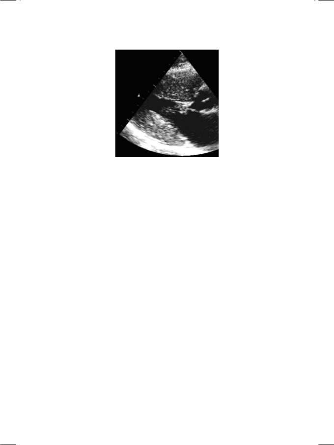
From the history and physical examination, this patient was clearly having biventricular heart failure. ECG showed low QRS voltages with non-specific T-wave changes but no Q waves. There was no tachyarrhythmia or conduction block. X-ray chest findings were interstitial pulmonary edema and small bilateral pleural effusion, but the heart size was normal. ECHO revealed relative small ventricular chambers with bilateral atrial dilatation. The ventricular free walls and the interventricular septum were thick and gave a “granular sparkling” ground-glass appearance (Fig. 13.1). There was also thickening of the mitral and tricuspid valve leaflets with minimal valvular regurgitation. The left ventricular ejection fraction was normal but the transmitral Doppler showed a tall E-wave with rapid deceleration and a small A-wave.
There were several unusual clinical findings in this case. There was biventricular heart failure but no clinical or radiological evidence of cardiomegaly.
60 |
|
Section 4 The Cardiomyopathies |
|
|
|
Figure 13.1: ECHO showing bright echogenicity with small ventricles and large atria
Prominent a waves in the JVP and presence of S4 on auscultation indicate atrial contraction against a stiff, noncompliant right ventricle. Failure of the JVP to fall during inspiration (or paradoxical rise) constitutes the Kussmaul’s sign and also indicates ventricular diastolic restriction. Despite evidence of ventricular hypertrophy on ECHO, the voltages of the QRS complexes on the ECG were low. This indicates that myocardial infiltration was the cause of ventricular thickening in this case and not myocardial hypertrophy, as is observed in systemic hypertension and aortic stenosis. ECHO revealed relatively small ventricles and large atria, findings that are typically seen in mitral stenosis with pulmonary hypertension and secondary tricuspid regurgitation. However, in our case there was no evidence of mitral stenosis.
This patient therefore has a restrictive ventricular filling abnormality or left ventricular diastolic dysfunction (LVDD) or heart failure with preserved ejection fraction (HFPEF). Clinically speaking, the most frequent causes of LVDD are systemic hypertension and coronary artery disease, where the mitral inflow Doppler signal shows a slow relaxation pattern with the E wave shorter than the A wave. These conditions were clearly absent in our case. Therefore, the most probable diagnosis in this case is restrictive cardiomyopathy.
There are several causes of restrictive cardiomyopathy (RCMP). The most prominent cause of RCMP is endomyocardial fibrosis (EMF). Another reason is hypereosinophilic syndrome, the so called Loeffler’s endocarditis. A variety of systemic infiltrative disease such as amyloidosis, sarcoidosis and malignancy can present with RCMP. Connective tissue disorders especially scleroderma can also lead to myocardial restriction. Finally, RCMP may be observed in storage diseases like hemochromatosis and glycogen storage disorders (Table 13.1).
Amyloidosis is an important cause of restrictive cardiomyopathy. Besides cardiac involvement, there may be other systemic manifestations of amyloid deposition. Macroglossia with periorbital ecchymosis is pathognomic of amyloidosis. Neurological manifestations are sensory neuropathy, carpal tunnel syndrome and autonomic neuropathy with postural hypotension. Hepatomegaly may be due to amyloid infiltration or due to congestive heart failure. Renal
Case 13 Restrictive Cardiomyopathy |
|
61 |
|
|
|
Table 13.1: Causes of restrictive cardiomyopathy
Endomyocardial
Endomyocardial fibrosis
Hypereosinophilic syndrome
Infiltrative
Amyloidosis
Sarcoidosis
Malignancy
Gaucher’s disease
Non-infiltrative
Idiopathic
Scleroderma
Storage diseases
Hemochromatosis
Glycogen storage disorder
involvement may lead to nephrotic syndrome, which exacerbates the pedal edema of heart failure.
It is often challenging to differentiate restrictive cardiomyopathy from constrictive pericarditis and cardiac catheterization may be required for their distinction. The diagnosis of pericardial constriction is suggested if there is past history of tuberculosis, cardiac surgery or radiotherapy. Also, the X-ray chest shows linear pericardial calcification and ECHO may reveal pericardial thickening with multiple parallel lines, casting a bright reflection. Computed tomography (CT) or magnetic resonance imaging (MRI) may be required to demonstrate pericardiac thickening. Moreover, in constrictive pericarditis, the cardiac chamber size, myocardial thickness, valve structure and ejection fraction are all normal (Table 13.2). Clinically speaking, constrictive pericarditis presents with sole or predominant right heart failure while restrictive cardiomyopathy often presents with biventricular failure.
Table 13.2: Differences between constrictive pericarditis and restrictive cardiomyopathy
|
Constrictive pericarditis |
Restrictive cardiomyopathy |
|
|
|
Pericardium |
Thick |
Normal |
|
|
|
Myocardium |
Normal |
Thick |
|
|
|
Ventricles |
Normal |
Obliterated |
|
|
|
Atria |
Normal |
Dilated |
|
|
|
LV function |
Normal |
Mildly impaired |
|
|
|
MV and TV |
Normal |
Regurgitation |
|
|
|
MV inflow |
Abrupt halt |
Slow relaxation |
|
|
|

62 |
|
Section 4 The Cardiomyopathies |
|
|
|
MANAGEMENT ISSUES
Diuretics reduce dyspnea and edema and they are the mainstay of the medical management of restrictive cardiomyopathy. However, vigorous diuresis should be avoided since it would compromise ventricular filling, reduce cardiac output and even cause syncope. It is crucial to maintain sinus rhythm and to cardiovert atrial fibrillation by electrical (DC shock) or pharmacological (amiodarone) means. This is because atrial fibrillation leads to loss of atrial contribution to ventricular filling and shortens the diastolic filling time. Digoxin is best avoided for rate control and heart failure treatment, due to the high risk of ventricular arrhythmias in the presence of myocardial disease. Beta blockers and verapamil improve diastolic dystunction and control tachyarrhythmias. Specific therapies for restrictive cardiomyopathy are steroids for sarcoidosis and scleroderma as well as iron chelation therapy with desferrioxamine for hemochromatosis.
RECENT ADVANCES
The diagnosis of cardiac amyloidosis can now be confirmed by tissue biopsy. Amyloid shows birefringence when stained with Congo red and viewed under polarized light. Biopsy material can be obtained either from abdominal fat pad or from rectal and gingival sites. Endomyocardial biopsy is only performed if the above tissue biopsies are inconclusive and yet the clinical suspicion is strong.
Of late, cardiac MRI imaging with gadolinium enhancement has shown high sensitivity and specificity for amyloidosis. Recently, chemotherapy targeted against clonal plasma cells, that produce monoclonal light chains, has been tried as a treatment modality in cardiac amyloidosis. Also, sequential cardiac and stem cell transplant has been optimistically attempted.
C A S E
14 Hypertrophic
Cardiomyopathy
CASE PRESENTATION
A 48-year old man was brought to the emergency room by his office colleagues, because he had fainted while climbing stairs. There was no history of tonic-clonic jerks, frothing at the mouth or tongue bite. On regaining consciousness, the patient admitted having had two such episodes in the past, one while playing cricket with his son and the other while changing a flat-tyre of his car. There was no sensation of palpitation before these syncopal episodes. There was no past history of exertional chest pain or breathlessness, although at times he did feel light-headed at the end of the day. He did not suffer from hypertension or diabetes but he did smoke occasionally and took alcohol over the weekends. His father had passed away due to sudden cardiac death at the age of 42 and one of his elder cousin brothers was on treatment for heart disease.
On examination, the patient was not tachypneic and there was no sweating. The pulse rate was 88 beats/min. and regular, but two distinct peaks were felt in systole. Vigorous pulsations were seen over the carotid arteries. The BP was 118/78 mm Hg in the right arm. There was no anemia, cyanosis or sign of congestive heart failure. The apex beat was forceful and normal in location but two separate impulses were felt on palpation. The S1 and S2 were normal with a prominent S4 audible in late diastole. A short systolic murmur was appreciated at the upper left sternal border that peaked in mid-systole and ended abruptly. There was no ejection click or palpable thrill and the murmur did not radiate to the neck. The lung fields were clear.
CLINICAL DISCUSSION
From the history and physical examination, this patient had syncope due to left ventricular outflow tract (LVOT) obstruction. The cause of obstruction possibly had a familial basis. ECG showed tall R waves with T wave inversion, in the lateral precordial leads, but there was no S-T segment shift or presence of Q waves. The rhythm was sinus and the Q-T interval was not prolonged. The X-ray chest did not reveal any cardiomegaly or sign of pulmonary edema.
On ECHO, the left ventricle cavity was small in size with a normal ejection fraction. There was significant hypertrophy of the left ventricular walls, with hypertrophy of the interventricular septum (IVS) exceeding that of the LV posterior wall (LVPW). The anterior mitral leaflet (AML) impinged on the
64 |
|
Section 4 The Cardiomyopathies |
|
|
|
Figure 14.1: ECHO showing asymmetrical septal hypertrophy impinging on the left ventricular outflow tract
hypertrophied septum during systole (Fig. 14.1). The aortic valve was structurally normal. On Doppler, an LV outflow tract gradient was demonstrated, proximal to the aortic valve. The high velocity jet had a typical concave appearance. On colour flow mapping, a mitral regurgitation jet was seen entering the left atrium. These ECHO findings are typical of hypertrophic obstructive cardiomyopathy (HOCM).
In retrospect, there were several pointers towards the diagnosis of HOCM, in the patient’s history and physical examination. History of syncope with an ejection systolic murmur raised the possibility of aortic stenosis, but there was no ejection click, palpable thrill or radiation of murmur to the neck. Moreover on ECHO, the aortic valve leaflets were structurally normal and the outflow tract gradient was located proximal to the aortic valve. A positive family history of sudden cardiac death may be due to congenital channelopathies that cause ventricular tachyarrhythmias but the patient denied palpitation and the ECG showed sinus rhythm with a normal Q-T interval and no evidence of WPW syndrome. Moreover in most channelopathies, the heart is structurally normal. Atherosclerotic coronary artery disease is a prominent cause of premature sudden cardiac death which may run in families. However, coronary artery disease is rarely if ever associated with left ventricular hypertrophy, unless there is severe hypertension. Syncopal episodes are also uncommon, unless there is severe calcific aortic stenosis.
A double-peaked radial pulse that consists of a percussion wave and a tidal wave, is known as pulsus bisferiens. It indicates rapid early ejection of the left ventricle, followed by slow delayed emptying, after recoil of the vascular bed. Pulsus bisferiens is typical of HOCM but also occurs in combined aortic valve disease (stenosis plus regurgitation). The strong and bifid apex beat indicates forceful atrial contraction preceding ventricular systole. A prominent S4 sound in presystole also carries the same significance. These are both clinical signs of reduced left ventricular compliance due to myocardial hypertrophy.
Case 14 Hypertrophic Cardiomyopathy |
|
65 |
|
|
|
Figure 14.2: ECHO showing isolated hypertrophy of the left ventricular apex
Hypertrophic cardiomyopathy is inherited as an autosomal dominant trait, caused by mutation of genes that encode for sarcomere proteins of the cardiac myocyte. It is characterized by left ventricular hypertrophy (LVH), systolic anterior motion (SAM) of the anterior mitral leaflet and dynamic left ventricular outflow tract obstruction (LVOTO). The left ventricular hypertrophy (LVH) is typically (but not exclusively) asymmetrical, involving the septum more than the free wall. Other causes of LVH such as severe aortic stenosis or uncontrolled hypertension are typically absent. This asymmetrical septal hypertrophy (ASH) superficially resembles the LVH observed in athletes. There is associated diastolic dysfunction of the left ventricle. In a specific variant known as apical cardiomyopathy, there is prominent hypertrophy of the ventricular apex without outflow obstruction (Fig. 14.2). The ECG typically shows deep inverted T waves in the precordial leads.
Systolic anterior motion (SAM) of anterior mitral leaflet (AML) is the hallmark of hypertrophic obstructive cardiomyopathy (HOCM). During later part of systole, the AML moves anteriorly to coapt with the IV septum (Fig. 14.3). This occurs due to drag or Venturi effect caused by high velocity in the LV outflow tract (LVOT). SAM occurs during late stage of systole when the LV cavity is smaller and causes dynamic LVOT obstruction. Therefore, the term hypertrophic obstructive cardiomyopathy (HOCM) is more appropriate. If no LVOT obstruction is demonstrable, the terminology used is non-obstructive idiopathic hypertrophic subaortic stenosis (IHSS).
Figure 14.3: ECHO showing systolic anterior motion of anterior mitral leaflet