

новая папка / [libribook.com] 50 Cases in Clinical Cardiology_ A Problem Solving Approach 1st Edition
.PdfCase 31 Atrial Myxoma |
|
145 |
|
|
|
Table 31.1: Differences between atrial myxoma and atrial thrombus
|
Atrial myxoma |
Atrial thrombus |
|
|
|
Site |
Atrial septum |
Posterior wall |
|
|
|
Attachment |
Pedunculated |
Free |
|
|
|
Shape |
Lobulated |
Rounded |
|
|
|
Echogenicity |
Echolucent |
Echogenic |
|
|
|
Prolapse in MV |
Often |
Rare |
|
|
|
Mitral valve |
Normal |
Diseased |
|
|
|
In retrospect, the mid-diastolic murmur in our case was not due to mitral stenosis but caused by the atrial myxoma obstructing the mitral valve (ball-valve effect). Also, the third heart sound preceding the murmur was not the opening snap of the valve but the ‘tumour-plop’ of the myxoma. On M-mode scan of the mitral valve, there is an early echo-free zone, before the myxoma obstructs the valve. This echo-free zone is not seen in case of mitral stenosis.
In general, cardiac tumours are rare. Secondary metastases are far more common than primary neoplasms of the heart. Metastasis from lung and breast are the commonest due to their overall prevalence and direct extension to the heart (Table 31.2). Majority (75%) of primary cardiac tumours are benign of which nearly half (50%) are myxomas. Other benign tumours are rhabdomyoma andfibroelastoma.Majorityofthemalignanttumoursarethesarcomassuchasa rhabdomyosarcoma and fibrosarcoma.
Table 31.2: Various types of cardiac tumours
Primary (rare) |
Benign (common) |
Myxoma |
|
|
|
|
|
Rhabdomyoma |
|
|
|
|
|
Fibroelastoma |
|
|
|
|
Malignant (rare) |
Angiosarcoma |
|
|
|
|
|
Fibrosarcoma |
|
|
|
|
|
Rhabdomyosarcoma |
|
|
|
Secondary (common) |
Metastasis |
Lung, breast |
|
|
|
|
|
Stomach, ovary |
|
|
|
|
|
Lymphoma |
|
|
|
A myxoma is a friable and gelatinous tumour of connective tissue origin, related to germ-line mutation. It is more common (75%) in women than men and is generally diagnosed in the fourth or fifth decade of life. It occurs 3 times more often in the left atrium than in the right atrium. Although usually single and
2 to 8 cm in size, myxomas may also be multiple, familial and part of a clinical syndrome with neurofibromas, naevi and lentigines.

146 |
|
Section 10 Intracardiac Masses |
|
|
|
NAME syndrome: Naevi, Atrial myxoma, Myxoid neurofibroma, Ephilides
LAMB syndrome: Lentigines, Atrial Myxoma, Blue naevi
Myxomas may present with any or all of the triad of symptoms which are constitutional, obstructive and embolic in nature. Systemic symptoms may be vague and include low grade fever, lethargy, arthralgias and weight loss.
Obstructive symptoms are due to mitral inflow occlusion and resemble the symptoms of mitral stenosis such as pulmonary congestion, hemoptysis and syncope. Finally, fragments of myxoma tissue may embolize to remote vascular locations.
PERTINENT INVESTIGATIONS
Transesophageal echocardiography (TEE) may provide vital additional information, over and above that obtained from the transthoracic approach. This pertains to the size, site, attachment of the myxoma and its valvular involvement. This information serves as a guide for surgical resection. Advanced imaging techniques such as computed tomography (CT) and magnetic resonance imaging (MRI), provide further useful anatomical information.
MANAGEMENT ISSUES
Surgical resection of the myxoma is the only definitive form of treatment and most myxomas are amenable to surgery. Complete full thickness resection is preferable, in order to prevent recurrence. Myxomas are removed even if they do not cause any symptoms, in order to avoid embolic events. The atrial septum to which the myxoma is often attached, is reconstructed using a pericardial patch.
Long-term surveillance by annual follow-up ECHO is recommended to detect recurrence.
RECENT ADVANCES
Newer sophisticated imaging techniques such as computed tomography (CT) and magnetic resonance imaging (MRI) can improve upon the anatomical information about myxomas provided by echocardiography (ECHO). More recently,contrastECHOhasbeenusedtodifferentiatemyxomasfromthrombion the basis of their vascularity. Intravenous injection of microbubbles will perfuse the tumour but not the thrombus.
|
|
C A S E |
|
|
|
|
|
|
|
|
|
||
|
|
|
|
|
||
|
|
32 |
Atrial |
|
|
|
|
|
|
|
|
|
|
|
|
|
|
Thrombus |
|
|
|
|
|
|
|
|
|
|
|
|
|
|
|
|
CASE PRESENTATION
A 32-year old married woman, a domestic servant by profession, came to a general hospital with sudden weakness of the right hand and inability to walk, since the last 10 hours. There was no history of recent febrile illness, trauma to the head or purulent discharge from the ear. However, she always felt tired and short of breath and she had curtailed her working hours for the past 1 year. She also experienced episodic palpitations and dizzy spells, ever since the birth of her second child, 3 years back. She recollected being extremely breathless in the last trimester of her pregnancy and her obstetrician strictly advising her not to bear any more children. The patient also distinctly remembered having been incapacitated by severe joint pains for over a month, when she was 15 years of age.
On examination, she was anxious and tachypneic. Her tongue and palms were pale but there was no cyanosis or icterus. The pulse was irregular, low in volume at a rate of about 90 beats/min. while the heart rate was around 110 beats/min. The BP was 104/72 mm Hg in the right arm and all peripheral pulses were palpable. The JVP was raised and there was mild pitting edema over the ankles. The apex beat was normal in character and a left parasternal heave was felt. The S1 was variable in intensity and the P2 was loudly audible. A low-pitched mid-diastolic rumble was heard over the cardiac apex, which did not accentuate in pre-systole. The lung fields were clear on auscultation. On neurological examination, power in the right hand and at the elbow was grade 3 while power in the right knee was grade 4. Sensations were preserved and the deep tendon reflexes were diminished on the right side. The was also a right supranuclear facial nerve palsy and slight slurring of speech.
CLINICAL DISCUSSION
From the history and physical examination, this woman had rheumatic heart disease with mitral stenosis and atrial fibrillation. In all likelihood, she had an embolic stroke due to a dislodged fragment of a left atrial thrombus, which occluded the left middle cerebral artery and caused hemiparesis. An irregular pulse, with a pulse rate deficit compared to the heart rate, are signs of atrial fibrillation. The S1 is variable in intensity, because of a beat-to-beat variation of the diastolic filling period. In the presence of atrial fibrillation, the middiastolic murmur does not undergo presystolic accentuation, because the atrial contribution to ventricular filling is absent.
148 |
|
Section 10 Intracardiac Masses |
|
|
|
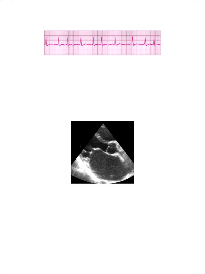
Figure 32.1: ECG showing atrial fibrillation with fast ventricular response
As expected, the ECG showed atrial fibrillation with a fast ventricular response (Fig. 32.1). Additionally, there was evidence of right ventricular hypertrophy and right-ward deviation of the QRS vector. X-ray chest findings were cardiomegaly, straightening of the left heart border with a prominent atrial appendage as well as signs of interstitial pulmonary edema. On ECHO, the left ventricle was normal but the left atrium and right ventricle were dilated. The mitral valve leaflets were thickened and showed limited excursion with diastolic doming (Fig. 32.2). The aortic valve was structurally normal. No mass or thrombus was demonstrated in any cardiac chamber. The pulmonary artery pressure, as estimated from the tricuspid regurgitant jet, was elevated.
Figure 32.2: ECHO showing thickened mitral leaflets, diastolic doming and dilated left atrium
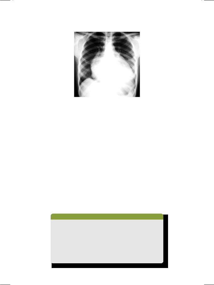
Enlargement of the left atrial appendage is typical of mitral valve disease, be it stenosis or regurgitation, especially when due to rheumatic heart disease. Probably acute rheumatic carditis damages the left atrial wall and its appendage, causing them to bulge when the atrial filling pressure rises. Enlargement of the left atrial appendage causes straightening of the left heart border on chest X-ray (Fig. 32.3). The “4-bump heart” is formed above downward by the aortic knuckle, pulmonary artery, left atrial appendage and the left ventricular lateral wall.
The risk of thromboembolism in mitral stenosis is very high, particularly if atrial fibrillation is present and more so if it is intermittent. Mitral stenosis can be safely assumed to be the cause of cerebral infarction, even if a left atrial thrombus is not demonstrable. A thrombus that is too small for detection, thrombus in the atrial appendage or one that has already embolised, may be missed on
Case 32 Atrial Thrombus |
|
149 |
|
|
|
Figure 32.3: X-RAY showing cardiomegaly with straightening of left heart border
transthoracic echo. Transesophageal echocardiography (TEE) vastly improves the detection of a thrombus in the left atrial cavity or its appendage. Occasionally, an ECHO may show a very large atrial thrombus, which is potentially fatal if it suddenly obstructs the mitral valve. Such a ‘ball-valve’ thrombus is an indication for urgent surgical intervention.
A left atrial thrombus sometimes needs to be differentiated from a left atrial myxoma, since both can cause distal embolism. Thrombus usually arises from the posterior atrial wall or floats freely, while myxoma is usually pedunculated and is attached to the inter-atrial septum. Thrombus is rounded in shape and uniform in echogenicity, while myxoma is often lobulated with variable echogenicity due to areas of hemorrhage, necrosis or calcification. Finally, thrombus uncommonly prolapses into the diseased mitral valve while myxoma commonly prolapses into the normal mitral valve. The differences between atrial thrombus and atrial myxoma are given in Table 32.1.
Besides an atrial thrombus and an atrial myxoma, other structures that may be sometimes seen in the left atrium are supravalvular ring (cor triatriatum), dilated coronary sinus, anomalous pulmonary veins, large mitral leaflet vegetation and reverberation artefacts from a calcified mitral annulus.
Table 32.1: Differences between atrial thrombus and atrial myxoma
|
Atrial thrombus |
Atrial myxoma |
|
|
|
Site |
Posterior wall |
Atrial septum |
|
|
|
Attachment |
Free |
Pedunculated |
|
|
|
Shape |
Rounded |
Lobulated |
|
|
|
Echogenicity |
Echogenic |
Echolucent |
|
|
|
Prolapse in MV |
Rare |
Often |
|
|
|
Mitral valve |
Diseased |
Normal |
|
|
|

150 |
|
Section 10 Intracardiac Masses |
|
|
|
MANAGEMENT ISSUES
In a patient of mitral stenosis and atrial fibrillation with a thrombo-embolic stroke, the first priority is anticoagulation. Low molecular-weight heparin(LMWH) such as enoxaparin is preferred because of predictable pharmacokinetics, without the need of prothrombin time (PT) and aPTT monitoring. An oral anticoagulant like warfarin is initiated almost simultaneously, since it requires 3 to 5 days to produce optimal anticoagulation. Oral anticoagulation is required on a long-term basis in a patient of mitral stenosis with history of thromboembolism.
Other cardiovascular drugs are used to reduce the symptomatology of mitral stenosis. Diuretics reduce pulmonary congestion, especially in those with concomitant mitral regurgitation. Digoxin, beta-blockers and verapamil control the ventricular response in atrial fibrillation and improve left ventricular diastolic filling. Percutanenous balloon mitral valvuloplasty (BMV) in not advocated in patients with a demonstrable left atrial thrombus. Mitral valve replacement is required for critical stenosis (mitral valve area < 1 cm2).

|
|
C A S E |
|
|
|
|
|
|
|
|
|
||
|
|
|
|
|
||
|
|
33 |
Ventricular |
|
|
|
|
|
|
|
|
|
|
|
|
|
|
Thrombus |
|
|
|
|
|
|
|
|
|
|
|
|
|
|
|
|
CASE PRESENTATION
A 67-year old gentleman was brought to the cardiology department by his son for a follow-up visit, 1 week after discharge from the hospital.The patient had sustained an anterior wall myocardial infarction 2 weeks ago, for which he received thrombolytic therapy. However, the streptokinase infusion was stopped at the half-way stage, because the patient developed hypotension. Thereafter, his course in the hospital was by-and-large uneventful, barring a extrasystolic ventricular bigeminy lasting 36 hours, which was treated with lignocaine infusion. Upon returning home, he did not experience any angina, dyspnea, palpitation or syncope. Medications prescribed to him were aspirin 150 mg, clopidogrel 75 mg, ramipril 5 mg, atorvastatin 20 mg and metoprolol 50 mg.
On examination, the patient was alert, comfortable and not tachypneic. The pulse was fair in volume, at a rate of 92 beats/min. and few ectopic beats were noticed. The BP was 110/66 mm Hg in the right arm. All peripheral pulses were well palpable. The JVP was not raised and there was no hepatomegaly or ankle edema. On precordial examination, the apex beat was diffuse in nature and displaced towards the left axilla. The S1 and S2 heart sounds were normal but a soft S3 was audible in diastole. There was no murmur or pericardial rub audible. Few inspiratory crackles were heard over the lower lung fields posteriorly.
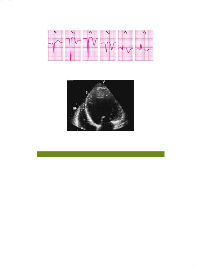
ECG showed sinus rhythm at a rate of 92 beats/min. and an occasional unifocal ventricular premature beat was seen. No couplet or bigeminal rhythm was noticed. There was complete attenuation of the R waves with deep Q waves in the precordial leads V1 to V4. Small q waves were also seen in leads V5 and V6 (Fig. 33.1). There was coving of the S-T segment with inversion of the T wave in all the precordial leads. The Q-T interval was normal.
ECHO showed a dilated left ventricle with depressed systolic function (ejection fraction 35%). The mid and distal segments of the interventricular septum, the left ventricular apex and the distal segment of the lateral wall were hypokinetic but no dyskinetic area was identified. A well-defined rounded mass was seen arising from the ventricular apex and protruding into the left ventricular cavity (Fig. 33.2). The echogenicity of the mass was variable and it did not move in synchrony with the ventricle. The posterior mitral leaflet failed to reach the plane of the mitral annulus and the leaflet coaptation point was distally located in the ventricle. The aortic valve leaflets were mildly thickened but had normal excursion without systolic doming. No pericardial effusion was noted.
152 |
|
Section 10 Intracardiac Masses |
|
|
|
Figure 33.1: ECG showing R wave attenuation, deep Q waves with S-T segment coving and T wave inversion
Figure 33.2: ECHO showing a dilated, hypokinetic left ventricle with a mural thrombus arising from the apex
CLINICAL DISCUSSION
From the history and physical examination as well as from the ECG and ECHO findings, this elderly gentleman had sustained a recent extensive anterior wall myocardial infarction. He had now developed left ventricular systolic dysfunction and a ventricular thrombus. Two weeks after acute myocardial infarction (MI) usual complications include post-MI angina, malignant ventricular arrhythmias and left ventricular failure. Sometimes, a ventricular thrombus may form on an infarcted and scarred myocardial segment or within a left ventricular aneurysm. This patient did not demonstrate any dyskinetic myocardial segment and there was no outward bulge of the left ventricular cavity. A fragment of the ventricular thrombus may break away and travel through the systemic circulation to cause embolization in any distal vascular bed.
A dilated left ventricle with reduced systolic wall motion and stagnated blood flow, is an ideal setting for ventricular thrombus formation. A ventricular thrombus may form on a dyskinetic, infarcted and scarred myocardial segment or within a left ventricular aneurysm. Idiopathic dilated cardiomyopathy is another important reason for ventricular thrombus formation.
A pedunculated ventricular thrombus appears as a well-defined, rounded, mobile stalked mass, that protrudes into the ventricular cavity. The mobility of the thrombus is not synchronous with the left ventricular free wall motion. It is

Case 33 Ventricular Thrombus |
|
153 |
|
|
|
more likely to embolize if the echodensity is variable due to areas of necrosis. A mural ventricular thrombus is a flat, laminated mass, contiguous with the ventricular wall with which it moves synchronously. It is more echogenic than the adjacent myocardium and less likely to embolize than a pedunculated thrombus. Thrombus always has a clear identifiable edge while an artefact caused by stagnated blood has a hazy appearance. On color flow mapping, the flow stops abruptly at the edge of a thrombus but not at the edge of an artefact.
Left ventricular thrombus is a masquerader of several other mass lesions. Causes of a mass in the left ventricle are rhabdomyoma, false tendon and prominent papillary muscle. Mural thrombus can be distinguished from localized myocardial thickening by the fact that myocardium thickens during systole while a thrombus does not. Thrombus can be differentiated from a cardiac tumor by the fact that adjacent wall motion is almost always abnormal in case of thrombus and often normal in case of tumor.
MANAGEMENT ISSUES
Presence of a ventricular thrombus is an established indication for oral anticoagulant therapy. Other clear indications for anticoagulation are mechanical prosthetic heart valve, left atrial thrombus with mitral stenosis and ventricular aneurysm. When an oral anticoagulant like warfarin is initiated, it takes 3 to 5 days for the onset of full therapeutic effect. In this interim period, heparin therapy may be given. Unfractionated heparin requires monitoring but it is cost effective. Low molecular weight heparin (LMWH) is more expensive, but it does not require prothrombin time (PT) or aPTT monitoring.
It is now widely recommended that every myocardial infarction patient must receive aspirin, a statin, an ACE-inhibitor and a beta-blocker. Our patient had already been prescribed all these drugs. A diuretic can be added to reduce cardiac work-load, if pulmonary congestive symptoms develop. An aldosterone antagonist such as spironolactone or eplerenone can also be used. These drugs, besides reducing cardiac work-load, improve ventricular remodelling and lower cardiovascular mortality.