

новая папка / [libribook.com] 50 Cases in Clinical Cardiology_ A Problem Solving Approach 1st Edition
.Pdf
|
|
C A S E |
|
|
|
|
|
|
|
|
|
||
|
|
|
|
|
||
|
|
6 |
|
Mitral |
|
|
|
|
|
|
|
|
|
|
|
|
|
Stenosis |
|
|
|
|
|
|
|
|
|
|
|
|
|
|
|
|
CASE PRESENTATION
A 23-year old woman of low socio-economic status came to a general hospital with shortness of breath, fatigue and palpitations since 2 years and fever with productive cough for the last 1 week. She denied any chest pain, wheezing or hemoptysis. At the age of 12, she had a prolonged febrile illness with joint pains and ever since, she had been receiving monthly injections of penicillin.
On examination she was tachypneic with pallor but there was no cyanosis or icterus. Pulse rate was 96 beats/min. regular, with a BP of 100/70 mm Hg. Her temperature was 1000 F. The JVP was not raised, thyroid gland was not enlarged and there were no palpable lymph nodes. There was no evidence of pharyngo-tonsillitis, swollen joints or petechial spots over the skin, eyes or finger-tips. The apex beat was tapping in nature with a left parasternal heave. The S1 was loud and the P2 was also accentuated. A low-pitched mid-diastolic rumbling murmur was heard over the cardiac apex. The murmur was preceded by an opening snap and accentuated just before systole. There were scattered rhonchi and crepts over the lung fields.
CLINICAL DISCUSSION
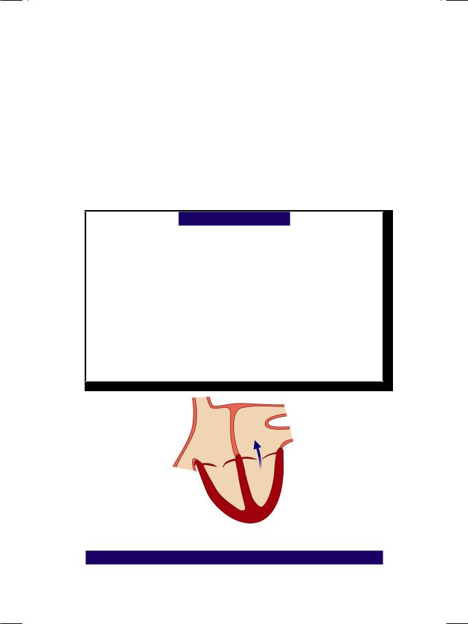
From the history and physical examination, this young woman in all probability had rheumatic heart disease with mitral valve stenosis (Fig. 6.1). The S1 is loud
Figure 6.1: Mitral valve stenosis

26 |
|
Section 2 Mitral Valve Diseases |
|
|
|
since the mitral valve leaflets are distant from each other at the end of diastole and snap together loudly. Other reasons for a loud S1 are sinus tachycardia and a short P-R interval, where the diastole is short. The mid-diastolic murmur of mitral stenosis (MS) is best heard with the patient in the left lateral decubitus position, using the stethoscope bell.
The length of the murmur correlates with the severity of stenosis. The murmur undergoes presystolic accentuation due to atrial contribution to ventricular filling. In mild MS, the murmur may be only presystolic. If MS is associated with atrial fibrillation the S1 is variable in intensity due to variable duration of diastole. Also, presystolic accentuation is lost due to lack of atrial contribution to ventricular filling. Presystolic accentuation is also absent in a calcified valve and after commissurotomy. Severe mitral stenosis may be silent due to low cardiac output and the fact that the right ventricle underlies most of the precordium because of clockwise cardiac rotation.
The opening snap heralds the onset of ventricular diastolic filling and the end of isovolumic relaxation. It indicates pliability of the valve, suitability for valvotomy and is absent in a heavily calcified valve or after commissurotomy. An early opening snap after A2 (short 2A-OS interval), indicates higher left atrial pressure and more severe MS. Besides mitral stenosis, other reasons for a middiastolic murmur are acute rheumatic valvulitis (Carey-Coomb murmur), aortic regurgitation (Austin-Flint murmur), left atrial myxoma (prolapse into mitral orifice) and left-to-right shunt (increased transmitral inflow).
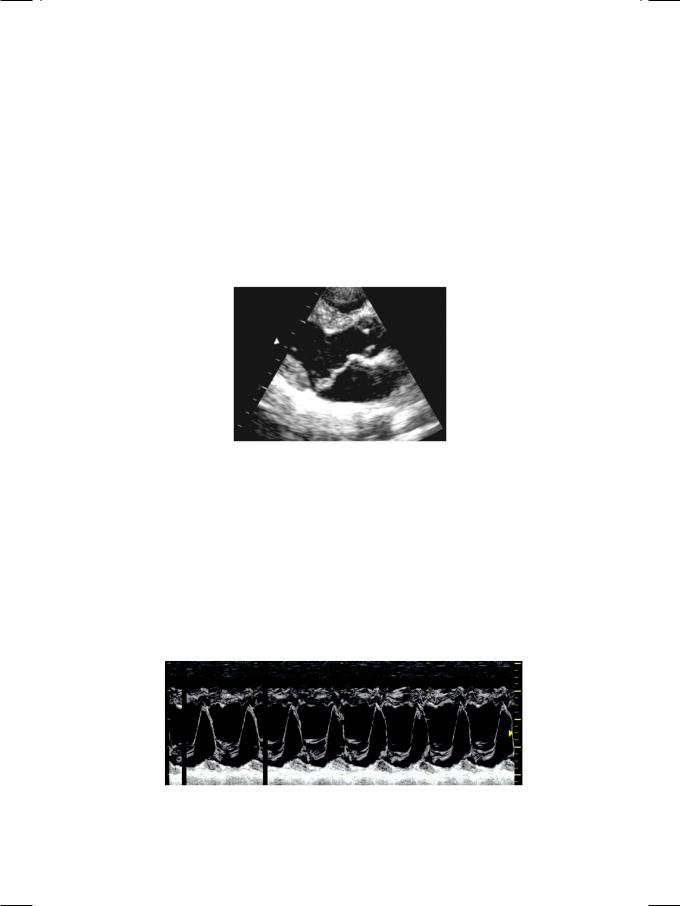
Figure 6.2: M-mode ECHO showing reduced excursion of AML with paradoxical motion of PML
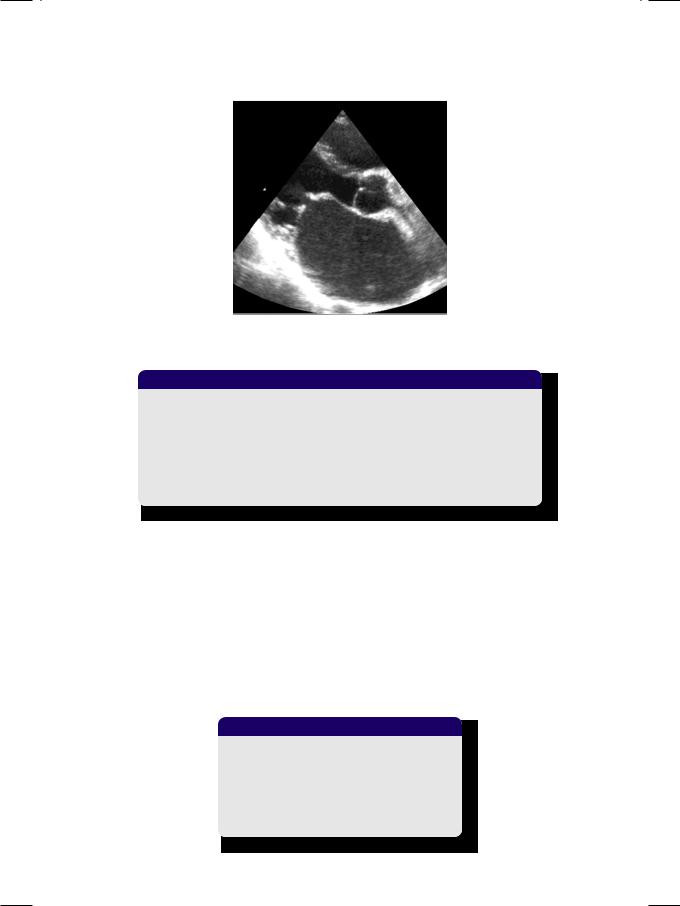
ECG showed sinus rhythm, P mitrale, right ventricular hypertrophy with a rightward QRS axis. X-ray chest findings were straightening of the left heart border with pulmonary congestion. On M-mode ECHO, excursion of the anterior leaflet was reduced with paradoxical excursion of the posterior leaflet (Fig. 6.2). On 2-D ECHO, the left atrium and right ventricle were dilated. The mitral valve leaflets were thickened with limited excursion and restricted valve opening. The anterior mitral leaflet showed diastolic doming (Fig. 6.3). There was no abnormality of the aortic valve and the estimated pulmonary artery pressure was elevated.
The severity of mitral stenosis can be gauged and classified, according to several calculated indices from echocardiography. These are the pressure gradient across the valve (PG), the time taken for the PG to fall to half (P½t; pressure half-time), the pulmonary artery pressure (PAP) estimated from the peak tricuspid velocity (TR Vmax) and the mitral valve area (MVA) calculated by planimetry (Table 6.1). However, there are some fallacies associated with these
Case 6 Mitral Stenosis |
|
27 |
|
|
|
Figure 6.3: 2-D ECHO showing restricted opening with diastolic doming and dilated left atrium
Table 6.1: Assessment of the severity of mitral stenosis
|
Mild |
Moderate |
Severe |
|
|
|
|
PG (mm Hg) |
< 5 |
5-10 |
>10 |
|
|
|
|
P½ t (msec) |
<150 |
150-220 |
>220 |
|
|
|
|
TRVmax (m/sec) |
<2.7 |
2.7-3.0 |
>3.0 |
PAP (mm Hg) |
<30 |
30-50 |
>50 |
|
|
|
|
MVA (cm2) |
>1.5 |
1.0-1.5 |
<1.0 |
calculations. The peak velocity and pressure gradient depend on the heart rate and stroke volume. Measurement of mitral valve area may be fallacious due to heavily calcified leaflets, subvalvular pathology and prior commissurotomy.
Rheumatic heart disease is the predominant cause of mitral stenosis. 80% of cases are women in the 2nd or 3rd decade of life, from the developing countries. Rare causes are congenital MS (parachute valve), mucopolysaccharidosis (Hurler’s syndrome), severe annular calcification and connective tissue disorders (Table 6.2). Complications of MS are pulmonary congestion, respiratory infections, hemoptysis, right heart failure and systemic thrombo-embolism due to atrial fibrillation associated with left atrial thrombus.
Table 6.2: Causes of mitral stenosis
• Rheumatic heart disease (commonest) • Congenital parachute valve
• Mitral annular calcification • Connective tissue disorder
• Hurler’s mucopolysaccharidosis

28 |
|
Section 2 Mitral Valve Diseases |
|
|
|
PERTINENT INVESTIGATIONS
Since rheumatic heart disease is the predominant cause of mitral stenosis, all patients who are febrile should be investigated for rheumatic fever, infective endocarditis and respiratory tract infection. Relevant investigations are Hb, TLC, DLC, ESR, urine R/E, CRP value and ASLO titre. Throat-swab culture for betahemolytic Streptococcus and bacterial blood cultures may be included.
Transthoracic ECHO is central to the diagnosis and assessment of mitral stenosis. Besides determining the severity of MS, it can assess left atrial size, left ventricular function and estimate pulmonary artery pressure. Transesophageal echocardiography (TEE) allows better assessment of the subvalvular and commissural architecture and improves the detection of a left atrial thrombus.
MANAGEMENT ISSUES
All patients of mitral stenosis should be offered penicillin prophylaxis against rheumatic fever, until they attain the age of 40 years. A broad-spectrum antimicrobial agent should be administered prior to any invasive dental or surgical procedure, to guard against infective endocarditis. Diuretics reduce pulmonary congestion, especially in patients with associated mitral or aortic regurgitation. Rate control with digoxin, beta-blocker or verapamil improves left ventricular diastolic filling, in patients who are in sinus rhythm and controls the ventricular response, in whom the rhythm is atrial fibrillation. Since atrial fibrillation in patients with mitral stenosis is associated with a high risk of thrombo-embolism, these patients should also be on a long-term oral anticoagulant agent like warfarin.
Percutaneous balloon mitral valvuloplasty (BMV) is the procedure of choice in symptomatic severe mitral stenosis, provided a transesophageal echocardiography (TEE) does not reveal subvalvular fusion, commissural calcification, atrial thrombus or more than mild MR (Table 6.3). Recurrence of symptoms after valvotomy is more often due to induced mitral regurgitation rather than restenosis. If valvotomy cannot be performed because of the aforementioned reasons, mitral valve replacement (MVR) is undertaken. During MVR, if atrial fibrillation is present, left atrial radiofrequency ablation (RFA) and appendage ligation are also performed.
Table 6.3: Indications for valvotomy in MS
The absence of concomitant:
• Subvalvular fusion • Left atrial thrombus • Immobility of leaflets
• Commissural calcification
• Moderate/severe regurgitation
RECENT ADVANCES
Although transesophageal echocardiography (TEE) has vastly improved acquisition of detailed information over the transthoracic approach, multi-slice computed tomography (CT) is being increasingly used to accurately assess the area and the precise nature of the mitral valve.
|
|
C A S E |
|
|
|
|
|
|
|
|
|
||
|
|
|
|
|
||
|
|
7 |
|
Mitral |
|
|
|
|
|
|
|
|
|
|
|
|
|
Regurgitation |
|
|
|
|
|
|
|
|
|
|
|
|
|
|
|
|
CASE PRESENTATION
A 46-year old woman of stocky built, was asked to see a cardiologist by her family doctor. She complained of exertional shortness of breath and had a cardiac murmur. She was a known case of hypertension, adequately controlled by medication. Her breathlessness had been felt since the last 4 months but had significantly worsened over the preceding 2 weeks. There was no history of exertional chest pain or of palpitation and fainting. She did not smoke or consume alcohol but she seldom exercised and did not follow the diet-plan suggested by her physician.
On examination, she was overweight without any clinical signs of cortisol excess or hypothyroidism. The pulse was regular, good in volume at a rate of 92 beats/min. The BP was 136/88 mm Hg in the right arm. There was no sign of heart failure. The apex beat was hyperdynamic, ill-sustained heaving in nature and displaced towards the left axilla. The S1 was soft. A2 was loud and a soft S3 was audible in early diastole. A soft blowing pansystolic murmur was audible over the mitral area, that radiated towards the axilla. Also, a short diastolic rumble was heard over the cardiac apex. Few basilar rales were auscultated over the lower lung fields.
Figure 7.1: Mitral regurgitation
CLINICAL DISCUSSION
From the history and physical examination, this woman had mitral regurgitation (Fig. 7.1) with possibly associated mitral stenosis and evidence of left ventricular
30 |
|
Section 2 Mitral Valve Diseases |
|
|
|
dilatation. The S1 was soft since the mitral leaflets are close to each other at the end of diastole and snap together softly. The A2 was loud due to associated systemic hypertension. The displacement of the apex beat and audible S3 sound are indicative of left ventricular diastolic overload. The S3 is a low-pitched sound that follows the A2. It indicates abrupt halting of left ventricular filling. Physiological S3 is appreciated in mitral regurgitation, left-to-right shunt and high cardiac output states. Pathological S3 is audible in aortic regurgitation and left ventricular systolic dysfunction.
The pansystolic murmur of mitral regurgitation typically radiates to the left axilla (and sometimes even to the left scapula), if the anterior mitral leaflet is diseased. It radiates to the base of the heart, if the posterior mitral leaflet is involved. This differentiates it from the pansystolic murmur of tricuspid regurgitation or a ventricular septal defect. The accompanying diastolic rumble does not necessarily indicate concomitant mitral stenosis and is due to torrential flow across the valve, which is the sum of normal atrial blood volume and the regurgitant volume. It is worth mentioning that the murmur of acute or severe MR is not pansystolic but early systolic, because less turbulence is generated by the large valve orifice. Moreover, the rapid rise of left atrial pressure impedes regurgitation during later systole. The murmur of mitral annular calcification is also typically early systolic.
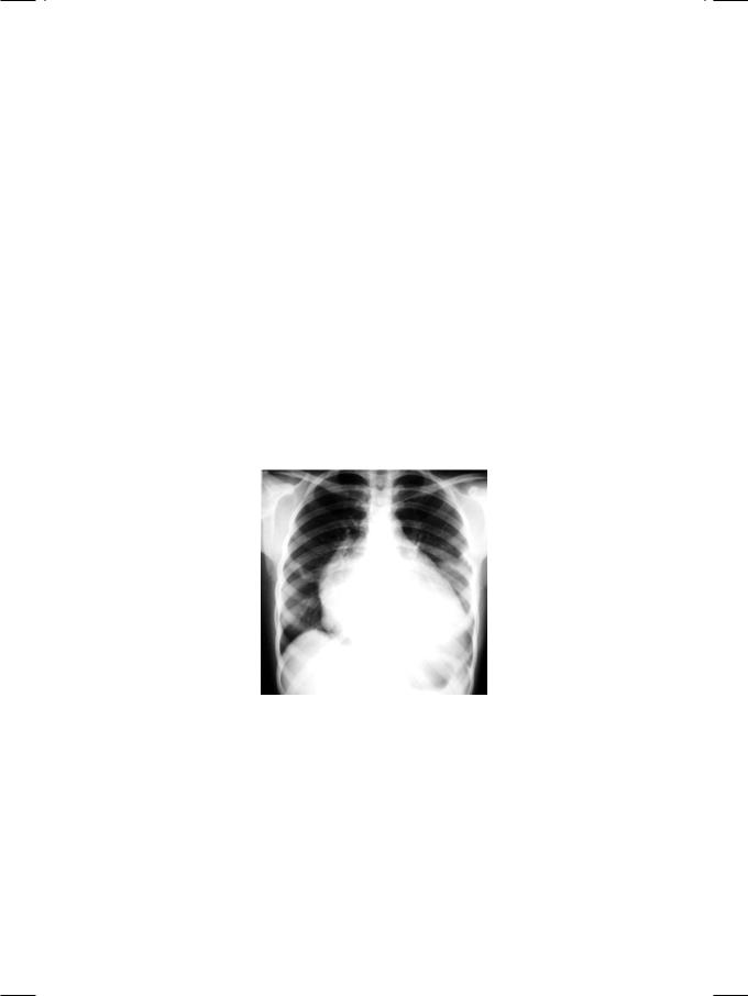
Figure 7.2: X-ray showing cardiac enlargement straightening of left heart border
ECG of the patient showed tall R waves in left precordial leads with upright T waves, indicating left ventricular diastolic overload. X-ray chest findings were moderate cardiomegaly, pulmonary congestion and straightening of the left heart border (Fig. 7.2). The “4-bump” left heart border is due to a large left atrial appendage, which is damaged during acute carditis and bulges due to high left atrial pressure in rheumatic mitral regurgitation (MR). On ECHO, the left ventricle was dilated and hyperkinetic with an ejection fraction of 55% and left atrial enlargement. Importantly, in functional MR due to annular dilatation secondary to cardiomyopathy, there is global hypokinesia of the left ventricle
Case 7 Mitral Regurgitation |
|
31 |
|
|
|
or a regional wall motion abnormality. In acute MR due to ruptured chordae tendinae, there is minimal left ventricular dilatation. The mitral valve leaflets were thickened and fibrotic indicating rheumatic MR. Other potential abnormalities of the mitral valve in case of MR are mitral leaflet redundancy and prolapse, flail leaflet, annular calcification or vegetations in case of endocarditis.
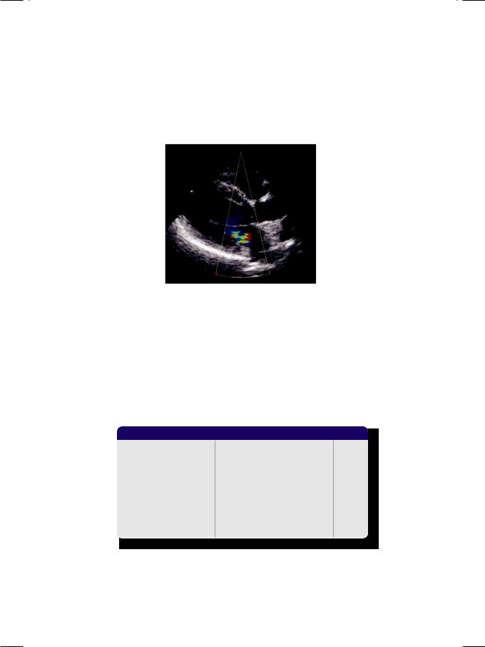
Figure 7.3: ECHO showing a color-flow map in the left atrium
On colour flow mapping, a regurgitant jet was seen entering the left atrium (Fig. 7.3). The volume of MR (ml/beat), the MR jet area (cm2) and the percentage of left atrial area (%) filled by the jet are used to gauge the severity of MR (Table 7.1). However, there are some fallacies associated with these calculations. The spatial profile of the MR jet does not truly reflect the regurgitant volume. It depends upon the shape of the valve orifice, the angle of the jet, left ventricular filling pressure and the size of the left atrium. The MR jet may be “eccentric” or “off-centre” in case of leaflet prolapse, papillary muscle rupture and paraprosthetic leak.
Table 7.1: Assessment of the severity of mitral regurgitation
|
Mild |
Moderate |
Severe |
|
|
|
|
MR jet area (cm2) |
<4 |
4-8 |
>8 |
Percent of LA area (%) |
<25 |
25-50 |
>50 |
|
|
|
|
MR Volume (ml/beat) |
<30 |
30-59 |
>60 |
|
|
|
|
LV dysfunction |
Mild-to-moderate |
Severe |
|
|
|
Ejection fraction (%) |
30-60 |
<30 |
|
|
|
LV diameter (mm) |
40-55 |
>55 |
There are several causes of mitral regurgitation (MR). Usual causes of primary MR are rheumatic heart disease, mitral leaflet prolapse, papillary muscle dysfunction, infective endocarditis and mitral annular calcification. Uncommon causs include congenital endocardial cushion defects, endomyocardial fibrosis

32 |
|
Section 2 Mitral Valve Diseases |
|
|
|
Table 7.2: Causes of mitral regurgitation
Chronic MR
• Mitral valve prolapse
• Dilated cardiomyopathy • Endomyocardial fibrosis • Rheumatic heart disease • Mitral annular calcification • Connective tissue disorder
• Papillary muscle dysfunction
Acute MR
• Acute myocardial infarction • Infective endocarditis
• Blunt chest trauma.
and connective tissue disorders. Occasionally, secondary MR is due to mitral annular dilatation, as a result of dilated or ischemic cardiomyopathy. Acute MR can result from rupture of papillary muscle or chordae tendinae in case of myocardial infarction, infective endocarditis or after blunt trauma to the chest (Table 7.2).
MANAGEMENT ISSUES
Patients of rheumatic MR below the age of 40 should receive penicillin prophylaxis to protect them from acute rheumatic fever. Those undergoing a dental or surgical procedure, need antibiotic prophylaxis against infective endocarditis. Vasodilators such as ACE inhibitors or ARBs combined with a diuretic relieve pulmonary congestion, especially if systemic hypertension is present. Digoxin and an anticoagulant are used if there is concomitant atrial fibrillation. Secondary MR is reduced by lowering preload and afterload, which decrease the diameter of the mitral annulus.
Surgical operations for MR are mitral valve repair and valve replacement. Repair of the valve or annuloplasty is preferable if the valvular anatomy is suitable, since it preserves left ventricular geometry and spares the patient from the problems of anticoagulation. Replacement of the valve is indicated if the valvular anatomy is severely distorted.
RECENT ADVANCES
Mitral regurgitation in the context of ischemic heart disease is a clinical challenge. The MR is not due to an intrinsic valvular abnormality, but the result of ventricular dilatation with annular enlargement, papillary muscle dysfunction and dysfunctional ventricular remodeling with increased sphericity. These patients can be offered restrictive annuloplasty, which involves insertion of an undersized annular ring to improve leaflet apposition. Therefore, patients who are to undergo coronary artery bypass graft (CABG) surgery should be adequately assessed preoperatively for mitral regurgitation.

|
|
C A S E |
|
|
|
|
|
|
|
|
|
||
|
|
|
|
|
||
|
|
8 |
|
Mitral Valve |
|
|
|
|
|
|
|
|
|
|
|
|
|
Prolapse |
|
|
|
|
|
|
|
|
|
|
|
|
|
|
|
|
CASE PRESENTATION
A 26-year old married woman visited a renowned cardiologist for coronary angiography, with the complaints of episodic chest pain and palpitation. The chest pain was precordial in location and described as sharp and pricking. There was no feeling of tightness in the chest or suffocation and the pain did not radiate to the left arm or lower jaw. The palpitation was accompanied by restlessness, dryness of mouth and dizziness and sometimes caused “black-out” with fainting. The episodes of chest pain and palpitation were unrelated to physical exercise, taking meals or change in body posture, but usually related to some emotional upheaval. She had first experienced these symptoms during her final high-school exams at the age of 18, but the frequency of episodes had significantly increased, ever since her arranged marriage 3 months back. There was history of repeated sore-throat during childhood, but she never had a prolonged febrile illness with painful joints.
On examination, the patient had a slender body habitus with an anxious look on her face. The extremities were warm with sweaty palms and a fine distal tremor. There was no anemia, cyanosis or edema and the JVP was not raised. The thyroid gland was not enlarged and there were no eye-signs of Grave’s disease. The pulse was rapid, good in volume at a rate of 96 beats/min with a BP of 140/84 mm Hg. The precordium was hyperdynamic with a pectus excavatum sternal deformity. The apex beat was normal in location and there was no parasternal heave. The S1 was loud, S2 was normally split and no S3 or S4 was heard in diastole. A high-pitched systolic murmur was audible between the cardiac apex and the left sternal border. The murmur started well after the S1 and had a typical honking character. It was associated with a sharp clicking sound in mid-systole. The lung fields were clear on auscultation.
CLINICAL DISCUSSION
From the history and physical examination, this young anxious lady had atypical cardiac symptoms with a mid-systolic click and a mid-systolic murmur. The most likely diagnosis in this case is mitral valve prolapse (MVP). The mid-systolic click is a high-pitched sharp sound produced by sudden tensing of the redundant mitral leaflet. At times, multiple clicks are appreciated. This is often followed by a midor late-systolic murmur that typically has a whooping or honking character.
34 |
|
Section 2 Mitral Valve Diseases |
|
|
|
Another cause of a mid-systolic murmur is papillary muscle dysfunction. The click and murmur can vary with alteration of left ventricular (LV) volume, by change in patient posture. During standing or Valsalva manoeuvre, when the LV volume is reduced, the click moves closer to S1 and the murmur becomes louder. Conversely, during squatting when the LV volume increases, the click moves closer to S2 and the murmur becomes softer.
In mitral valve prolapse syndrome, the SI is loud for several reasons. The high adrenergic activity increases the heart rate and shortens diastole. The wide excursion of the myxomatous, redundant valve leaflet increases the force of mitral valve closure. Finally, merger of the non-ejection click with the S1 , increases the intensity of the latter.
Figure 8.1: ECHO showing buckling of mitral leaflets, above the plane of mitral annulus
ECG of the patient showed sinus rhythm at the rate of 92 with few atrial ectopic beats and T wave inversion in leads LIII, aVF, V5, V6. The X-ray chest was unremarkable. On ECHO, the left ventricle was normal in size and systolic function, but the left atrium was mildly dilated. The anterior mitral leaflet was thick and redundant, with systolic buckling above the plane of the mitral annulus into the left atrium (Fig. 8.1). On M-Mode scan, there was abrupt posterior displacement of both leaflets in systole, giving a “hammock-like” appearance (Fig. 8.2). On colour flow mapping, an eccentric regurgitant jet was seen entering the left atrium.
Figure 8.2: M-mode scan showing posterior motion of mitral leaflets (“hammock” appearance)