

новая папка / [libribook.com] 50 Cases in Clinical Cardiology_ A Problem Solving Approach 1st Edition
.PdfCase 10 Aortic Regurgitation |
|
45 |
|
|
|
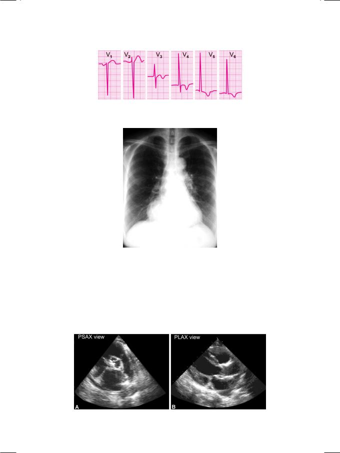
Figure 10.2: X-ray showing cardiomegaly with pulmonary congestion
Figure 10.3: ECHO showing regurgitant jet
in the outflow tract
Table 10.2: Assessment of the severity of aortic regurgitation
|
Mild |
Moderate |
Severe |
|
|
|
|
AR jet depth upto |
LVOT |
Mid–LV |
LV apex |
|
|
|
|
AR jet width (% of LVOT) |
<25 |
25-65 |
>65 |
|
|
|
|
AR volume (ml/beat) |
<30 |
30-59 |
>60 |
|
|
|
|
AR: Aortic regurgitation on Doppler
LVOT: Left ventricular outflow tract
The causes of aortic regurgitation can be classified into the causes of aortic valve disease and the causes of aortic root dilatation. Valvular diseases are congenital bicuspid aortic valve, rheumatic heart disease and connective tissue disorders. Aortic root dilatation occurs in uncontrolled hypertension, Marfan’s syndrome and ankylosing spondylitis. Acute AR can result from aortic dissection, annulo–aortic ectasia, infective endocarditis or after blunt trauma to the chest (Table 10.3).

46 |
|
Section 3 Aortic Valve Diseases |
|
|
|
Table 10.3: Causes of aortic regurgitation
Valvular AR
• Bicuspid aortic valve
• Rheumatic heart disease • Connective tissue disorder
Root dilatation
• Marfan syndrome
• Ankylosing spondylitis • Systemic hypertension
Acute AR
• Chest wall trauma • Dissection of aorta
• Infective endocarditis
PERTINENT INVESTIGATIONS
In addition to echocardiography, cardiac computed tomography (CT) or magnetic resonance imaging (MRI) may be required for the detailed assessment of the aortic valve as well as the proximal aorta. Radionuclide ventriculography may be required for the detailed assessment of left ventricular volume, geometry and systolic function. Exercise testing may be considered in an asymptomatic person to assess his functional capacity. Coronary angiography is indicated to assess the coronary circulation and the need for bypass surgery at the time of aortic valve replacement. At the time of angiography, a root aortogram should also be performed to assess the proximal aorta.
MANAGEMENT ISSUES
As far as the medical management of AR is concerned, diuretics and vasodilators are the mainstay of therapy. The diuretics reduce blood pressure, left ventricular volume and the symptoms of heart failure. Vasodilators reduce afterload, increase forward flow and decrease the regurgitant volume. Additionally, they control systolic blood pressure which is nearly always an issue in these patients. Aortic valve replacement (AVR) is the surgical procedure of choice in patients of AR who are symptomatic or have left ventricular dilatation (Table 10.4). Aortic root repair or replacement may be combined with AVR, if there is a significant degree of aortic dilatation. AVR may be combined with coronary artery bypass graft (CABG) surgery, if the latter is indicated.
Table 10.4: Indications for surgery in aortic regurgitation
• Symptomatic acute moderate to severe AR
• Symptomatic chronic moderate to severe AR
• Asymptomatic chronic AR of any degree with left ventricular dysfunction and dilatation
LV dilatation : end-systolic diameter >55 mm end-diastolic diameter >75 mm
LV dysfunction : LV ejection fraction <50%

Case 10 Aortic Regurgitation |
|
47 |
|
|
|
RECENT ADVANCES
Aortic valve replacement with a mechanical or a bioprosthetic valve is associated with either the need for a long-term anticoagulant (requires monitoring and carries risk of bleeding) or the late failure of a bioprosthetic valve. Therefore, there is now growing interest in aortic valve repair, if the valvular anatomy is suitable. However, the long-term feasability of this technique needs further evaluation.

|
|
C A S E |
|
|
|
|
|
|
|
|
|
||
|
|
|
|
|
||
|
|
11 |
Aortic |
|
|
|
|
|
|
|
|
|
|
|
|
|
|
Sclerosis |
|
|
|
|
|
|
|
|
|
|
|
|
|
|
|
|
CASE PRESENTATION
A 77-year old elderly gentleman was brought by his son to his regular physician, for a routine periodic medical check-up. The patient had been hypertensive for the last 35 years and presently he was taking amlodipine 5 mg and metoprolol 50 mg daily. About 3 years back, he was also prescribed isosorbide mononitrate 40 mg and aspirin 75mg daily, because his ECG showed T wave inversion in the lateral leads. The patient did complain of breathlessness on climbing stairs but there was no history of angina, orthopnea or paroxysmal nocturnal dyspnea. Over the last three months, he also felt dizzy and light-headed on standing up from the lying position, but there was no history of palpitation or syncope.
On examination, the pulse was of good volume with few missed beats and the BP was 154/76 mm Hg, with a heart rate of 66 beats/min. The patient was conscious, cooperative and in no distress. The JVP was not raised but there was minimal pitting edema over both his ankles. The apex beat was slightly displaced towards the axilla and heaving in character. Systolic pulsations were observed over the aortic area and in the suprasternal notch. The S1 was normal, A2 was loud but no gallop was audible. A harsh systolic murmur was heard over the upper left sternal edge that radiated towards the neck. The murmur was not preceded by an ejection click or accompanied by a palpable thrill. A different soft systolic murmur was heard over the cardiac apex that radiated towards the left axilla. The lung fields were clear on auscultation.
CLINICAL DISCUSSION
From the history and physical examination, this elderly hypertensive gentleman had aortic root dilatation, with probably left ventricular outflow tract (LVOT) obstruction and also mitral regurgitation. ECG showed tall R waves with inverted T waves in the lateral precordial leads (Fig. 11.1). There were no significant Q waves or S-T segment shift, but few unifocal ventricular premature beats were observed. X-ray chest findings were mild cardiomegaly, dilated ascending aorta and a prominent aortic knuckle (Fig. 11.2).
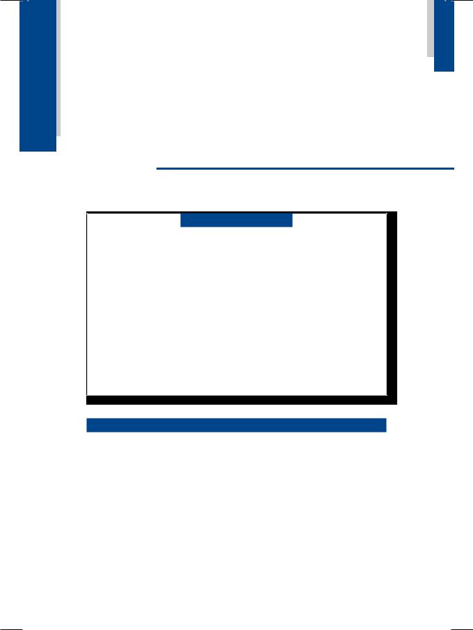
ECHO revealed normal sized left ventricular cavity, with an ejection fraction of 45%. There was mild concentric hypertrophy, but no wall motion abnormality of any ventricular segment. The aortic valve annulus and leaflets showed bright
Case 11â Aortic Sclerosis |
|
49 |
Figure 11.1: ECG showing tall R waves with inverted T waves in lateral leads
Figure 11.2: X-ray showing dilated ascending aorta with prominent aortic knuckle
echo-reflectivity (Fig. 11.3) with some restriction of leaflet excursion, but no systolic doming. The posterior segment of the mitral valve annulus and the base of the posterior leaflet also showed high echogenicity. On colour flow mapping, a mosaic jet was seen entering the left atrium across the mitral valve. On Doppler study, a peak systolic velocity (Vmax) of 2.5 m/sec was seen across the aortic valve with a calculated pressure gradient (PG) of 25 mm Hg.
Figure 11.3: ECHO showing calcification of aortic valve leaflets (A) and annulus (B)

50 |
|
Section 3 Aortic Valve Diseases |
|
|
|
||
|
|
|
|
|
|
|
|
|
|
|
|
|
|
|
|
|
|
|
|
Table 11.1: Causes of aortic root dilatation |
|
|
|
|
|
|
• |
Post-stenotic |
Aortic stenosis |
|
|
|
|
|
• |
Medial necrosis |
Marfan’s syndrome |
|
|
|
|
|
• |
Aorto-arteritis |
Takayasu’s disease |
|
|
|
|
|
• |
Atherosclerotic |
Elderly hypertensive |
|
|
|
|
|
• |
Collagen disorder |
Ankylosing spondylitis |
|
|
|
|
|
|
|
|
|
|
|
|
|
|
|
|
|
|
Dilatation of the proximal aorta or aortic root is not uncommon in elderly hypertensive subjects due to atherosclerosis. The aortic valve annulus however, is normal in diameter. Aortic dilatation also occurs in aortic valve stenosis due to the high velocity jet through the narrow, distorted valve impinging on the aortic wall. This is known as post-stenotic dilatation. Sometimes, aortic root dilatation is due to medial necrosis (Marfan’s syndrome), aorto-arteritis (Takayasu’s disease) or a connective tissue disorder (Ankylosing spondylitis). In these cases, the aortic valve annulus is also dilated and aortic regurgitation is often associated (Table 11.1).
In elderly hypertensive patients, besides aortic root dilatation, calcification of the aortic valve with or without stenosis is sometimes observed. This entity is referred to as aortic valve sclerosis. Aortic valve calcification may also be accompanied by mitral annular calcification (MAC). The calcified mitral annulus sometimes causes mild to moderate mitral regurgitation and rarely even mitral stenosis. Aortic sclerosis occurs in 25% subjects above the age of 65 years. It leads to aortic stenosis more often in those about 80 years of age with a severely calcified valve and in the presence of chronic kidney disease. There is a 50% risk of myocardial infarction, stroke or death within 5 years, due to multiple cardiovascular risk factors and severe atherosclerosis.
The systolic murmur of aortic stenosis is sometimes preceded by an ejection click,accompaniedbyapalpablethrillandfollowedbyamuffledA2 duetoreduced ejection volume. On the other hand, with the murmur of aortic sclerosis, there is no click or thrill and the A2 is loud because of associated systemic hypertension. A history of dizziness or syncope should always altert us to the possibility of left ventricular outflow tract (LVOT) obstruction. Alternatively, syncope can occur due to a tachyarrhythmia or because of sinus node dysfunction. In a patient of aortic sclerosis, angina pectoris may be multifactorial. One, there may be coronary artery stenosis. Two, the calcification of the valve may cause coronary ostial occlusion. Three, angina may be due to the increased myocardial oxygen demand, as a consequence of left ventricular hypertrophy.
MANAGEMENT ISSUES
The medical management of aortic valve sclerosis includes adequate control of hypertension and other cardiovascular risk factors. This patient was already taking amlodipine and metoprolol. Ramipril may be added since it is known to facilitate regression of left ventricular hypertrophy. Diuretics should be avoided if there is history of syncope. A statin can be added to the ongoing aspirin since both

Case 11 Aortic Sclerosis |
|
51 |
|
|
|
these agents are proven to prevent cardiovascular events. Elderly hypertensive patients often have associated orthostatic symptoms and they should be advised adequate fluid intake and gradual change of posture.
Aortic valve replacement (AVR) may be considered in symptomatic moderate to severe aortic stenosis especially with left ventricular dysfunction. At the time of AVR, coronary angiography should be performed to assess the status of the coronary vasculature. Conversely, all patients to be taken up for coronary artery bypass graft (CABG) surgery should be assessed for aortic stenosis and AVR can be performed at the time of operation.
RECENT ADVANCES
The technique of percutaneous aortic valve replacement (AVR) has been recently refined and it is now clearly feasible in candidates at high risk of a surgical procedure under general anesthesia.

S E C T I O N
4
The
Cardiomyopathies

C A S E
12 Dilated
Cardiomyopathy
CASE PRESENTATION
A 48-year old man presented to the emergency room with recent worsening of breathlessness, over the past few days. His dyspnea was worse at night and he required two or more pillows under his head to catch some sleep. Yet his nocturnal breathlessness woke him up frequently. For the past 4 months he had experienced increasing fatigue on walking and on climbing stairs. He was not a diabetic or hypertensive and denied any chest pain, palpitation or syncope. He did not smoke but took 5-6 large pegs of alcohol on most days of the week, for the last 30 years. None of his family members had history of heart disease.
On examination, he was tachypneic and diaphoretic. Pulse was rapid and feeble with a changing volume in alternate beats and the extremities were cold. His heart rate was 120 beats/min with a BP of 106/74 mm Hg. The JVP was clearly elevated and there was also pitting edema over his ankles. The liver edge was palpable 6 cm below the right costal margin. The apex beat was diffuse and displaced towards the axilla. The S1 and S2 were normal and there was a soft pansystolic murmur over the mitral area with an audible gallop sound in early diastole. There were bilateral basilar rales over the lower one-third of both lung fields.
CLINICAL DISCUSSION
Form the history and physical examination, this patient was obviously in congestive cardiac failure. The heart failure was systolic in nature, of the lowoutput variety and biventricular in origin. ECG showed sinus tachycardia, narrow QRS complexes and non-specific T-wave inversion, without any S-T segment shift or Q-waves. X-ray chest findings were moderate cardiomegaly, Kerley B lines, hilar congestion, and cephalized vessels, with pulmonary edema and a small right-sided pleural effusion (Fig. 12.1). ECHO revealed a dilated left ventricle (ejection fraction 25-30%) with reduced septal and posterior wall motion (global hypokinesia). Excursion of the mitral valve leaflets was decreased and the E-point septal separation was increased (Fig. 12.2). There were no regional wall motion abnormalities or abnormal architecture of the cardiac valves. There was mild mitral regurgitation and mild right ventricular dilatation. There are three main diagnostic possibilities in this case. These are: