

новая папка / [libribook.com] 50 Cases in Clinical Cardiology_ A Problem Solving Approach 1st Edition
.PdfCase 45 Ventricular Premature Beats |
|
205 |
|
|
|
Cardiac conditions in which VPCs are observed are:
•Coronary Artery Disease
Ischemia Infarction
Reperfusion
•Congestive heart failure
Hypertension Cardiomyopathy Ventricular aneurysm
•Mitral Valve Prolapse Syndrome
•Digitalis Treatment and Intoxication
•Cardiac Surgery and Catheterization.
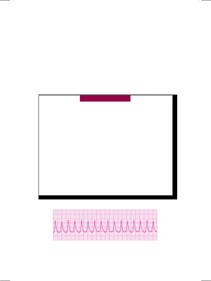
Figure 45.2: ECG showing a premature beat that exhibits the R-on-T phenomenon
A VPC that occurs very prematurely (very short coupling interval) superimposes on the T wave of the preceding sinus beat and is said to exhibit the ‘R-on-T’ phenomenon (Fig. 45.2). This represents the occurrence of ventricular stimulation,duringthevulnerablephaseortheperiodofsupernormalexcitability and is likely to precipitate ventricular fibrillation. The ‘R-on-T’ phenomenon is observed in these situations:
•VPCs after acute myocardial infarction
•Very premature stimuli during external pacing
•Electrical cardioversion during digitalis therapy
•VPCs with underlying Q-T interval prolongation.
VPCs may be asymptomatic or associated with palpitation and a sensation of “missed beats’. Awareness of VPCs is due to the post-VPC compensatory pause andincreasedforceofcontractionofthebeatfollowingtheVPC.Neckpulsations may be felt due to atrial systole occurring with a closed tricuspid valve, since the atria and ventricles are activated almost synchronously.
MANAGEMENT ISSUES
The management of ventricular premature beats is governed by the severity of ectopy,symptomsofthepatientandthepresenceofstructuralheartdisease.Few isolated beats without symptoms and in the absence of heart disease are mostly

206 |
|
Section 13 Cardiac Arrhythmias |
|
|
|
left alone. If symptoms are present, causative factors need to be corrected. This includes alleviating anxiety, withdrawal of adrenergic drugs and reduction of smoking and beverage intake. If these measures do not suffice, beta-blockers are the drugs of choice to treat VPCs associated with anxiety, mitral valve prolapse and thyrotoxicosis.
VPCsoccurringwithin24hoursofmyocardialinfarctionindicatereperfusion andcarryabetterprognosisthanthosethatappearafter24hours.Lidocaineand amiodarone are the drugs of choice to treat VPCs after myocardial infarction, heart surgery or after cardiac catheterization. Patients who have structural heart disease in the form of left ventricular hypertrophy (LVH), dilated cardiomyopathy
(DCMP) or arrhythmogenic right ventricular dysplasia (ARVD), almost always deserve an antiarrhythmic drug such as amiodarone. When a patient of congestiveheartfailureondigitalisdevelopssignificantVPCs,ithastobedecided on clinical grounds whether digitalis should be continued to manage the heart failure or withdrawn in view of drug intoxication. If digitalis is to be withdrawn, the antiarrhythmic drug of choice for digitalis induced VPCs is phenytoin sodium which should be used in conjunction with standard therapy of digitalis intoxication.
AntiarrhythmicdrugsmaybeprescribedtocontrolVPCsbutbeforeinitiating treatment, the following issues need to be addressed:
•Aggravation of bradycardia and left ventricular dysfunction
•Proarrhythmic effects and likelihood of other tachyarrhythmias
•Potential systemic side-effects of long-term antiarrhythmic therapy
•Inappropriate drug usage, since the cause-and-effect relationship between VPCs and fatal arrhythmias has not been established.
|
|
C A S E |
|
|
|
|
|
|
|
|
|
||
|
|
|
|
|
||
|
|
46 |
Ventricular |
|
|
|
|
|
|
|
|
|
|
|
|
|
|
Tachycardia |
|
|
|
|
|
|
|
|
|
|
|
|
|
|
|
|
. |
CASE PRESENTATION |
A 66-year old woman was brought to the emergency department with history of recurrent syncope preceded by palpitation and dizziness. She had sustained an anterior wall myocardial infarction 8 months back and also complained of exertional breathlessness with paroxysms of nocturnal dyspnea. She did not receive thrombolytic therapy and had not undergone primary angioplasty because she presented to the hospital, over 24 hours after the onset of chest pain. The patient was practically home-bound with limited physical activity and did not complain of exertional angina. An echocardiogram was performed a month after her myocardial infarction, which revealed a large wall motion abnormality involving the mid and distal septum, ventricular apex and the distal lateral wall. The left ventricular ejection fraction was 25%.
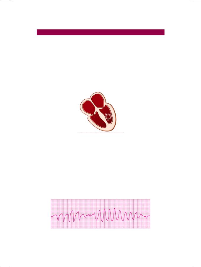
On examination, the patient was fully conscious and cooperative but tachypneic. The pulse was fast and irregular, low in volume, at a rate of 96 beats/min. with “missed beats”. The BP was 110/66 mm Hg with a respiratory rate of 28/min. The JVP was not raised and there was no pitting edema over the ankles. The cardiac apical impulse was diffuse and displaced towards the axilla. Besides S1 and S2, an S3 gallop sound was audible in early diastole. A soft holosystolic murmur was also heard at the cardiac apex. Bilateral basilar rales were audible over the lung fields. Along with a 12-lead ECG, a rhythm strip of lead LII was obtained, which showed an alarming but transient abnormality (Fig. 46.1).
Figure 46.1: ECG strip showing monomorphic ventricular tachycardia
208 |
|
Section 13 Cardiac Arrhythmias |
|
|
|
ECG INTERPRETATION
The ECG rhythm strip showed an abrupt onset of irregular tachycardia, at a rate of about 200 beats/min., with R-R interval of 7 to 8 mm. The QRS complexes were bizarre and wide, exceeding 0.14 sec in width, but did not conform to a bundle branch block pattern. The P and T waves were not discernable. These findings are consistent with the diagnosis of ventricular tachycardia. Ventricular tachycardia is due to enhanced automaticity of a latent ventricular pacemaker, that fires impulses rapidly. Alternatively, it is based on a closed re-entrant circuit around a fixed anatomical substrate in the ventricular myocardium (Fig. 46.2). Ventricular tachycardia may be sustained (lasting >30 sec) or non-sustained (lasting <30 sec). It is classified as monomorphic (similar QRS complexes) or polymorphic (variable QRS complexes) in nature.
Figure 46.2: Diagram to illustrate ventricular re-entrant circuit
Polymorphic ventricular tachycardia is characterized by phasic variation of the QRS direction. A series of ventricular complexes are first up-pointing then down-pointing and this phenomenon occurs in a repetitive continuum (Fig. 46.3). Since this gives the appearance of rotation around the isoelectric line, it is called “Torsades de pointes”, a ballet term which literally means “twisting around a point”. Torsades de pointes is generally, associated with prolongation of the Q-T interval. The prolonged Q-T interval favours the occurrence of a ventricular premature beat that coincides with the T-wave of the preceding beat (R-on-T phenomenon) and initiates the ventricular tachycardia.
Figure 46.3: ECG showing polymorphic ventricular tachycardia

Case 46 Ventricular Tachycardia |
|
209 |
|
|
|
Ventricular tachycardia superficially resembles a supraventricular tachycardia conducted aberrantly to the ventricles. Features in favour of a supraventricular tachycardia are clock-like regularity, triphasic QRS morphology, normal QRS axis, stable hemodynamic parameters and response to carotid sinus massage.
CLINICAL DISCUSSION
A series of three or more successive ventricular ectopic beats constitutes a ventriculartachycardia.Aventriculartachycardiathatlastsformorethan30secondsand requires cardioversion for termination is called sustained ventricular tachycardia. A non-sustained ventricular tachycardia lasts for less than 30 seconds and ends spontaneously. Ventricular tachycardia is considered repetitive or recurrent if three or more discrete episodes are documented, while chronic ventricular tachycardia is that in which recurrent episodes occur for over a month. Sustained ventricular tachycardia (scar VT) is often based on structural heart disease wherein a fixed anatomical substrate facilitates a re-entrant mechanism. Causes of sustained ventricular tachycardia are:
•Myocardial scar
Infarction Aneurysm
•Myocardial disease Myocarditis
Cardiomyopathy
•Congestive failure
Ischemic Hypertensive
•Valvular abnormality Mitral valve prolapse
Rheumatic heart disease.
Symptoms due to ventricular tachycardia are palpitation because of fast heart rate and angina due to increased oxygen demand and shortened coronary filling time. Lack of atrio-ventricular synchrony can cause dyspnea (pulmonary edema) and syncope (low cardiac output). The hallmark of ventricular tachycardia is the dissociation between atrial systole and ventricular systole (A-V dissociation). On auscultation, the intensity of S1 is variable because of changing diastolic filling period. Cannon a waves are observed in the neck veins due to atrial contraction against a closed tricuspid valve. An atrial impulse may occasionally find the ventricle receptive to depolarization and is able to “capture” the ventricle. A capture beat (complete capture) or a fusion beat (incomplete capture) is irrefut- able evidence of A-V dissociation. Markers of electrical instability in survivors of acute myocardial infarction are:
•Documented ventricular arrhythmias on 24-hour Holter monitoring
•Reproducible tachycardia on programmed electrical stimulation (PES)
•Late depolarizations on signal averaged electrocardiogram (SAECG).

210 |
|
Section 13 Cardiac Arrhythmias |
|
|
|
MANAGEMENT ISSUES
The management of ventricular tachycardia (VT) depends upon the etiology, the nature of heart disease and the hemodynamics of the patient. Ventricular tachycardia due to sympathetic stimulation (catecholaminergic VT), caused by stress, exercise or adrenergic drugs and in the absence of structural heart disease, responds well to beta-blockers. In the presence of heart disease and if hemodynamics are stable, chemical cardioversion with antiarrhythmic drugs is initiated. An intravenous bolus of lignocaine or amiodarone is followed by a maintenance infusion. Oral amiodarone is later continued indefinitely to prevent recurrence. Alternative agents are sotalol, propafenone, flecainide and ibutilide. If the hemodynamics are unstable due to hypotension, heart failure, or ongoing ischemia, electrical cardioversion with DC shock is the procedure of choice. An initial energy of 50 to 100 Joules is followed up with higher voltage, until sinus rhythm is restored. If there is circulatory shock to begin with, upto 360 Joules are used. Once sinus rhythm is restored, prophylactic pharmacological therapy is initiated and continued indefinitely.
The management of polymorphic ventricular tachycardia with Q-T prolongation is quite different. An infusion of magnesium sulphate or isoproterenol is used for chemical cardioversion. If the patient responds to beta-blockade, either long-term antiadrenergic therapy is initiated or cervical sympathetic ganglionectomy is offered. If the hemodynamics are unstable with hypotension or shock, electrical cardioversion is the procedure of choice, as per the protocol mentioned above. Alternatively, overdrive ventricular pacing is done, which is often successful. To patients with history of recurrent syncope or family history of sudden cardiac death (SCD) and to survivors of cardiac arrest, an implantable cardioverter defibrillator (ICD) device is offered.

S E C T I O N
14
Coronary Artery
Disease

|
|
C A S E |
|
|
|
|
|
|
|
|
|
||
|
|
|
|
|
||
|
|
47 |
Chronic Stable |
|
|
|
|
|
|
|
|
|
|
|
|
|
|
Angina |
|
|
|
|
|
|
|
|
|
|
|
|
|
|
|
|
. |
CASE PRESENTATION |
A 38-year old gentleman paid a visit to the cardiologist, with the complaint of retrosternal heaviness during brisk walking. The discomfort was described as a feeling of tightness in the chest and was associated with a sensation of suffocation and choking.The chest pain radiated towards the left shoulder, down the inner aspect of the left arm as well as to the neck and lower jaw. There was no history of dyspnea, palpitation, excessive sweating or syncope. The discomfort typically occurred when the patient undertook any form of physical activity, soon after a meal. He denied any chest pain at rest or during sleep. The frequency and severity of his episodic chest discomfort had not increased over the last three months. The patient was not taking any cardiovascular medication and had never undergone a blood glucose or lipid analysis. He smoked about 8 to 10 cigarettes a day for the last 20 years and took 5 to 6 alcoholic drinks over weekends. He did not follow any particular diet plan or exercise regime. His executive job entailed long hours of working and caused considerable mental stress. There was history of premature coronary artery disease as well as of diabetes mellitus, in several of his family members.
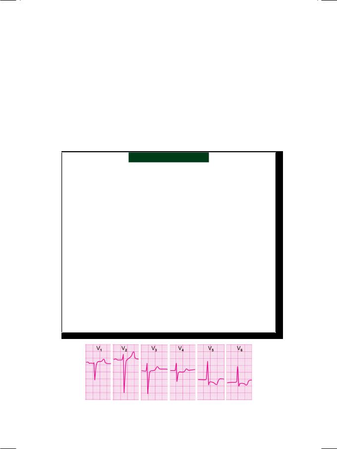
On examination, the patient was alert and in no distress. He was modestly overweight with a body mass index (BMI) of 27kg/m2. The pulse rate was 84 beats/ min. with a BP of 134/82 mm Hg and there were no signs of heart failure. Findings on general examination were xanthelasma on the upper eyelids, arcus senilis around the cornea and acanthosis nigricans over the nape of neck. The precordium was unremarkable with a normally located apex beat. There was no S3 or S4 gallop, murmur or friction rub upon auscultation. The lung fields were clear without any rhonchi or crepts. An ECG was obtained (Fig. 47.1).
Figure 47.1: ECG showing S-T segment depression with T wave inversion in the lateral chest leads
214 |
|
Section 14 Coronary Artery Disease |
|
|
|
ECG INTERPRETATION
The ECG showed normal sinus rhythm with normal P wave morphology and no prolongation of the P-R interval or Q-T interval. The QRS complexes were narrow, without significant Q waves or attenuation of the R wave height. A 2 mm horizontal depression of the S-T segment was observed in leads V4, V5 and V6. The T wave was also inverted in leads V5 and V6. These findings are consistent with the diagnosis of lateral wall myocardial ischemia, a common feature in a patient who has angina pectoris.
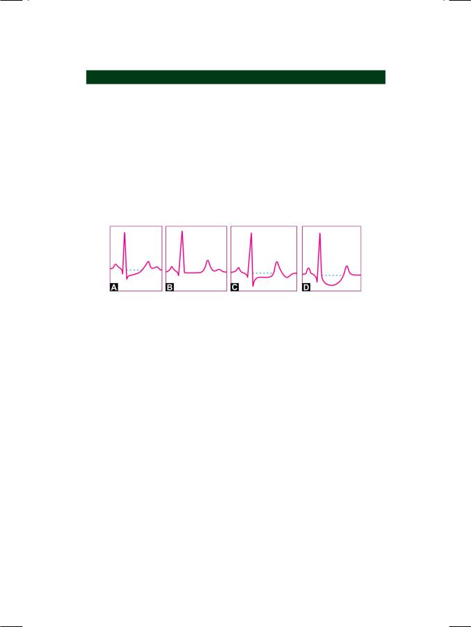
Coronary artery disease is the most important cause of S-T segment depression. The degree of S-T segment depression (greater than 1 mm) correlates with the severity of coronary insufficiency. Besides being depressed, the morphology of the S-T segment, with increasing severity of myocardial ischemia, can be classified as shown in Figure 47.2.
Figure 47.2: Nature of S-T segment depression with increasing severity of ischemia
A.Isolated J point depression (upsloping S-T segment)
B.Horizontality of S-T segment (sharp ST-T junction)
C.Plane S-T depression (horizontal S-T depression)
D.Sagging depression (hammock-like S-T segment).
Depression of the S-T segment constitutes the most useful criterion for the positivity of the stress ECG test, performed by exercising on a treadmill or bicycle ergometer. The degree of positivity of the stress test (mild, moderate or severe) can be gauged from several parameters of S-T segment depression. The magnitude of S-T segment depression and its shape are associated with the severity of coronary artery disease. Additionally, early appearance of S-T depression in the exercise period and longer persistence into the recovery period correlate with the positivity of the stress test.
Angina pectoris is described as a feeling of retrosternal heaviness or tightness, with a sensation of suffocation or choking. The pain is of a crushing or squeezing nature along with restlessness, sweating, palpitation, shortness of breath or extreme weakness. The pain may radiate to both shoulders and arms, down the inner aspect of left arm or to the neck and lower jaw. In contrast, the pain arising from the musculo-skeletal system of the chest is described as a dull ache or a sharp pricking sensation. The pain increases on deep inspiration, turning to one side, or on bending forwards. The pattern of radiation and accompaniments that characterize angina pectoris, are typically absent in muscular chest pain.
Stable angina is caused by an imbalance between the supply of and demand for oxygenated blood to the myocardium. The commonest cause is obstructive