

21.Bourdillon PD, Fineberg N. Comparison of iced and room temperature injectate for thermodilution cardiac output. Cathet Cardiovasc Diagn 1989;17:116–120.
22.Williams JE Jr., Pfau SE, Deckelbaum LI. Effect of injectate temperature and thermistor position on reproducibility of thermodilution cardiac output determinations. Chest 1994; 106:895–898.
23.Jansen JRC et al., Monitoring of the cyclic modulation of cardiac output during artificial ventilation. In: Nair S, editor. Critical Care and Pulmonary Medicine. New York: Plenum; 1980. p 59–68.
See also CARDIAC OUTPUT, FICK TECHNIQUE FOR; CARDIAC OUTPUT, INDI-
CATOR DILUTION MEASUREMENT OF; CORONARY ANGIOPLASTY AND GUIDEWIRE DIAGNOSTICS; MICROPOWER FOR MEDICAL APPLICATIONS; THERMISTORS.
CARDIOPULMONARY BYPASS. See HEART-LUNG
MACHINES.
CARDIOPULMONARY RESUSCITATION
EDWARD GRAYDEN
Mayo Health Center
Albertlea, Minnesota
INTRODUCTION
Cardiopulmonary resuscitation (CPR) may be defined as the emergency restoration of vital functions in a person who has undergone a life-threatening event. The term ‘‘cardiopulmonary resuscitation’’ is actually misleading since the goal of all CPR is to return the victim to appropriate cerebral function; cardiopulmonary resuscitation is the vehicle by which the rescuer attempts to reach this goal. The process of resuscitation may be viewed as a continuum where at one end of the spectrum psychomotor skills of CPR may be initiated by a lay bystander who might be the first rescuer on the scene of an accident, witness to someone choking on food at a restaurant, or perhaps is present when a family member succumbs to a heart attack. Cardiopulmonary resuscitation may also be viewed in a more general and organizational sense to encompass the entire process of the emergency response to victims. The education and training of the public and first responders in basic life support, such as policeman and firefighters, is the cornerstone in an attempt to reduce sudden death through lifesaving skills. Training in basic life support focuses on providing the rescuer with the ability to recognize emergencies, activate the Emergency Medical System (EMS, 911), maintain an airway, provide effective rescue breathing and cardiac circulation. American Heart Association sponsored programs also focus on prevention of risk through education of the public regarding the etiologies of coronary artery disease, myocardial infarction (heart attack), and cerebrovascular disease (stroke). Information presented through these programs attempts to modify lifestyle patterns and behaviors, such as smoking, known to cause or exacerbate these events. The new focus in community emergency response is in the training of laypersons in the use of the Automatic External Defibrillator
CARDIOPULMONARY RESUSCITATION |
35 |
(AED). Documentation of successful resuscitation in communities with high proportions of the public trained in CPR and use of an AED reach 49% in out-of-hospital victims known to have suffered ventricular fibrillation (a terminal cardiac dysrhythmia that is a common endpoint in the progression toward death) (1,2). Currently, there has been significant progress made in making these automatic defibrillators present in communities and in public places, such as shopping centers, sporting event facilities and mass transportation. The American Heart Association ‘‘ABCs’’ of CPR (airway, breathing, circulation) have now been supplanted with the ‘‘ABCDs’’ (airway, breathing, circulation, defibrillation). The progression of CPR continues into Advanced Cardiac Life Support (ACLS) supervised by a physician and consists of BLS as well as sophisticated adjuncts to provide oxygenation and ventilation, intravenous access with administration of drugs that support circulation, monitoring of cardiac rhythms with rapid interpretation of dysrhythmias and subsequent maneuvers to terminate or suppress these harmful cardiac electrical abnormalities, and postresuscitation care.
This article will first review the history of CPR followed by a detailed analysis of the pulmonary and cardiac physiology relevant to the application of these resuscitative functions. An overview of Emergency Cardiac Care (ECC) will be undertaken to enlighten the reader about the organizational process guiding CPR. The actual mechanism of BLS and ACLS will be then addressed with a brief overview of defibrillators. Finally, the salient points of this article will be summarized and future directions of resuscitation will be explored.
HISTORICAL PERSPECTIVE
Restoration of life to the dying has been a common action from antiquity to the present time. Ancient attempts at artificial respiration have been described by the prophet Elisha in the Bible (3). Galen was able to observe the inflation of a dead animal’s lungs in the second century, but there has been no recording of this significant finding applied to early attempts at resuscitation (4). Resuscitation methods during this time were futile—such as applying hot materials to the abdomen or whipping the victim; animal bladders were expanded with smoke and then the outlets of these bladders placed into the dying person’s rectum (5). Centuries later Paracelsus, a Swiss physician (1493–1591), first reported the use of a fireplace bellows to ventilate a dying patient. In 1740, the Paris Academy of Sciences recommended the instillation of air into a victim through a mouth-to-mouth technique and within 4 years Tossach used this method successfully to revive a person (4). Ironically, this technique was lost, only to be rediscovered some 200 years later. During the eighteenth century, multiple new attempts at artificial respiration occurred. The ‘‘Inversion Method’’ practiced in Europe and America was used for drowning whereby the victim was hung upside-down in an effort to drain water from the lungs and many successful attempts have been recorded for this maneuver. The ‘‘Barrel Method’’ as well as the ‘‘Trotting
36 CARDIOPULMONARY RESUSCITATION
Horse Method’’ were also used at this time consisting of rotating the prone drowning victim over a barrel that alternated chest compression (expiration) and chest relaxation (inspiration) or placing the drowned individual prone on a horse, with the bouncing incurred during the trot inducing the same rhythmic compression and relaxation (5). The realization that alternating compression and relaxation of the chest could induce expiration and inhalation, respectively, led to direct manual efforts by the rescuer. DeHaen in 1783 first described a chest compression, arm-lift combination (6). Leroy reported the first use of the supine victim ventilation position 1830 and later in this century ( 1860–1870s) Silvester’s, Howard’s, and Schafer’s prone methods of manual compression became popular and persisted into the twentieth century. The familiar Schafer–Emerson–Ivy ventilation method of scapular compression combined with pelvic-lift emerged in the United States at the beginning of this century.
The efficacy of these various methods of manual artificial respiration was resolved in the 1950s by Gordon, who performed experiments upon fresh corpses prior to rigor mortis and then on volunteers who underwent general anesthesia and paralysis by curare. Ventilatory volumes were measured and the ‘‘push–pull’’ maneuvers that caused active inspiration and expiration were at least twice as effective as the Schafer method or other procedures that only produced either active inspiration or expiration (7–9). The Holger–Nielson method (prone back-pressure, armlift) for resuscitation became the standard of care.
At the time of these scientific studies attempting to clarify manual methods of artificial respirations, Elam elected to evaluate the physiology of mouth-to-mouth ventilation. As an anesthesiologist, Elam had serendipitously performed mouth-to-mouth ventilation to paralyzed polio patients, for as long as several hours. Though mouth-to- mouth or mouth-to-nose ventilation had been know to have been practiced by midwives for the newborn, the question posed by this physician was, ‘‘What was the mechanism involved in the success of exhaled-air ventilation?’’ (10). The answers to this question came from a series of experiments where volunteers allowed themselves to be paralyzed while awake, and then ventilated by mouth-to-mouth, mouth-to-mask, or mouth-to-endotracheal tube by Elam and his colleagues until the paralyzing agent was allowed to wear off. Blood gas values were analyzed and the conclusion was that normal physiological parameters could be maintained by exhaled-air ventilation (11). This landmark study brought forth the subsequent challenge to the current back-pressure, arm-lift mode of artificial ventilation. In an effort to answer the question of which form of artificial oxygenation and ventilation would prove superior, a series of controlled experiments was then conducted by Elam and Safar. The various lung volumes with blood gas analysis for the back-pressure, arm-lift was compared with mouth-to-mouth ventilations. These two methods were used on awake, paralyzed volunteers and patients without any mask, endotracheal tubes or adjunctive airway support! These experiments also investigated the mechanisms of soft tissue airway obstruction and the effectiveness of head-tilt and jaw-thrust in maintaining the airway in rescue breathing (the jaw-thrust was first
described in Germany by Esmarch and Heiberg in the nineteenth century). The data and conclusions of these studies were published and within one year a dramatic change was made within the American and International Red Cross, global medical associations and the Armed Forces. Modern resuscitation through mouth-to-mouth oxygenation and ventilation was born through these landmark investigations (12–20). ‘‘Airway, Breathing’’ of the ‘‘ABCs’’ for current CPR principles had been founded.
The advent of electrical energy production in the eighteenth century made possible the first recorded successful defibrillation by Squires in 1775; a landmark publication came later in 1809 when Burns hypothesized that effective resuscitation would occur with the combination of artificial ventilation and electric shock (6). Even though a primitive ‘‘shock instrument’’ was fabricated by Aldini (6) in the 1830s, there did not appear to be any significant research into electrical cardiac excitation until much later in the century. The miraculous discovery of anesthesia in the 1840s unfortunately led to catastrophic complications. Documentation of the first case of cardiac arrest was reported in 1848 when a child died under chloroform anesthesia while having a superficial procedure completed (21). As this type of complication became more commonplace, research began to focus upon cardiac physiology and mechanisms to restore the normal heart rhythm and function. Open-chest cardiac compression was first reported by Schiff in 1847 during unsuccessful attempts to circulate blood in dogs and 2 years later Niehans reported an emergency attempt at open cardiac compression in a patient who arrested during an induction of general anesthesia using chloroform. Cardiac contractions reoccurred for a brief time prior to the patient’s death (21). Interestingly, in 1847 Boehm reported the first study of closed-chest cardiac compressions in cats (22). The chest was compressed with a rhythmic motion and a cardiac pressure was sustained. In the next 10 years, Koenig and Maass reported eight successful closed-chest cardiac compressions in humans (23) secondary to anesthetic-initiated cardiac standstill; one of these resuscitations lasted for more than 1 h (24). Unfortunately, the open-thorax mode of direct cardiac massage was to be the predominant form of attempted circulatory support for the next 60 years despite these reports.
Alternating current, brought forth by the investigations of Tesla, was first reported by Prevost and Batelli to stop dog heart fibrillation in 1899 (25). Intense research into terminal cardiac dysrhythmias and electrical termination of these lethal rhythms was started in the United States by Kouwenhoven, a professor of electrical engineering, in 1928. The funding for this project was undertaken by the Consolidated Edison Company because of the numerous fatalities induced by electrocution of its employees. Termination of ventricular fibrillation through electrical countershock was confirmed and the effects of both alternating and direct current were investigated in the dog open heart model. By 1933, this group had described the principles necessary for successful open heart alternating current (ac) defibrillation (26). In 1939, the Russians Gurvich and Yuniev were the first to describe successful external defibrillation and reported that direct current
(dc) countershock was superior to ac generated currents. They reported that a capacitor discharge applied to the exterior of the dog’s chest would stimulate a cardiac rhythm if only applied no later than 1.5 min after the induction of ventricular fibrillation; however, they noted that the time to successful defibrillation could be extended to as long as 8 min by the application of external chest compressions. There was no description as to how these chest compressions were done (27). Unfortunately, their report was not available to western researchers until 1947 and substantiation of the benefits of dc versus ac would not be made for a number of years.
The research of Kouwenhoven at the Johns Hopkins Hospital continued in defibrillation experiments and in 1958 Knickerbocker, a research fellow, made an astute observation; during a defibrillation experiment he noted a pressure wave form being generated by the application of external electrodes on the dog’s thorax (28). During a later, but similar study, Knickerbocker had a dog unexpectedly start to fibrillate and since defibrillation electrodes were not immediately available, he employed the same type of pressure upon the dog’s sternum that he had found to generate a systolic pressure. After 5 min of chest compression, the animal was successfully defibrillated into a normal sinus rhythm. A surgeon, Dr. James Isaacs, who was also conducting experiments in the same laboratory, became aware of this incident and had the foresight to encourage new research by this group into the generation of circulatory blood pressures by external cardiac massage (29). During these subsequent studies, arterial-venous pressure gradients were found to be generated and carotid artery flow was documented. Data that was reproducible indicated that if chest compressions were initiated within 1 min of ventricular fibrillation and continued for as long as 20 min, dogs could be resuscitated by defibrillation and appeared to have no deficits in central nervous system function. Further experimentation on dogs led to the conclusion that the optimum location for chest compressions was on the distal one-third of the sternum with a force of between 35 and 45 newtons (30). Even though postmortem studies revealed numerous injuries, such as rib fractures to these animals, the life-saving benefits were very apparent. Soon the practicality of closed-chest compressions became evident when Kouwenhoven and Isaacs made these laboratory observations available to the surgical staff and, in the same year, a 2-year old child was successfully resuscitated in the operating room at Johns Hopkins Hospital. An organized approach directed at patient resuscitation followed, resulting in 118 cases of successful restoration of life by chest compression following documented ventricular dysrhythmia (31).
Further collaboration at this time by Safar, Elam, and Kouwenhoven resulted in the basic tenets of modern CPR. Since external cardiac chest compressions were found not to produce adequate tidal volumes from airway obstruction (32), control of the airway confirmed by head-tilt data became the ‘‘A’’ in the ‘‘ABCs’’ of CPR. Exhaled air ventilation would become the ‘‘B’’ for rescue breathing. The addition of cardiac compressions, the ‘‘C’’ in the rudiments of basic cardiopulmonary resuscitation was then combined to produce what is now the standard protocol of care in basic
CARDIOPULMONARY RESUSCITATION |
37 |
life support. The final studies determined what ratio for breathing and chest compressions would be used; one rescuer CPR utilized 2 breaths for every 15 compressions while the addition of a second rescuer could increase the ratio to 1 ventilation per 5 chest compressions (33).
While the first open chest defibrillation in an operating room was reported by Beck at Case Western University in 1947, Zoll reported the first successful closed-chest or external defibrillation in humans (34). This early defibrillator utilized 60 Hz ac current of 1.5 A at a range of 120–150 V. A 6:1 isolation step-up transformer converted the 120-V line current to a range of 0–720 V with the duration of current set at 0.15 s by a condenser-relay circuit. The machine was capable of producing 12,000 W during this time interval. The copper electrodes were 7.5 cm in diameter. This paper described the successful countershock for terminating ventricular fibrillation in four patients. The advent of external cardiac defibrillation would now usher in modern cardiopulmonary resuscitation when conjoined with airway manipulation, rescue breathing, and closed cardiac chest compressions.
The historical evolution for understanding the mechanisms of cardiopulmonary resuscitation has been paradoxical; the physiology of rescue breathing appears to have been well understood versus the mechanisms of cardiac flow due to chest compressions. Positive pressure ventilation, of which mouth-to-mouth resuscitation is an example, utilizes different mechanical principles to expand the lungs versus normal breathing, but the gas exchange once in the alveoli is very similar. The action of chest compressions, however, has remained controversial. After the serendipitous finding of increased blood pressure upon application of defibrillator paddles, Kouwenhoven hypothesized that sternum compression of the heart against the spine forced blood out of the ventricles (28), but no hemodynamic studies supported this claim. Further research demonstrated an increased venous pressure equal to arterial pressure during chest compression that brought into question whether the heart ejected blood in the normal manner (35). A study 1 year later actually measured cardiac output in patients being resuscitated utilizing external cardiac compressions. The ejected blood was found to have flows approximately one-quarter of normal even though systolic blood pressures appeared to be adequate (36). An investigation of actual intravascular pressures during external cardiac compressions determined that left atrial (venous) pressure was very close to arterial pressure, which argued against a projectile expulsion of blood by the heart. The hypothesis of this study was that the requisite flow needed for organ perfusion was driven by the action of the cardiac valves. This action was thought to account for the arterialvenous pressure gradient to sustain oxygen delivery (37). The cardiac compression–cardiac flow hypothesis was further contested with a series of studies generated by the observation that coughing by patients sustained blood pressure. Reports of successful resuscitation in documented ventricular fibrillation by coughing led to research that compared arterial pressures produced by chest compressions to that produced by cough. The conclusion was that improved hemodynamic parameters occurred with coughing CPR (38). Further interest into these mechanisms was
38 CARDIOPULMONARY RESUSCITATION
induced by a number of reports whereby trauma patients with a flail chest were not able to be resuscitated through closed-chest compressions; a flail chest results when the thoracic cage is compromised during rib fracture. Direct cardiac compression should be easier to produce since the ribs offer no resistance. Evidence appeared to support increased intrathoracic pressure rather than direct cardiac compression as the mechanism producing blood flow (39,40). Echocardiography was also utilized in several studies where CPR was initiated in humans; the cardiac valves were visualized and noted to be in the open position. Additionally, the left ventricle did not appear to be compressed, again lending credence to the ‘‘thoracic pump’’ theory of blood flow (41,42). Unfortunately, this theory could not account for coronary circulation blood flow or as to the mechanism of blood flow during disruption of intrathoracic pressure, such as when a pneumothorax (collapsed lung) occurs. Subsequent research utilizing very sophisticated instrumentation determined that, indeed, pressure gradients were generated with chest compressions in animals relative to aortic and thoracic venous vessels, data not supported by the thoracic pump theory. Contrast dye echocardiography demonstrated typical opening and closure of the mitral valve with projection of the contrast being propelled throughout the heart and then into the aorta (43,44). The momentum changed with these studies in elucidating the exact mechanism for blood flow, resupporting the cardiac compression hypothesis. What is currently hypothesized today is that both mechanisms seem to operate relative to resuscitation–generated cardiac ejection of blood. The key to understanding this paradox is that chest compressions involve two forces: compression and release of pressure upon the sternum. Compression of the heart forces blood through the atria and ventricles with flow generated, as evidenced by arterial and venous pressure gradients. Release of sternum pressure appears to augment venous return, supporting the thoracic pump theory. Therefore, it appears at this time that the current literature supports both mechanisms in CPR generated blood flow (45).
PULMONARY PHYSIOLOGY
Pulmonary function provides for the oxygenation of tissues and the removal of carbon dioxide from cell metabolism; human’s survival is dependent on this function. It is by no coincidence that the first two actions of cardiopulmonary resuscitation, airway establishment and then rescue breathing, must be accomplished prior to chest compressions. Resuscitation is hopeless unless oxygenation and ventilation can be established. It is easiest to appreciate pulmonary function as a progression of air transport from the airway into the lungs, with an overview of lung mechanics and the molecular basis for oxygen and carbon dioxide transport.
After a volume of air is breathed through the oral or nasal passages, this inspired gas passes to the lungs by way of the trachea, bronchi, and bronchioles. Muscular tone in the soft palate and pharynx maintain this anatomical area of the airway. The trachea is supported by numerous
cartilaginous rings. At the bronchiole and alveolar level, transpulmonary pressures are responsible for patency. Cardiopulmonary resuscitation of the unconscious victim demands that the first action taken by the rescuer is to make sure that the airway is open. The usual cause is obstruction of the airway by the tongue or soft tissues. Maneuvers to open the airway are the first line treatment in CPR when a person is found to be unresponsive.
The lungs function by expanding through a negative pressure pump mechanism causing inspiration of air by two mechanisms. The diaphragm, a large muscle located at the lung bases, contracts increasing the subatmospheric pressure and thus producing a pressure gradient relative to ambient air. Movement of the rib cage acts in conjunction with the diaphragm, as lung expansion occurs during elevation of the ribs. Normally, the ribs are positioned in a superior–inferior dimension; as the thoracic cage is raised, the ribs move in an anterior–posterior direction, increasing the intrathoracic lung compartment by 20%. The lung expansion through this mechanism also acts to produce a subatmospheric gradient, drawing air into the lungs. This occurs because the lung volumes increase at a more rapid rate than gas flow through the airway. As energy is utilized to cause this expansion, expiration during normal breathing is simply the result of the elastic recoil of the lungs and air is expelled, as now the pressure gradient reverses. During episodes of rapid oxygen metabolism, the work of breathing increases and thus the rapidity of chest wall movement requires a forceful expiration. The abdominal musculature functions in this manner to compress the diaphragm. It should be apparent that pressure–volume relationships establish the adequacy of lung mechanics. Transmural pressures, that is, the difference between the interior of the lung minus the lung exterior (or the pleural space, which separates the lung from the chest wall), define the various lung volumes as well as being a measure of elastic forces on the lung (the force tending to cause lung collapse). The slope of the P–V curve at any point represents the lung compliance; in the normal adult lung this averages 200 mL of air/cm of water, that is, when transpulmonary pressure increases by 1 cm of water, the lungs expand by 200 mL. Lung compliance is not only affected by the elastic force of the lung tissue, but also by the forces generated by surface tension in lung and pleural fluids. This surface tension elastic force is reduced in the lung by surfactant, a complex molecule primarily composed of phospholipids, which has hydrophilic and hydrophobic moieties.
When a rescuer determines that a person is unconscious and begins CPR, the airway is first opened and then rescue breathing is attempted. As mouth-to-mouth ventilations are instituted, now the lungs are expanded by positive pressure, quite different than the previously described normal mechanism. The intraalveolar as well as intrapleural pressure will rise above atmospheric pressure. The diaphragm is progressively pushed toward the abdomen in contradistinction to this muscle’s upward or cephalad movement with contraction. Upon expiration, the intrapleural pressure, which is positive, decreases to subatmospheric pressure upon end-expiration and the diaphragm also moves away from the abdomen. When
|
5 |
|
|
|
|
|
|
|
|
|
|
|
|
|
Inspiratory |
|
|
|
|
|
|
|
|||||
|
|
|
|
|
|
|
|
||||||
|
|
|
|
|
|
|
|
|
|||||
|
|
Inspiratory |
|
|
|
||||||||
|
|
reserve |
|
|
|
||||||||
|
|
|
capacity |
|
|
|
|||||||
|
|
volume |
|
|
|
|
|||||||
L |
3.0 |
|
|
Vital |
|
|
|||||||
|
|
|
|
|
|
|
|
|
|||||
|
|
|
|
|
|
|
|||||||
Volume, |
|
|
|
|
|
|
|
capacity |
|
|
|||
|
|
|
|
|
|
|
|
Total |
|||||
|
2.5 |
|
|
|
|
|
|
Expiratory |
|
|
|||
|
|
|
|
|
|
|
|
|
|||||
|
|
|
Tidal |
|
|
|
lung |
||||||
|
|
Functional volume |
|
reserve |
|
|
capacity |
||||||
|
1.25 |
residual |
|
volume |
|
|
|
|
|||||
|
|
|
|
||||||||||
|
|
|
|
|
|
|
|
||||||
|
0 |
capacity Residual |
|
volume |
|
|
|
|
|
|
|
||
|
|
|
|
|
|
|
|
|
|||||
|
|
|
|
|
|
|
|
|
|
|
|
|
|
|
|
|
|
|
|
|
|
|
|
|
|
|
|
|
|
|
|
|
|
|
|
|
|
|
|
|
|
|
|
|
|
|
|
|
|
|
|
|
|
|
|
Figure 1. The dynamic lung volumes that can be measured by simple spirometry are the tidal volume, inspiratory reserve volume, expiratory reserve volume, inspiratory capacity, and vital capacity. The static lung volumes are the residual volume, functional residual capacity, and total lung capacity. Reprinted from Anesthesiology, 4th ed., Benumof: Respiratory Physiology and Respiratory Function During Anesthesia, p. 590, 1981, with permission from Elsevier Science.
positive pressure ventilation is employed in other clinical settings, such as with ventilator therapy, a constant concern is that with any damage to the lungs, gases will be propelled into the pleural space. If there is no egress of these gases, a ball-valve mechanism ensues, and the increasing positive pressure in this pleural space will compress the lung, causing hypoxemia and death (pneumothorax).
The lungs are subdivided into four static volumes and four capacities (Fig. 1). A capacity is the combination of two or more lung volumes; capacities are helpful in describing the pulmonary function and disease processes. A device called a spirometer, invented in 1846 by Huntchinson for amusement purposes, is used for these measurements (46). The original machine was a watertight bell emersed in a water tank and connected by tubing to the patient’s airway. As this bell moves with inhalation or exhalation, an attached writing instrument marks these volumes on a chart. The current spirometers utilize a bellows or piston with electronic circuitry. All measurements are representative of the average adult man. These volumes and capacities are 20–25% less in women.
1. |
Tidal Volume: The volume of air either inspired or |
|
|
expired with a normal breath; This is 500 mL; these |
|
|
are minimum volumes that are typically attempted |
|
|
in rescue breathing. |
|
2. |
Inspiratory Reserve Volume: This is the maximum |
|
|
volume of air that can be inspired after a normal tidal |
|
|
volume; it is |
3000 mL. |
3. |
Expiratory Reserve Volume: The maximum volume |
|
|
of air that can be ejected after expelling the tidal |
|
|
volume; it is |
1100 mL. |
4.Residual Volume: The volume of air remaining in the lungs after a maximal expiration; this volume is
1200 mL.
The four lung capacities consist of the following:
1.Inspiratory Capacity–Tidal Volume plus Inspiratory Reserve Volume: This volume represents the
CARDIOPULMONARY RESUSCITATION |
39 |
maximum amount of air that can be inspired after a normal expiration and represents 3500 mL.
2.Functional Residual Capacity–Expiratory Reserve Volume plus Residual Volume: The volume of air
in the lungs after a normal expiration; 2300 mL.
3.Vital Capacity–Inspiratory Reserve Volume plus Tidal Volume plus Expiratory Reserve Volume: This volume is the maximum amount of air that can be expelled after a maximum inspiration and is
4600 mL.
4.Total Lung Capacity–Vital Capacity plus Residual Volume: The maximum volume of air that can be expired after greatest possible inspiration.
The minute respiratory volume is equal to the tidal volume as a product of the respiratory rate. Since the normal tidal volume is 500 mL and the normal respiratory rate is 12–15 breaths/min, the minute respiratory volume is 6– 7.5 L/min. The inspired and expired volumes are not quite equal since the volume of oxygen absorbed through the alveoli is slightly greater than the volume of carbon dioxide that is expired. Only the inspired air that reaches the alveoli can participate in oxygenating the blood. There is a portion of a normal inspiration that does not reach the alveoli and this volume of gas is referred to as dead space ventilation. Anatomic dead space refers to the volume of gas from the nose, mouth, and trachea to the respiratory bronchioles. This volume averages 2.2 mL/kg. Thus in a normal tidal volume of 500 mL, only 350 mL of air and thus 72 mL of oxygen, is available for gas exchange. The tidal volume and the respiratory rate have a profound effect upon the total alveolar ventilation. This fact has been reflected in the revisions of CPR literature over the years. Suppose patients all have the same total minute ventilation of 5000 mL. The first patient has only a small tidal volume of 150 mL and is breathing 33 times/min, producing a minute ventilation of 5000 mL. Recall that not all of the air in a breath reaches the alveoli; dead space is 150 mL. The total dead space ventilation would be equivalent to the total minute ventilation. The actual alveolar ventilation would be zero. This patient will become hypoxic very quickly. The second patient has a tidal volume of 250 mL and is breathing at a rate of 20 times/min. The total minute ventilation will be again 5000 mL. The alveolar ventilation will be 2000 mL. The third patient has a tidal volume of 500 mL and a breathing rate of 10 times/ min; again the total minute ventilation is 5000 mL, but in this case the actual alveolar ventilation is 3500 mL. The conclusion that should be drawn from these examples is that the efficiency of ventilation is greater when the tidal volume is increased versus the equivalent change in
respiratory rate relative to total alveolar ventilation. The composition of air that one breathes changes sig-
nificantly from the atmosphere to the alveolus. At sea level, nitrogen produces a partial pressure of 597 mmHg and composes 78% of room air. Oxygen has a partial pressure of 159 mmHg and represents almost 21% of the total for atmospheric gas. Carbon dioxide and water make up the remaining partial pressures and percentages. Once the air is humidified by the nasal and oral airways, water vapor

40 CARDIOPULMONARY RESUSCITATION
comprises 47 mmHg and increases to 6% of the mixture with a corresponding reduction for nitrogen and oxygen. The alveolar air has a reduction in both nitrogen (569 mmHg, 75%) and oxygen (104 mmHg and 13%). In the clinical setting, the alveolar oxygen tension is an extremely useful measurement to evaluate the variables in pulmonary mechanics and gas exchange. The ideal alveolar gas equation is useful approximation and is expressed as follows:
PAO2 ¼ ½ðPB PH2OÞðFIO2Þ& |
PACO2 |
þ F |
R |
Where PAO2 is the partial pressure of oxygen in the alveoli; PB is the barometric pressure; PH2O is the partial pressure of the water vapor in the alveoli at 37 8C; FIO2 is the partial pressure of oxygen; PACO2 is the partial pressure of alveolar carbon dioxide; R is the ratio between the volume of carbon dioxide diffusing from the pulmonary blood to the alveoli and the oxygen diffusing from alveoli into pulmonary blood. Approximately 200 mL/min of carbon dioxide versus 250 mL of oxygen exchange, so the ratio 0.8. F is a small correction factor that can be ignored clinically. Therefore, for example, suppose that a patient has been medicated with opioids after a painful operation and the alveolar partial pressure rises to 65 mmHg since these drugs reduce the respiratory sensitivity to carbon dioxide. The barometric pressure is 760 mmHg.
Therefore,
65 PAO2 ¼ ½ð760 47Þ&ð0:21Þ 0:8
PAO2 ¼ 68 mmHg
These figures have a profound influence upon oxygenation in resuscitation. A simplified example will enlighten the reader; from the previous review of lung volumes, the total lung capacity is 5000 mL. If roughly 20% of the atmosphere is oxygen, then 20% of the total lung volume, 1000 cm3, will contain oxygen. As mentioned earlier, the basal metabolic rate for oxygen consumption is 250 mL/ min. Therefore, the quotient of the 1000 cm3 relative to the oxygen consumption of 250 mL/min yields 4 min until hypoxia ensues from lack of oxygen. This is reason why time is so critical for the rescuer; unfortunately, the brain is the most oxygen-sensitive organ in the body and cerebral function diminishes rapidly after this critical four minutes. In ACLS, supplemental oxygen is immediately made available to the victim. Given the previous example, if 100% oxygen is administered without entrainment of room air (and nitrogen), now the total lung volume of oxygen would be 5000 cm3. At the same basal metabolic rate for oxygen utilization, 250 mL/min, theoretically the patient could remain apneic for 20 min before hypoxia would ensue! Practically, this does not occur because of the metabolic byproduct of carbon dioxide diffusing into the alveoli as well as the tremendously increased energy requirements caused by the ventricular dysrhythmias; however, the point to be made here is how the atmospheric composition of gases can easily be altered by the addition of supple-
mental oxygen to improve the mortality and morbidity of cardiopulmonary resuscitation.
Alveolar ventilation is the ultimate endpoint with respect to lung mechanics. Air must be transmitted throughout the respiratory passages until oxygen can be absorbed by the blood. As a person inspires a normal tidal volume, the contained oxygen reaches the terminal bronchioles. Interestingly there is no organized flow of gas from this point to the alveoli; the oxygen traverses the respiratory bronchiole and alveolar duct into the alveolus for gas exchange by simple diffusion. Once the oxygen reaches the alveolar membrane, the diffusing capacity, which averages 21 mL/min per mmHg, causes the 250 mL of oxygen to traverse the respiratory membrane since the driving oxygen pressure difference is 12 mmHg. The basic metabolic rate for oxygen utilization is equal to 250 mL/min. Therefore, during quiet respiration, with normal tidal volumes, oxygen intake is appropriate for oxygen utilization. When physical work or exercise increases the metabolic requirements for oxygen, the diffusing capacity can increase threefold in a young healthy adult male. The egress of carbon dioxide through the alveolar membrane is also crucial for survival. The diffusing capacity has never been measured accurately for carbon dioxide due to the rapidity with which this gas passes from red blood cell to alveolus; however, since the diffusion coefficient of carbon dioxide is 20 times that of oxygen, a range of between 400 and 1200 mL/min per mmHg would be expected for this gas.
OXYGEN AND CARBON DIOXIDE TRANSPORT
Once oxygen diffuses through the alveolar membrane and enters the venous pulmonary blood, it is primarily carried in combination with hemoglobin encased in the red blood cells and secondarily in solution. Hemoglobin is a tetramer molecule consisting of four amino acid polypeptide chains and four heme groups. The globin, or protein portion, consists of two pairs of identical alpha chains and, in the adult hemoglobin, two beta chains. The locus for the alpha chains is located on chromosome 16. The alpha chain is always present; however, there may be some variety in the non-alpha chain. Fetal hemoglobin, for example, has two gamma chains, which increases the hemoglobin binding of oxygen, increasing the efficiency of maternal oxygen transport across the placenta. The four heme moieties are located in the center of each globin molecule. Heme is synthesized from glycine and succinyl coenzyme A to form a tetrapyrrol ring. Subsequent enzymatic reactions produce a protoporphrin and, finally, ferrous iron is inserted into the center of this ring as a function of mitochondrial synthesis. Since there are four heme-combining sites in each hemoglobin molecule, a maximum of four oxygen molecules can attach to the receptors. When all four receptor sites are combined with oxygen, the hemoglobin has a 100% saturation. If only three molecules of oxygen are bound, the hemoglobin is 75%, and so forth. Oxyhemoglobin is hemoglobin that has oxygen bound to the heme sites (HbO2); unbound hemoglobin is termed ‘‘reduced hemoglogin’’ or ‘‘deoxyhemoglobin’’ (Hb). The key principle to
Arterial oxygen
ml/min |
|
ml/min |
|
|
|
|
Saturation |
|
|
|
|
|
|
|
|
|
|
|
|
|
|
|
|
|
|
|
|
|
|
|
||||||||||||
Available |
|
Supply |
|
|
Content |
(%) |
|
|
|
|
|
|
|
|
|
|
|
|
|
|
|
|
|
|
|
|
|
|
|
|
||||||||||||
|
|
|
1000 |
|
|
|
|
|
|
ml/L |
|
|
|
|
|
|
|
|
|
|
|
|
|
|
|
|
|
|
|
|
|
|
|
|
||||||||
800 |
|
|
|
|
200 |
|
100 |
|
|
|
|
|
|
|
|
|
|
|
|
|
|
|
|
|
|
|
|
|
|
|
a |
|||||||||||
|
|
|
|
|
|
|
|
|
|
|
|
|
|
|
|
|
|
|
|
|
|
|
|
|
|
|
|
|||||||||||||||
|
|
|
|
|
|
|
|
|
|
|
|
|
|
|
|
|
|
|
|
|
|
|
|
|
|
|
|
|
|
|
|
|
|
|
|
|
|
|||||
600 |
|
|
|
800 |
|
|
|
160 |
|
|
80 |
|
|
|
|
|
|
|
|
|
|
|
|
|
|
|
|
|
|
|
|
|
|
|
|
|
|
|
|
|||
|
|
|
|
|
|
|
|
|
|
|
|
|
|
|
|
|
|
|
|
|
|
|
|
|
|
|
|
|
|
|
|
|
|
|
|
|||||||
|
|
|
|
700 |
|
|
|
|
|
|
|
|
|
70 |
|
|
|
|
|
|
|
|
|
|
|
|
v |
|
|
|
|
|
|
|
|
|
|
|
|
|
|
|
|
|
|
|
|
|
|
120 |
|
|
|
|
|
|
|
|
|
|
|
|
|
|
|
|
|
|
|
|
|
|
|
|
|
|
|
|
|
|
|||||
400 |
|
|
|
600 |
|
|
|
|
|
60 |
|
|
|
|
|
|
|
|
|
|
|
|
|
|
|
|
|
|
|
|
|
|
|
|
|
|
|
|
||||
|
|
|
|
|
|
|
|
|
|
|
|
|
|
|
|
|
|
|
|
|
|
|
|
|
|
|
|
|
|
|
|
|
|
|
|
|||||||
|
|
|
|
500 |
|
|
|
80 |
|
|
|
50 |
|
|
|
|
|
|
|
|
|
P50 |
|
|
|
|
|
|
|
|
|
|
|
|
|
|||||||
|
|
|
|
|
|
|
|
|
|
|
|
|
|
|
|
|
|
|
|
|
|
|
|
|
|
|
|
|
|
|
|
|||||||||||
200 |
|
|
|
400 |
|
|
|
|
|
|
40 |
|
|
|
|
|
|
|
|
|
|
|
|
|
|
|
|
|
|
|
|
|
|
|
|
|
|
|
|
|||
|
|
|
|
|
|
|
|
|
|
|
|
|
|
|
|
|
|
|
|
|
|
|
|
|
|
|
|
|
|
|
|
|
|
|
|
|
||||||
0 |
|
|
|
300 |
|
|
|
|
|
|
|
|
|
30 |
|
|
|
|
|
|
|
|
|
|
|
|
|
|
|
|
|
|
|
|
|
|
|
|
|
|
|
|
|
|
|
|
|
|
|
|
|
|
|
|
|
|
|
|
|
|
|
|
|
|
|
|
|
|
|
|
|
|
|
|
|
|
|
|
|
|
|
|
|||
|
|
|
|
|
|
|
|
|
|
|
|
|
|
|
|
|
|
|
|
|
|
|
|
|
|
|
|
|
|
|
|
|
|
|
|
|
|
|
|
|
||
|
200 |
|
|
|
|
40 |
|
|
|
20 |
|
|
|
|
|
|
|
|
|
|
|
|
|
|
|
|
|
|
|
|
|
|
|
|
|
|
|
|
||||
|
|
|
|
|
|
|
|
|
|
|
|
|
|
|
|
|
|
|
|
|
|
|
|
|
|
|
|
|
|
|
|
|
|
|
|
|
|
|||||
|
|
|
|
100 |
|
|
|
|
|
0 |
|
|
|
10 |
|
|
|
|
|
|
|
|
|
|
|
|
|
|
|
|
|
|
|
|
|
|
|
|
|
|
|
|
|
|
|
|
|
|
|
|
|
|
|
|
|
|
|
|
|
|
|
|
|
|
|
|
|
|
|
|
|
|
|
|
|
|
|
|
|
|
|
|
|||
|
|
|
|
0 |
|
|
|
|
|
|
|
|
0 |
|
|
|
|
|
|
|
|
|
|
|
|
|
|
|
|
|
|
|
|
|
|
|
|
|
|
|
|
|
|
|
|
|
|
|
|
|
|
|
|
|
|
|
|
|
|
30 |
50 |
70 |
90 |
110 |
|||||||||||||||||||||
|
|
|
|
|
|
|
|
|
|
|
|
|
|
10 |
||||||||||||||||||||||||||||
Oxygen partial pressure (mmHg)
Figure 2. The oxygen–hemoglobin dissociation curve. Four different ordinates are shown as a function of oxygen partial pressure (the abscissa). In order from right to left, they are: saturation (%), O2 content (mL of O2/0.1 L) of blood; deoxygen (O2) supply to the peripheral tissues (mL/min); and O2 available to the peripheral tissues (mL/min), which is the O2 supply minus
200 mL/min that cannot be extracted below a partial pressure of 20 mmHg. Three points are shown on the curve: a, normal arterial: n, normal mixed venous; and P50, the partial pressure (27 mmHg) at which hemoglobin is 50% saturated. Reprinted from Anesthesiology, 4th ed., Benumof: Respiratory Physiology and Respiratory Function During Anesthesia, p. 596, 1981, with permission from Elsevier Science.
understand is that oxygen binding to hemoglobin is directly related to the partial pressure of oxygen. As the inhaled air reaches the alveoli and participates in gas exchange, hemoglobin becomes fully saturated with oxygen relative to the partial pressure at the alveolar membrane. Oxygen delivery and unbinding occurs at the tissue partial pressure. The initial binding of the first oxygen molecule to hemoglobin facilitates the further binding of the second molecule, and in turn, these first two molecules facilitate further binding of the third oxygen molecule. This interaction occurs until the fourth oxygen molecule is bound, and this characteristic of changing oxygen affinity of hemoglobin is reflected in a sigmoid curve when the percent saturation of hemoglobin is plotted against the partial pressure of oxygen (Fig. 2).
The curve has a steep and flat portion. The steep slope of the curve reflects the rapid combination of oxygen with hemoglobin as the partial pressure increases. Beyond 60 mmHg, the curve flattens, reflecting very low increases in saturation relative to increases in oxygen partial pressures. The clinical significance of this flat portion of the curve can be observed by noting that a fall from 100 to 60 mmHg only decreases the oxygen saturation from near
100–90%. This zone of the curve provides for a safe range of minimal saturation and decreases relative to great decreases in partial pressure during oxygen loading. Furthermore, increasing the partial pressure beyond 100 mmHg of O2 does not really oxygenate the blood to any
CARDIOPULMONARY RESUSCITATION |
41 |
significant degree; since the hemoglobin is fully saturated, only the dissolved plasma oxygen will increase.
Another significant property of hemoglobin is the fact that the oxygen affinity of this molecule changes with intracellular pH (Bohr effect). As the end product of metabolism, carbon dioxide is present at the tissue level and is converted to a weak acid by the red blood cell catalyst, carbonic anhydrase. This weak acid ionizes to hydrogen ion and lowers the intracellular pH, which decreases the oxygen affinity of hemoglobin, and thus facilitates the unloading of oxygen at the tissue level where it is precisely needed. Since reduced hemoglobin is a weaker acid than hemoglobin, the hydrogen ions are bound and thus deoxyhemoglobin returns to the lungs, where the reverse situation occurs. Carbon dioxide is reconverted in the red blood cell, and with the diffusion of this CO2 into the alveoli, the pH rises and the affinity of hemoglobin increases for oxygen.
PULMONARY CIRCULATION
Pulmonary blood flow begins with ejection of venous blood from the right ventricle into the pulmonary arteries. Successive arterial branching occurs so that at the level of the alveolar circulation the capillaries lie in intimate contact with the alveoli allowing for a very efficient and exceedingly large surface area for gas exchange. Since the pulmonary arterial pressure is only 20% or so of the systemic circulation, with a mean pressure of 18 mmHg, these arterioles do not require significant amounts of smooth muscle. Thus the walls of these vessels are extremely thin, allowing for the diffusion of oxygen and carbon dioxide. This characteristic makes these capillaries very susceptible to distortion relative to alveolar pressure. Since the arterial pressure is so low, alveolar pressure may at times exceed pulmonary capillary pressure and this transmural pressure will cause these tiny vessels to collapse. In the upright lung, this situation occurs where pulmonary blood flow pressure is minimal, that is, at the superior aspect of the lungs. This pressure gradient scenario may be observed in Fig. 3.
In zone 1, where pulmonary pressure can fall below alveolar pressure, the potential exists for no flow to occur in the capillary. Any situation that decreases systemic blood pressure and thus pulmonary blood flow such as hemorrhage, or increases alveolar transmural pressure, such as might positive pressure ventilation encountered in rescue breathing, might cause this change. The alveolar pressure exceeds pulmonary arterial pressure and, in turn, pulmonary venous pressure.
In zone 2, the pulmonary arterial pressure increases due to the elevated hydrostatic pressure as a function of position relative to the column of blood. The alveolar pressure exceeds pulmonary arterial pressure in this zone; however, the pulmonary venous pressure relative to alveolar pressure is low and thus the gradient in this zone is the difference between arterial and alveolar pressure. The analogy to this unique lung region has been described as the vascular waterfall effect (47). The elevation of the river above the dam is described as pulmonary arterial pressure
42 CARDIOPULMONARY RESUSCITATION
The Four Zones of the Lung
1. Collapse
Zone 1
|
|
PA |
> P pa > Ppv |
Ppa |
= PA |
|
|
|
2. Waterfall |
|
|
|
|
|
|||
|
Zone 2 |
|
|
|
|
|
||
Arterial |
Alveolar Venous |
Ppa > PA > Ppv |
|
|
|
|
|
|
Ppa |
PA |
|
|
|
|
|
|
|
Ppas |
|
|
|
|
|
|
|
|
3. Distention |
|
|
Distance P |
pv |
= P |
A |
||
|
|
|
|
|
||||
|
|
|
|
|
|
|
||
Zone 3
Ppa > Ppv > PA
4. Interstitial pressure
Zone 4
Ppa > PISF > Ppv > PA
Blood flow 
Figure 3. The Four Zones of the Lung. Schematic diagram showing distribution of blood flow in the upright lung. In zone 1, alveolar pressure (PA) exceeds pulmonary artery pressure (Ppa), and no flow occurs because the intraalveolar vessels are collapsed by the compressing alveolar pressure. In zone 2, arterial pressure exceeds alveolar pressure, but alveolar pressure exceeds venous pressure (Ppv). Flow in zone 2 is determined by the arterial–alveolar pressure difference (Ppa–PA) and has been likened to an upstream river waterfall over a dam. Since Ppa increases down zone 2 and PA remains constant, the perfusion pressure increases, and flow steadily increases down the zone. In zone 3, pulmonary venous pressure exceeds alveolar pressure, and flow is determined by the arterial–venous pressure difference (Ppa–Ppv), which is constant down this portion of the lung. However, the transmural pressure across the wall of the vessel increases down this zone so that the caliber of the vessels increases (resistance decreases), and therefore flow increases. Finally, in zone 4 pulmonary interstitial pressure becomes positive and exceeds both pulmonary venous pressure and alveolar pressure. Consequently, flow in zone 4 is determined by the arterial interstitial pressure difference (Ppa–PISF). Reprinted from Anesthesiology 4th ed., Benumof: Respiratory Physiology and Respiratory Function During Anesthesia, p. 578, 1981, with permission of Elsevier Science. Diagram modified and reprinted with permission from West JB: Ventilation/Blood Flow and Gas Exchange, 4th ed., Blackwell Scientific Publishers, Oxford, 1970.
and the dam height analogous to alveolar pressure. The downstream river is equivalent to pulmonary venous pressure. Pulmonary blood flow is relative only to the difference between the height of the river upstream and the elevation of the dam. The distance that the water falls over the dam is immaterial to flow rate. Since the alveolar pressure tends to remain constant throughout this zone, but the pulmonary alveolar pressure increases secondary to the gravity, flow increases linearly. Zone 2 circulation is unique in that ventilation and cardiac changes may alter flow dynamics, shifting these relationships into a momentary zone 1 or 3 picture.
The dynamics in zone 3 are straightforward. Here pulmonary venous pressure exceeds alveolar pressure and blood flow is governed by the arterial–venous gradient, which occurs in the systemic circulation. Blood flow never ceases and all capillaries remain patent, with the
additional feature of decreasing alveolar pressure maximizing vessel diameters and decreasing pulmonary vascular resistance. The rate of pleural pressure rises as a function of the transmural pressure gradient between lung apex and base; this pressure does not increase as rapidly as the pulmonary artery–venous difference that optimizes blood flow.
Zone 4 is ordinarily not present in normal lung physiology. Some pathological process is required to increase fluid pressure between cells where pulmonary venous and alveolar pressure is exceeded. Conditions such as iatrogenic fluid overload, pulmonary embolism, high levels of negative pleural pressure encountered with airway obstruction in a spontaneously breathing patient, or thoracentesis maneuvers causing profound negative pleural pressures (48,49) may cause this situation. Pulmonary arterial pressures exceed interstitial pressures, which, in turn exceeds venous and alveolar pressures. Since interstitial pressures are greater than venous pressures, regional blood flow is decreased relative to zone 3, and flow is governed by the pulmonary arterial-to- interstitial gradient.
In conclusion, it should be evident that both alveolar ventilation and pulmonary blood flow have a variable distribution throughout the lung. The lung base not only receives more blood flow than the apex but, because the compliance of the basal alveoli is greater than the apical alveoli, the lung base receives a greater amount of the tidal volume. Since the blood flow gradient is steeper than the ventilation gradient, the base is relatively overperfused and thus hypoventilated; the reverse situation occurs in the apex where the lung is overventilated and hypoperfused. These conditions have a profound effect upon endorgan oxygen transport. The first scenario refers to physiologic shunt blood flow; should absolutely no ventilation occur, a true shunt occurs. Decreased ventilation relative to perfusion increases alveolar carbon dioxide and thus, as seen in the alveolar gas equation, alveolar oxygen concentration will decrease. The oxygen content of the systemic arterial blood is decreased and thus oxygen transport to the tissue results in hypoxemia. A ventilated alveoli that is not perfused, as in zone 1, does not participate in gas exchange. Alveolar carbon dioxide decreases and alveolar oxygen increases due to the absence of blood flow. This situation is termed ‘‘alveolar dead space ventilation’’. The composition of alveolar gas is essentially equal to atmospheric gas. The extremes of alveolar dead space ventilation and shunt are ends of a continuum in lung ventilation and perfusion dynamics. Ventilation and perfusion ratios will vary throughout the lung both on an anatomical and physiological basis. The total effective gas exchange can thus be seen as the complex interplay between lung mechanics, ventilation, perfusion, and molecular interactions.
CARDIAC PHYSIOLOGY
The heart is an extremely efficient pump, which results in the progressive pulsatile ejection of blood to the organs. The heart is composed of four chambers: two atria and two ventricles. As blood enters the right atrium from the large veins, passive flow continues into the right ventricle. The right atrium then contracts, forcefully ejecting
the remaining 25% of this blood into the right ventricle. After a delay, this right ventricular blood flow is directed into the pulmonary arteries. A progressive reduction in vessel size results in a capillary meshwork intimately in contact with the alveoli whereby the gas exchange mechanisms function. Pulmonary venous blood, now oxygenated and devoid of carbon dioxide, enters the left atrium. This blood is pumped to the left ventricle, and into the systemic circulation where the cycle is continuously repeated.
Cardiac muscle has some similarities to skeletal muscles, but also some very significant differences as well. Cardiac muscle is arranged in a striated latticework with actin and myosin filaments, which lie adjacent to one another and contract in the same manner as skeletal muscle. However, cell membranes separate these fibers yet allow ionic diffusion between these membranes or intercalated disks. Thus during a chemical depolarization resulting in an action potential, unimpeded progression of this electrical current flows with minimal resistance throughout the heart. The intercalated disks allow for the heart to actually act as two separate systems. The two atria are electrically excited as a unit, as are the ventricles. The anatomical division of atria and ventricles by nonconducting fibrous tissue does not allow conduction to occur between the atrial and muscle in an unorganized manner. A very specialized conduction system ensures that atria and ventricles are depolarized in a progressive manner.
The atrioventricular valves close during ventricular contraction (systole) preventing the backflow of blood into the atria. The tricuspid valve lies between the right atrium and right ventricle; the mitral valve is located between the left atrium and left ventricle. As blood is ejected out of the right and left ventricles, the semilunar valves open; the pulmonary and aortic valves, respectively, then close during cardiac relaxation (diastole) to prevent blood from returning from the pulmonary and systemic circulation. Note that the first arterial branches off the aorta are the coronary arteries.
The specialized conducting system of the heart that produces a progressive, rhythmical contraction of atria and ventricles has several components. The sinus node provides the genesis for cardiac depolarization. This specialized cardiac muscle is located in the right atrium just below and lateral to the superior vena caval ostium. This strip of tissue, 15 mm long, connects directly to the atrial musculature. Generation of action potentials in the sinus node progresses directly to the entire atria causing a unified contraction of all muscle fibers at once. The resting membrane potential of the sinus node fibers is ca. 60 mV compared with the ca. 90 mV for cardiac muscle. This difference in the sinus node electronegativity is due to the fact that sodium ions with their positive charge progressively ‘‘leak’’ intracellularly. A progressive rise in threshold voltage occurs until ca. 40 mV; opening of the rapid sodium channels at this point then produces the initial cardiac depolarization. The sustained contraction of the cardiac muscles is due to the secondary influx of calcium ions, followed by the influx of potassium ions, which exchange with the outward diffusion of the sodium ions.
|
|
CARDIOPULMONARY RESUSCITATION |
43 |
||
|
|
|
|
Sinus |
|
|
|
|
|
nodal fiber |
|
|
+20 |
Threshold for |
|
Ventricular |
|
(mV)potential |
−40 |
|
muscle fiber |
|
|
discharge |
|
|
|
||
|
0 |
|
|
|
|
|
|
|
|
|
|
Membrane |
−80 |
“Resting potential” |
|
|
|
|
|
|
|
||
|
|
|
|
|
|
|
0 |
1 |
2 |
3 |
|
|
|
|
Seconds |
|
|
Figure 4. Rhythmical sinus node action potential compared with ventricular muscle fiber. Reprinted from Textbook of Medical Physiology, 10th ed., Guyton and Hall, p. 108, 2000, with permission from Elsevier Science.
This last ion counterexchange of potassium for sodium limits the induced hyperpolarization of the cell allowing repolarization. This phenomenon of ‘‘leaky’’ sodium channels produces the rhythmic excitation, which initiates the cardiac cycle. The rate of this sinus node depolarization is controlled by the autonomic nervous system through the interaction of the para-sympathetic (acetylcholine) and sympathetic (norepinephrine) fibers. Generally, the length of time for this activation is on the order of 10 ms. Drugs utilized in ACLS, such as atropine and epinephrine, affect the firing interval of the sinus node.
Once the atrial muscle fibers are activated, the action potentials cause a generalized contraction of all of these fibers at once, again due to the unique anatomy of the cardiac musculature. Activation of the left atrium occurs through the specialized fibers termed the ‘‘anterior interatrial band’’. The anterior, middle, and posterior internodal pathways transmits the pacemaker impulses to the atrioventricular node in 0.03 s. The AV node is essentially a junction box that has two unique features; a delay in the pacemaker action potential occurs here, which affords a delay in ventricular contraction so that the blood is allowed to empty from the atria to both ventricles and normally action potentials can only travel in one direction. This atrioventricular node is positioned in the right atrial posterior wall just behind the tricuspid valve. The delay in the ventricular depolarizing impulse is 0.13 s.
The final pathway for the activation of the ventricles occurs through the Purkinje fibers, which terminate in the left and right bundle branches. These branches run in the ventricular septum separating the right and left ventricle and then terminate into progressively smaller branches throughout the ventricular muscle. The Purkinje fibers act in contradistinction to the AV node; action potentials are transmitted at a velocity 100-fold allowing rapid excitation and contraction of both ventricles. Transit through the Purkinje fibers is only 0.03 s with the same approximate time necessary for complete ventricular muscle activation.
The electrical activity described in Fig. 5 can be measured at the skin such that electrical potentials are recorded as the ECG. A normal ECG consists of several

44 CARDIOPULMONARY RESUSCITATION
R
Segments |
|
PR |
|
|
ST |
|||||
|
|
|
|
|||||||
|
P |
|
|
|
T |
|||||
|
|
|
|
|
|
|
|
|
|
U |
|
|
Q |
S |
|||||||
Intervals |
PR |
|
|
|
ST |
|||||
|
|
|
|
|
|
|
|
|
|
|
|
|
|
|
QRS |
|
|
||||
|
|
|
|
|
|
|
|
QT |
|
|
|
|
|
|
|
|
|
||||
Figure 5. A normal electrocardiogram (ECG) cycle with wave segments and intervals. Reprinted from Anesthesiology, 5th Edition, Hillel and Thys: Electrophysiology, p. 1232, 2000, by permission from Elsevier Science.
waves of depolarization and repolarization. The P wave is produced from the summed action potentials generated during atrial depolarization. It is upright and, after returning to baseline, a pause is observed reflecting the progressive depolarization through the AV node. This P–R interval (actually the P–Q interval, but often the Q-wave is not visualized) begins at the initiation of the P wave and ends at the beginning of the QRS complex. The normal duration of the P–R interval is 0.12–0.21 s or three to five of the small squares on the ECG graph paper. During the P–R interval atrial depolarization occurs as well as the electrical activity generated in the AV node. The QRS complex, which consists of a Q wave, R wave, and S wave, represents the electrical activity causing ventricular depolarization. The Q wave is seen as a negative deflection from the baseline, which is followed by the large positively deflected R wave. The S wave follows the R wave and, like the Q wave, has a negative deflection. Often the Q wave and S wave may not be observed in the complex. The normal QRS duration is usually no >0.10 s or 2.5 of the small ECG squares. The S–T segment begins at the end of the S wave (commonly termed the ‘‘J point’’) and ends at the onset of the T wave. This interval is usually isoelectric, but can have a normal variance of ca. 0.5 to þ2.0 in the precordial leads (see below). The normal duration is <0.12 s and 2.5 ECG squares. The T wave is a repolarization wave and reflects potentials generated with ventricular recovery. This wave usually has a positive deflection of 0.5 mV, but often is not observed because of decreased amplitude. The T wave represents a continuum of absolute to a relative refractory period of ventricular depolarization. It is important to note that the electrical activity observed in the electrocardiogram represents electrical activation of the atrial and ventricular muscles and not the actual contractions themselves. A standardized method for recording ECGs consists of graph paper upon that positivity is reflected with upward deflections and negativity, downward deflections. Ten small divisions represent 1 mV. The large vertical lines represent 0.20 s with the smallest intervals representing 0.04 s. These voltages recorded at the skin are very small relative to the actual potentials of
110 mV at the heart. Proximity of the recording electrodes
as well as angular direction from the heart then will affect the size and shape of the ECG, respectively.
The standard electrocardiogram utilizes 12 leads (electrodes) to view the electrical activity of the heart. Six standard limb leads are combined with six chest (precordial) leads. Initial resuscitation events that require dysrhythmia interpretation usually view the bipolar leads I, II, III. These leads utilize two electrodes, one positive and one negative, to monitor the heart and record potential differences. Electrodes are applied through an adherent conductive gel to the left shoulder, right shoulder and left leg essentially forming a triangle (Einthoven’s triangle, Fig. 6). The ground lead is usually placed on the right leg. Lines that bisect the sides of the triangle have their origin at the heart, the center of the triangle, which is the zero axis of each side. In lead I, the left shoulder is connected to the positive electrode and the right shoulder electrode is negative; the recording is the potential difference between the left shoulder and right shoulder. In lead II, which is the most common limb lead to be monitored, the negative electrode is placed on the right shoulder and the positive terminal to the left leg. Depolarization in the heart follows this same electrical vector as lead II and thus optimizes P wave height and shape as well as QRS morphology. Lead III places the positive terminal on the left leg and the negative terminal on the left arm. These three leads are very similar to one another in that the P, Q, and T waves are positive. They are excellent for dysrhythmia interpretation; since electrical activity from atria to ventricles is displayed, waveform and time related changes are very apparent, both to the diagnostician and computer (such as in an automatic external defibrillator).
CARDIAC ARREST DYSRHYTHMIAS
The previous section presented information relevant to understanding and interpreting the normal ECG. This
Figure 6. Einthoven’s triangle.
CARDIOPULMONARY RESUSCITATION |
45 |
Figure 7. The fatal rhythm of ventricular fibrillation.
primer of basic electrocardiography will enable the reader to now have some ability to differentiate the lethal rhythms that cardiopulmonary resuscitation demands for optimizing treatment.
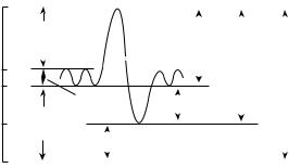
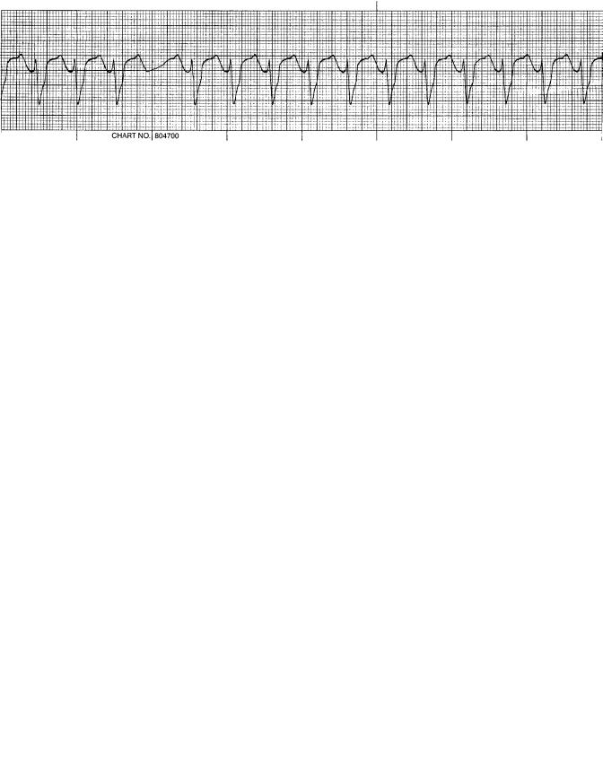
The fatal dysrhythmia of ventricular fibrillation (Fig. 7) is a common endpoint in cardiac arrest. There are no QRS complexes, P waves or T waves that are identifiable. Scientific evidence over the years has conclusively established that only early defibrillation has any hope of restoring a normal sinus rhythm to the heart and subsequent survival. The fibrillating heart has no ability to eject blood since there is no coordination of heart muscle and no progressive flow of blood from atria to ventricles to systemic circulation. Cardiac depolarization and repolarization within the ventricular muscle occurs in a chaotic manner; ventricular muscle is activated in an unorganized fashion. This electrical activity sustains a vicious cycle of reexcitation, never allowing the return of normal cardiac function. The ventricles neither relax nor contract and in this ventricular fibrillating state consume massive amount of energy. Since there is no ejection of blood, unconsciousness from lack of cerebral blood flow occurs within seconds and death ensues from hypoxia.
After a normal cardiac sequence there is a refractory period (as mentioned previously with the beginning of the T wave, which is at the end of the cardiac cycle), whereby this cardiac impulse cannot reexcite the heart until a new electrical stimulus is generated from the sinoatrial node. However, the underlying etiology of ventricular fibrillation appears to be due to electrical reentry or ‘‘circus movements’’, where the normal termination of depolarization does not occur. These abnormal electrical pathways may be generated in several ways: a shortened refractory period, decreased depolarization velocities, or increased distance for the normal electrical impulse to travel. Recall that the progression of depolarization takes place only in one direction and essentially travels almost in a circle with excitation of the ventricles. If this normal impulse reaches cardiac muscle that has already been depolarized, the refractory time will not allow another depolarization. Stimulation of cardiac muscle will not occur until the entire myocardium is ready to be energized as one unit. Suppose that one of the three abnormal conditions were present; any ventricular muscle that was not refractory could be stimulated to contract in an unorganized manner. In a clinical setting, many individuals have hypertension and develop enlarged hearts. A large ventricular muscle mass
would create an increased distance for the normal electrical impulse to follow, thus creating the potential for a ‘‘circus movement’’ to initiate reexcitation of muscle fiber. Rates of depolarization from the sinoatrial node through the AV node may result from blockade of this specialized system from a variety of causes. Electrolyte imbalance, as well as coronary artery disease, are common factors in inducing conduction block. Alterations in the sympathetic nervous system as well as drugs may act in sensitizing the heart, allowing more rapid conduction of impulses and increased susceptibility to fibrillation. Once the ventricular muscle begins this chaotic activity, a chain reaction phenomena begins: conduction velocities throughout the heart decrease, allowing even more time for reentrant depolarizations to occur and the actual muscle refractory time is decreased, increasing the opportunity for these impulses to propagate this dysrhythmia.
The cornerstone of cardiopulmonary resuscitation is early defibrillation. The previous American Heart Association mnemonic of CPR, the ‘‘ABCs’’, which consisted of Airway, Breathing, Circulation, has been changed to ‘‘ABCDs’’ to include defibrillation. After one shock, 60% of all victims succumbing to ventricular fibrillation will survive; after two shocks, 80% survive; after three shocks, 90% will be successfully resuscitated (48). Electrical countershock utilizing high-voltage current can inhibit defibrillation by instantaneously depolarizing all cardiac muscle tissue. The myocardium then is totally refractory to any reentry currents. The electrocardiogram will typically record asystole, or no evidence of electrical activity, from the heart for several seconds. Resumption of the normal cardiac pacemaker will resume, and organized contraction should reoccur. Time is of the essence, since as a heart continues in fibrillation, the rapid utilization of highenergy phosphates depletes this ‘‘fuel’’ for resumption of normal cardiac activity. It is obvious that delay in defibrillation induces a state whereby even successful technique in countershock will be not be able to sustain a normal cardiac rhythm due to the lack of substrate for myocardial energy consumption. The underlying philosophy of cardiopulmonary resuscitation now is early access to defibrillation for the victim.
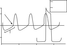
Pulseless ventricular tachycardia (Fig. 8) is the other malignant dysrhythmia that requires immediate external countershock. Unlike ventricular fibrillation, the electrocardiogram displays a rapid regular rhythm with a widened and abnormal appearing QRS complex. Usually,
46 CARDIOPULMONARY RESUSCITATION
Figure 8. Ventricular tachycardia.
the rate is very rapid, with a range of 100–250 beats/min, contrasted to the normal sinus rhythm rate of 60–100 beats/min. Three or more of these bizarre appearing complexes define this dysrhythmia. Usually, the P wave and T wave are obscured and thus the P–R interval cannot be measured. This rapid rate does not allow adequate filling of the ventricles, and causes a ventricular lack synchrony with the atria; the result is dramatic loss of cardiac output and blood pressure. Since there is no effective cardiac output and these dysrhythmias degenerate into ventricular fibrillation, external countershock is mandated. The sinus node functions normally in VT, whereby the atria are properly depolarized. There can be a retrograde depolarization of the atria from the ventricles in some instances of ventricular tachycardia and there will be a definite P wave associated with the abnormal QRS complex. Usually, these retrograde P waves have a negative (downsloping) peak. Another unusual feature of this dysrhythmia is that at certain time intervals the atria may be able to initiate an impulse completely through the AV node and Purkinje system at the instant where the ventricular-initiated depolarizations leave the conduction system vulnerable. The result is termed a ‘‘capture beat’’. If this normally conducted impulse occurs at the same time that a ventricular depolarization is generated, a fusion beat is generated, which appears as a cross between the normaland ven- tricular-originated complex. This dysrhythmia may either be paroxysmal or sustained and the shape of these QRS waves either monomorphic or polymorphic. Degeneration into ventricular fibrillation is a common course; for this reason countershock is required. The mechanism for pulseless ventricular tachycardia is hypothesized to be a
reenty depolarizing current due to delayed conduction. The site of occurrence for this aberrant mechanism would localize to the Purkinje system and ventricles.
An unusual form of ventricular tachycardia (Fig. 9) is termed ‘‘Torsade de Pointes’’ or twisting of points (49). Notice the QRS morphology viewing the rhythm strip from left to right. On first appearance, this dysrhythmia appears to be ventricular tachycardia. The QRS complexes are wide (versus the normal narrow QRS shape), but constantly changing in shape and amplitude yet there appears to be a rhythmic oscillation about the baseline. The depolarization wave appears to twist around the central axis or helix. This dysrhythmia is triggered by electrical potentials that either occur before or after the normal spontaneous depolarization of the heart or is associated with a prolonged QT interval. Should this dysrhythmia be associated with no evidence of effective blood flow, immediate countershock would be the treatment of choice after the ‘‘ABCs’’ of CPR have been accomplished. However, if Torsade de Pointes is misdiagnosed as ventricular tachycardia and a pulse is present, the potential exists for the wrong treatment and lethal results. This dysrhythmia usually occurs when there is an underlying prolongation of the QT interval. Since many of the antiarrhythmic agents will prolong this interval, it should be apparent that selection of one of these drugs to terminate a misdiagnosed ventricular fibrillation when Torsade is present has the potential to produce a nontreatable dysrhythmia. Antidepressants, antipsychotics, and electrolyte abnormalities (particularly hypokalemia and hypomagnesemia) will produce the underlying QT prolongation that serves as the catalyst for Torsade
Figure 9. Torsade de Pointes.
CARDIOPULMONARY RESUSCITATION |
47 |
Figure 10. Asystole.
to appear (50). Strategies for treating this rhythm are to correct the factors providing the substrate for dysrhythmia; magnesium supplementation, and correction of all electrolyte abnormalities as well as what is termed ‘‘overdrive pacing’’ to externally pace the heart to rates of between 100 and 120 beats/min (51).
Asystole (Fig. 10) is the terminal arrhythmia that represents a dying heart. Observation of the above example shows the absence of electrical activity (flat line), but occasionally P waves, or what is termed ‘‘ventricular escape beats’’, may be present. Usually, the presence of asystole means death for the victim, so rescuers are taught to view this rhythm in more than one lead since fine ventricular fibrillation may actually be the abnormal rhythm rather than asystole. This distinction is extremely important since VF needs to be immediately treated with defibrillation, whereas in asystole the possible causes need to be identified, and defibrillation is contraindicated for resuscitation. Why not shock the heart that is in asystole? The answer lies in the fact that rarely a victim may experience high levels of parasympathetic nerve input to the heart. This parasympathetic stimulation can produce complete cessation of the atrial and ventricular pacemaker. Defibrillation also produces an intense parasympathetic nerve discharge, which, in this specific situation could terminate any chances for the heart to recover normal pacing function (52). Evidence substantiates that defibrillation for asystole does not improve the survival rates in out-of-hospital arrest scenarios (53). There have been rare occasions reported in the literature where victims have positively responded to transcutaneous cardiac pacing for which many defibrillators now have this
capability. The heart is stimulated externally to produce an effective cardiac output. The caveat is that this maneuver can only be effective if there are only several minutes of asystole. A specific situation that lends itself to transcutaneous pacing is the asystole that occurs after defibrillation. This witnessed arrhythmia occurs immediately after some countershock attempts and thus there can be a window of < 1 min for pacing to be initiated. There may be certain types of patients who also might respond to asystolic pacing, such as those with Stokes– Adams–Morgagni syndrome, where intermittent atrioventricular block produces asystole or the hypoxia initiated P wave asystole (54).
The last terminal rhythm group that occurs frequently in resuscitation efforts is pulseless electrical activity (PEA). Observe the rhythm strip in Fig. 11; this is an example of a normal sinus rhythm. At first, one would question why a normal ECG would be included in emergency cardiac care and treatment. The key to understanding pulseless electrical activity is that the rhythm may appear to be entirely normal; however, upon further physical assessment of the victim, no pulse is detectable. Historically, cardiologists have referred to this state as electromechanical dissociation, where the normal atrial and ventricular depolarization progresses without any myocardial muscle contraction. Recent scientific evidence utilizing echocardiography and invasive pressure monitoring catheters have found that minute myofibril contractions do occur with depolarization; however, there is not enough effective projectile pressure generated to produce any external signs of perfusion, thus the term pseudoelectromechanical dissociation has been proposed (55,56).
Figure 11. Pulseless electrical activity.
48 CARDIOPULMONARY RESUSCITATION
Other rhythms are also included in this group; ventricular escape rhythms, postdefibrillation rhythms, and bradyasystolic rhythms have different QRS morphology than the normal sinus rhythm; however, the same underlying problem of no pulse and no organ perfusion is the defining issue. The key to resuscitation of the victim in PEA is to quickly recognize and treat the underlying problem that has created this scenario. Since there is no perfusing rhythm it should be obvious that the ‘‘ABC’’ basics must be immediately made available to the victim while the differential diagnosis prioritized to the specific situation is reviewed.
There have been a number of mnemonic memory aids suggested to recall the most common conditions provoking pulseless electrical activity. Two common lists are the ‘5 Hs’ and ‘5Ts’ and the ‘‘MATCH-HHH-ED’’ (57,58).
Hypovolemia; Hypoxia; Hydrogen ion excess (acidosis). Hyper or Hypokalemia (excess or loss of potassium ions). Hypothermia; ‘‘Tablets’’ (referring to medications or
drug overdose).
Tamponade (blood trapped around the heart chamber). Tension pneumothorax (trapped air causing lung overinflation and resultant loss of cardiac perfusion). Thrombosis (secondary to myocardial infarction) and Thrombosis (secondary to pulmonary embolism).
M—Myocardial injury or infarction A—Acidosis
T—Tension pneumothorax C—Cardiac tamponade H—Hypothermia H—Hypo or Hyperkalemia H—Hypoxia H—Hypovolemia E—Embolism
D—Drug overdose or chemical toxins.
The most common cause of PEA is hypovolemia: inadequate intravascular fluid volume. These fluid volume losses may be real, such as in acute hemorrhage or relative, wherein the capacitance vessels (veins) vasodilate, leaving inadequate vascular blood and fluids to compensate. A classic evolution to pulseless electrical activity would ensue from hemorrhage, producing normal electrical complexes, with progression to a tachycardia where the heart rate increases to compensate for pressure loss. Peak pressure for each cardiac ejection of blood (systolic pressure) would decrease, but increased constriction of the circulatory vessels would increase (diastolic pressure). As the blood pressure continues to drop to extremely low levels secondary to the hemorrhage, the sinus tachycardia continues. At this point, the astute clinician would consider this cause for PEA and immediately give the victim intravascular fluid followed by blood products to stabilize the victim. It is obvious that the definitive treatment in this case would be surgery; the goal of the resuscitative response is to maintain perfusion and oxygen delivery to the tissues. Time again is critical since without effective
cardiac flow, perfusion of the myocardium is nonexistent and hypoxia develops quickly. Progression toward ventricular fibrillation, asystole, and death are the end result. An example of relative hypovolemia is septic shock. In this situation, a bacterial infection produces endotoxins, which causes the resistance vessels to dilate. There is normal circulating blood volume, but increased vessel capacitance resulting in a progressive blood pressure drop. Due to the underlying pathology, the vessels are unable to adequately constrict and the evolution to cardiac dysfunction secondary to poor or absent coronary perfusion occurs, eventually producing the terminal rhythms of ventricular fibrillation and asystole. Treatment in this scenario consists of increasing the fluid volume as well as to utilize drugs that act to constrict the blood vessels.
Each one of the other conditions mentioned above that can cause pulseless electrical activity also has an immediate treatment response, after the appropriate steps in basic life support have been met. Thus while the victim is being oxygenated and ventilated, and perfusion accomplished through chest compressions, the clinician is rapidly focusing upon the conditions that would be most causative and then initiating treatment. In hypoxia, treatment consists of supplemental oxygen and adequate mechanical ventilation of the lungs. In cardiac tamponade or tension pneumothorax, needle decompression of the pericardial sac or lung is the required emergency management. Electrolyte imbalances require immediate infusion or neutralization of the ion. A preexisting acidosis might require exogenous sodium bicarbonate. An acute myocardial infarction (heart attack) causing PEA would need supplemental oxygen, pain management, fluid resuscitation and immediate infusion of coronary artery thrombolytics. Pulseless electrical activity is always associated with an underlying clinical condition that, when identified and treated early, has the potential to be reversed and thus effective perfusion restored to the victim.
MECHANISMS FOR COMMUNITY CPR
The American Heart Association is a volunteer organization that has as its goal the reduction of mortality and morbidity secondary to cardiovascular and cerebrovascular disease. The AHA established a scientific mission in 1963 to evaluate and promulgate standards of CPR. In 1971, the Emergency Cardiac Care Committee was founded. Since the first conference on CPR in 1966 sponsored by the National Academy of Sciences and the National Research Council, guidelines for CPR have been based upon the available scientific evidence. Since this time there have been six conferences, the last in 2000 consisting of resuscitation experts worldwide. All aspects of CPR have been evaluated, including the actual access for emergency care in the community, education of the layperson, and delivery of basic CPR and ACLS. ‘‘Emergency Cardiovascular Care’’ is the term used to describe the organized response to life-threatening emergencies for the adult, pediatric, and neonatal victim. Other ECC provisions for care include educating the layperson to recognize the signs and symptoms of myocardial ischemia and infarction, stroke, activation of the EMS system (911) as well as early
implementation of basic life support, defibrillation, and advanced cardiac life support with immediate victim transfer to the hospital. The goal of ECC is to save lives and this also includes educating the public regarding cardiovascular risk and how to maintain healthy lifestyles. The cornerstone of all emergency care is the layperson. Effective emergency care can only occur with public awareness of these events as well as prompt administration of CPR and use of the automatic external defibrillator. Once the EMS system has become involved with the victim, progression of care is dictated by a physician and ACLS protocol. The optimization of oxygenation and ventilation through supplemental oxygen and adjunctive respiratory devices, electrocardiographic monitoring with rhythm assessment and treatment, establishment of intravenous access for appropriate medications and postresuscitation management are all elements of ACLS.
The American Heart Association ‘‘Chain of Survival’’ is a crucial concept to be understood in the context of CPR and is taught to the layperson in the basic lifesaving courses. There are four critical links in this chain that require specific actions on the part of the public. In the event of an emergency the first link involves activating the emergency medical services system. Obviously, the layperson must be able to recognize that a true emergency exists, and this underscores the efforts by the community organizations to educate the public regarding signs and symptoms of heart attack, stroke, and loss of the airway. The key is unresponsiveness; anyone found unconscious should initiate the first link by way of immediately involving EMS through calling 911. Once an emergency medical dispatcher is contacted, immediate aid in the form of paramedicals is sent to the scene. The dispatcher is also taught how to aid the layperson in how to provide basic CPR, which is the second link in the chain. Only until the EMS personnel arrive and begin managing care for the victim does the dispatcher terminate the call. In some communities, ‘‘enhanced 911’’ is available in which a computer will provide the dispatcher with the address from which the call is made in case communication is difficult or there is a premature telephone disconnection. This second link of CPR is the most critical phase in the Chain of Survival since the rescuer provides oxygen to the victim and, if needed, circulates this oxygen by chest compressions to the brain, heart, and vital organs. If bystander CPR is initiated within 4 min after a victim collapses, the odds of survival, after discharge from the hospital, are doubled (59).
The third link in the chain is early defibrillation, either by the public use of the automated external defibrillator or by the paramedic on the scene. Early access to defibrillation for the victim of cardiac arrest will significantly improve the chance for survival: each minute of delay for defibrillation for the victim in ventricular fibrillation decreases the chance of survival by 7–10%, and if defibrillation is provided within the first 5 min, chance of survival is 50% (60). Unfortunately, if >12 min of delay from collapse to initial resuscitation is encountered, the survival rate only ranges between 2 and 5%, and intact neurological function is compromised (61).
Recently, gambling casinos have implemented access for defibrillators and for victims who received a shock
CARDIOPULMONARY RESUSCITATION |
49 |
within 3 min had a 74% survival rate since a low response time of between 2 and 3 min was documented (62). Since time to defibrillation is so critical, automatic external defibrillators have been made much more accessible to the public. These devices can now be found in large gathering places such as stadiums, golf courses, airports and airplanes, shopping malls, large grocery stores, and other facilities where people in great numbers tend to congregate. So important is the early access to defibrillation that a great majority of states have enacted legislation to encourage use of these devices. The Cardiac Arrest Survival Act provides legal immunity for the layperson and the public business or corporate entity that uses or provides an automatic external defibrillator for resuscitation, which is essentially an expansion of the ‘‘Good Samaritan’’ type legislation. This immunity should encourage active participation by the public for involvement in victim resuscitation. Public access defibrillation has been described as the second most significant advance, compared to CPR, in the pre-hospital rescue scenario.
The final fourth link in the Chain of Survival is early ACLS by highly trained paramedical personnel. Emergency medical technicians expand (EMTs) incorporate basic CPR with interpreting cardiac dysrhythymias and, if required, defibrillation. Emergency medical technicians expand the immediate life-saving care by providing supplemental oxygen, intubation and control of the airway, gaining intravenous access and administering pharmacologic medications while in contact with a physican. This process occurs at the scene, and once the victim is stabilized, advanced cardiac life support continues through transport to the hospital emergency room. The most significant impact of early ACLS is to prevent the catastrophic progression of lack of oxygenation and cardiac arrest rather than to treat the terminal conditions inherent in this process.
The rescuer, whether a lay person of EMT, who begins CPR in the ‘‘field’’ must continue BLS (63) until one of the following events occurs:
1.The victim begins to show signs of spontaneous ventilation and perfusion.
2.Care is transferred to another qualified BLS responder, EMT, or ALS medical providers; or to a physician who makes the determination that resuscitation should be terminated.
3.The rescuer cannot continue resuscitation due to exhaustion or to hazards that may jeopardize the rescuer’s life or the lives of others in the team.
4.An authentic no-CPR order is presented to the responders.
The determination to discontinue resuscitation depends on a stepwise evaluation of the efforts made during BLS and ACLS. A review of the process should ensure that each step in the resuscitation has been carried out in a flawless manner. Successful ventilation and intubation, intravenous access and the administration of appropriate medications as well as countershock should be achieved according to ACLS protocol. Electrocardiography evaluation should render a conclusion of no reversibility for the underlying agonal rhythm. Recently, the determination of end-tidal
50 CARDIOPULMONARY RESUSCITATION
carbon dioxide has been advocated as a potential predictor of death (64). During resuscitation end-tidal carbon dioxide reflects the adequacy of cardiac output generated during chest compressions. This study suggested that after standard ACLS protocols had been followed for 20 min, a persistent end-tidal carbon dioxide level of 10 mmHg or less predicted nonsurvival in the victim with electrical activity, but without a pulse.
CARDIOVASCULAR DISEASE
Every year 500,000 people are hospitalized for treatment of chest pain secondary to cardiac origin and 1.5 million victims will experienced a heart attack (65,66).
Some 500,000 people a year who have a myocardial infarction (heart attack) will die from this insult and 225,000 of these deaths will occur within the first hour after symptoms and prior to reaching a hospital (67,68). In 17% of the victims, chest pain is the first and only symptom (67). It is again significant that time to intervention for the patient experiencing a myocardial infarction is crucial to survival; treatment must be undertaken within the first several hours after the symptoms occur (70,71). Early treatment underscores the necessity for rapid recognition of a cardiac event, followed by rapid CPR, defibrillation,
ACLS and transport to the hospital.
The most common cause of a heart attack is ischemic atherosclerotic disease. The essential pathophysiology is the narrowing of the coronary artery lumens by deposits of fat-substrate such as cholesterol and lipids, which eventually retain calcium. The actual process of the accumulation of these plaques occurs very slowly, but has been demonstrated to begin at an early age. This same process affects the cerebral arteries as well and is the etiology for an ischemic stroke. As the coronary artery lumen narrows, a dynamic situation develops where blood flow and thus oxygen supply will not meet with increased demand for oxygen by the cardiac muscle fibers. Typically the coronary artery will have a circumference reduction of 70% for symptoms to occur. This condition of ischemia will produce a characteristic constellation of transient symptoms, referred to as angina pectoris. Chest pain is the most common sign of an acute cardiac ischemic event, occurring in 70–80% of the population (72). This pain appears to have several different components: transmission of dull, poorly localized pain occurs through the sympathetic visceral nerve fibers; a somatic pain generator produces the sharp and dermatomal aspects; and psychological input gives rise to the sense of impending doom (73,74). This cerebral input to the event may significantly exacerbate the ischemia since activation of sympathetic nervous system will increase heart rate and contractile force, further tipping the scale toward more energy consumption and thus oxygen demand.
Paradoxically, the majority of episodes of an acute coronary event (angina and or infarction) occur during periods of rest or mild to moderate exercise; profound physical exercise is associated with the minority of events (75). The victim will experience an intense, dull, crushing pressure sensation in the chest, most commonly behind the
breastbone (retrosternally) and/or pain in the back, arms, shoulders, or mandible. Often nausea, vomiting, sweating, and shortness of breath (dyspnea) may accompany the pain. There appears to be a circadian rhythm regarding the occurrence of angina and the progression to infarction. Two daily peaks in incidence have been noted with the first pattern beginning from awakening to about noon, and the second peak occurring in the early evening (76,77). There are atypical presentations to unstable angina or myocardial infarction that will delay access for the victim. This subset of the population may have only vague, mild discomfort, which can be confused with a myriad of medical complaints. Diabetics, women, and the elderly all have a higher incidence of nonclassical presentations for cardiovascular ischemia (78,79). Diabetics are prone to neurological dysfunction and thus may have no sensation of the pain associated with angina. A retrospective review found that 30% of first heart attacks in men and 50% of first infarctions in women did not present with classical signs and symptoms and were clinically not recognized (80). When oxygen demand decreases, such as when the physical activity is discontinued, the decreased oxygen supply secondary to the narrowed lumen will be adequate and the symptoms will usually resolve. Progression of the disease, however, results in a much more severe mechanism for ischemia. The plaque is predisposed to rupture and when this occurs, activation of the coagulation system releases mediators that form a clot or thrombus over the plaque, which can further limit blood flow, or catastrophically, stop all blood flow completely. If the partial occlusion from the thrombus is severe enough, what is termed ‘‘unstable angina’’ develops. Though there have been many definitions of unstable angina, the main characteristic is that this type of angina occurs at rest and is progressive or prolonged in nature. Nocturnal angina, again with the victim at rest, would be classified as unstable angina. The heart is consuming the least amount of oxygen yet there is a lack of supply due to lumen reduction from the plaque. Clot enlargement provides the mechanism for dislodgement of particles or emboli, which then travel downstream, lodging in the microvasculature and these individuals are at a very high risk for progression to irreversible cardiac damage.
Complete occlusion of the coronary artery results in a myocardial infarction. Deprivation of oxygen results in the death of cardiac myofibrils and induces irritability in the cardiac conduction system, setting the stage for the initiation of lethal dysrhythmias such as ventricular fibrillation. Where and how severe the damage is to the heart depends on what coronary artery has the occlusion. If the left main coronary artery has an acute total obstruction, mortality is very high since no blood flow will occur through the two distal branches, the left anterior descending and circumflex arteries. Blood flow will be blocked to the entire left ventricle, the septum between the left and right ventricle, and the bundle branches. Even if the patient receives timely CPR, and clot lysis, a significant amount of heart may be destroyed resulting in scar tissue and a drastic reduction in blood flow, resulting in what is termed ‘‘congestive heart failure’’. If the thrombus were to lodge and occlude the right coronary artery, hypoxia would occur in
the AV node, right ventricle, and in the majority of individuals, the posterior and inferior aspect of the left ventricle.
The continuum of acute cardiac injury, from unstable angina to myocardial infarction typically presents with characteristic electrocardiographic signs. Recall what the normal elements are to the ECG. During episodes of unstable angina where cardiac muscle demand for oxygen exceeds supply, ischemic changes to the ECG are seen as S–T segment depression, defined as to a 1 mm change from baseline on the standard graphpaper or changes in the T waves. When this S–T segment depression is downsloping, this is a sensitive sign of ischemia. The T waves may appear inverted or enlarged and symmetrical. When actual occlusion of the artery occurs, myocardial injury to tissues ensues and both muscle contraction and conduction are decreased from normal. The ECG change characteristic of injury is S–T segment elevation in contradistinction to changes of ischemia. When there is 1 mm above baseline for the S–T segment elevation, significant cardiac injury has occurred. This group of patients who exhibit S–T segment elevation on the ECG in two contiguous leads may be salvaged through reperfusion therapy. Restoration of blood flow and the course of injury is very dependent on early administration of thrombolytics or percutaneous transluminal coronary angioplasty (PTCA). The greatest improvement in mortality and morbidity occurs when reperfusion therapy is administered within the first 3 h after onset of symptoms (81,82). Conjoined with early CPR and defibrillaton, reperfusion therapy stands as one of the greatest advances in acute coronary syndromes. Current regimens include the fibrinolytics streptokinase and alteplase, as well as numerous other similar agents that act by inducing fibrinolysis through interactions with tissue plasminogen activator. The PTCA is a mechanical procedure whereby a catheter is guided through the coronary arteries into the area of stenosis, and then a balloon is inflated to expand the vessel. This procedure is restrictive in that only specialized centers have the capability to utilize this regimen, although it may be superior to thrombolytics.
Myocardial infarction defines the actual death of cardiac tissue. This is the end result of the process of ischemia with myocardial cell injury. The infarcted tissue area, again representative for the specific coronary artery occluded, will exhibit characteristics associated with loss of cellular life. Intracellular contents are released after loss of cell wall integrity. Some of these enzymes, such as creatine phosphokinase and the troponins, can be measured in the bloodstream to confirm infarction. The classic ECG changes for myocardial infarction are the presence of abnormal Q is waves. When a Q wave is or 1 mm in width and the height is >25% of the R wave height, the diagnosis can be made. An abnormal Q wave reveals the existence of dead cardiac tissue, but does not reveal anything about when the infarction happened. The assumption can be made for a recent infarction if the Q wave is associated with S–T segment changes and/or T wave changes. A non-Q wave infarction can also occur: There is myocardial cell wall dissolution with the release of cardiac enzymes, but only accompanied by S–T segment changes or T wave abnormalities. There is a lower mortality rate for non-Q wave heart attacks, but, unfortunately, an increased incidence of future reinfarction or
CARDIOPULMONARY RESUSCITATION |
51 |
death (83,84). Fibrinolytics are contraindicated in patients with non-Q wave infarction since the clot occlusion may be paradoxically aggravated by release of thrombin, which further activates platelets (85).
Prehospital intervention for the victim suffering an acute cardiac event is based upon the ‘‘Chain of Survival’’. Once the signs and symptoms of a heart attack are recognized, early access to the emergency medical system is imperative. A common problem encountered is denial either from the victim or the rescuer, which impedes response time. Once the EMS personnel arrive at the scene a pertinent medical history is obtained and physical examination completed. A complete 12 lead ECG is obtained and then transmitted to the physician who is dictating care. Oxygen is the first line treatment for anyone complaining of chest pain. It should be recalled that supplemental oxygen substantially increases the oxygen tension in the blood and significantly improves tissue oxygenation. A critical blood flow restriction may be palliated by improving oxygen supply in this manner. The administration of the drug nitroglycerin is quickly administered for the victim symptomatic for angina in conjunction with oxygen. Nitroglycerin is delivered sublingually for rapid absorption into the bloodstream. Nitroglycerin is effective in relieving the symptoms of angina in several ways: relaxation of venous smooth muscle occurs due to binding of specific vascular receptors. As relaxation of the venous capacitance vessels occurs, venous return to the heart is decreased, thereby relieving ventricular wall tension, which ultimately decreases ventricular work and oxygen consumption. The nitrates also dilate the large coronary arteries as well as increasing blood flow through collateral vessels, which improves ischemic blood flow (86,87). Aspirin is the third drug that should be administered immediately by either the BLS provider or EMT when symptoms suggest a cardiac event (and the victim is not allergic to aspirin). A regular tablet of aspirin (325 mg), when ingested, will cause an immediate anticlotting mechanism by way of platelet inhibition. There is evidence that suggests aspirin decreases coronary artery reocclusion and future coronary symptoms, with reduction of death and furthermore, the effects of aspirin appear to be additive to fibrinolyis (88). The fourth drug that is administered during episodes of chest pain secondary to unstable angina or myocardial infarction is morphine. Although morphine is a narcotic analgesic, it produces beneficial hemodynamic effects in addition to profound pain relief. Morphine causes decreased vascular tone in the venous capacitance vessels, thus reducing myocardial wall tension, much like nitroglycerin. The mechanism, however, is different in that the action appears to be mediated through central nervous system reductions in sympathetic tone (89). A convenient mnemonic has been utilized, ‘‘MONA’’, for recall of these four immediate effective therapies for pre-hospital treatment of the acute coronary syndrome.
CEREBROVASCULAR DISEASE
There has been a concerted effort in the Emergency Cardiovascular Care system to improve pre-hospital recognition of
52 CARDIOPULMONARY RESUSCITATION
the warning signs of stroke and provide rapid access to the Emergency Medical System. Public awareness of the issues regarding a ‘‘brain attack’’ has lagged relative to the exposure and education afforded cardiovascular disease. Stroke ranks third behind heart disease and cancer for morbity in the United States; 500,000 Americans a year suffer from a cerebrovascular accident and 125,000 of these victims will die (90). Until recently, stroke victims were only offered supportive and rehabilitative therapy for the complications experienced if they survived the initial insult. However, advances in fibrinolytic therapy, as in treatment of cardiovascular disease, dramatically improves outcome for the patient who has experienced an ischemic stroke (91). Fibrinolytic treatment reduces stroke disability and significantly improves quality of life after hospital discharge (92,93). The caveat is that the cerebrovascular accident must be recognized and treatment initiated in a timely manner; fibrinolytics need to be provided within 3 h after the onset of an ischemic stroke (94). Thus there is a narrow window of opportunity to limit cerebral damage, which underscores how important the role is for the public in providing immediate access to the EMS system for the victim.
The underlying cause for an ischemic stroke is comparable to the etiology for myocardial infarction. There is a disruption to cerebral blood flow due to the presence of an occlusive clot. The oxygen supply to the particular area of the brain supplied by the blocked artery does not meet the tissue demand and the same process of ischemia, injury, and cell death will occur. The thrombus, which occludes the vessel, is the end result of atherosclerotic changes to the artery. However, due to the unique anatomical positions of the cerebral arterial system, a blood clot formed elsewhere in the body can embolize to disrupt blood flow to the brain. Approximately 75–85% of all strokes are of this type, and defined as ischemic, and furthermore can be classified as to the arterial system that is affected. The two major arterial conduits to the brain are the carotid arteries and vertebrobasilar arteries, which affect the cerebral hemispheres or brain stem–cerebellum, respectively. Typically, a person who is at risk will develop what is termed ‘‘a transient ischemic attack’’ (TIA) prior to a full-blown stroke. Essentially, a TIA is a reversible mini-stroke that may affect specific brain function or eyesight and will last anywhere from minutes to hours (95). The TIA is a harbinger of a future ‘‘brain attack’’ much like unstable angina will forecast a heart attack. About 5% of those persons presenting with a TIA will end up with a stroke in 1 month; the risk will increase to 12% after 1 year and an extra 5%/year thereafter (96). Fortunately, the symptoms from a TIA will bring the patient into the medical system for evaluation whereby treatment regimens clearly reduce risk for ischemic stroke. The surgical procedure of carotid endarterectomy in which the carotid artery plaque is removed has been proven very beneficial for patients that have had a recent TIA and a >70% stenosis of the carotid artery (97). In those individuals who are not operable candidates, aspirin and the specific platelet inhibitor types of drugs have been shown to be successful in preventing subsequent stroke in patients presenting with TIA (98).
The minority of acute strokes are due to hemorrhage of cerebral artery. The bleeding may occur in the subarach-
noid space, which is in the superficial exterior aspect of the brain, or in the brain tissue itself, defined as an intracerebral hemorrhage. The common etiology to a subarachnoid hemorrhage is an aneurysm where the arterial wall weakens, and eventually a disruption occurs (99). In the case of a hemorrhage into the brain tissue itself, high blood pressure appears to be the major causative factor (100). While there are similar signs and symptoms in both types of stroke, there are also distinct differences in findings, which aids in the diagnosis. In general, the presentation for a subarachnoid hemorrhagic stroke is more severe with a very common complaint of an extremely painful headache, which tends to be global, and may have radiation of pain into the face or neck. This headache is often accompanied by mental status changes, nausea, vomiting, photophobia, or cardiac dysrhythmias. In a minority of patients, a prodromal episode of these symptoms may be caused by leakage of the aneurysm offering a warning sign (101). While the victim suffering from an intracerebral hemorrhage may also present with a severe headache, these patients tend to have a greater neurological insult with significantly depressed mental status function. The signs and symptoms of an ischemic versus a hemorrhagic stroke overlap and diagnosis may be difficult based upon the medical history and physical findings. Since radiological imaging offers the greatest aid in differentiating these two types of cerebrovascular accidents, time is very critical to clinch the diagnosis to offer the appropriate treatment. Fibrinolytics would obviously be a catastrophic therapy in the mistaken treatment of what appears to be an ischemic stroke when the etiology is a ruptured blood vessel that requires surgery.
The American Heart Association ‘‘Chain of Survival’’ that has been implemented and associated with cerebrovascular disease has been applied to the pre-hospital care of the stroke victim. Early recognition of a stroke and activation of the EMS system are paramount in initial therapy, which is often problematic, since, unlike a heart attack, stroke may be difficult to detect. While early defibrillation is not ordinarily indicated for the stroke victim, the possibility always exists that coincidental lethal dysrhythmias may be present during the initial presentation (102). The last link in the chain is early hospital care. The common theme regarding out-of-hospital management for cardiac or stroke victims is rapid entry for the victim into advanced life saving. The ‘7-D’ mnemonic has been recommended as an aid for care in the stroke patient: Detection; Dispatch; Delivery: Door; Data; Decision; Drug (103). Early detection, with an accurate recall of the initial signs of a stroke are critical to care and must be accomplished by the immediate family member or layperson, with immediate access to ‘‘Dispatch’’, the EMS personnel. An important point to emphasize is that the majority of strokes occur at home (104). Paramedics who arrive at the scene must confirm a rapid, tentative diagnosis through focused medical history and physical examination, and then ‘‘Deliver’’ the patient rapidly to the hospital. Once the patient is through the Emergency Department ‘‘Door’’ the medical history and physical examination are further refined along with radiography (computerized tomography). A ‘‘Decision’’ is made regarding whether fibrinolytic therapy is
indicated for an ischemic stroke and the ‘‘Drug’’ treatment is initiated. The drug therapy must be initiated within 3 h after the onset of an ischemic stroke.
The changes in mental status and/or sensorimotor function in a cerebrovascular accident may range from minor, almost unrecognizable changes, to loss of consciousness and seizures. A person may exhibit grades of confusion, with a progression to stupor or coma where the airway is obtunded and basic life support is required. Comprehension of language often occurs with inappropriate responses to simple questions. Physical manifestations are often present unilaterally. Paralysis in either the face, upper, or lower extremity may range from slight weakness to frank inability to exercise any muscular control. Since the face is always exposed, muscle weakness is exhibited by loss of tone and sagging of the muscles in facial expression. Difficulty writing (aphasia) or speaking (dysarthria) occurs due to loss of appropriate motor input from the brain. Loss of sensation is another common sign of a stroke (or a TIA). Visual disturbances, including blindness are much more obvious and usually involve only one eye. If the location of the ischemia is in the vertebrobasilar arterial system, centers of the brain controlling coordination are involved and signs such as gait disturbances (gait ataxia) are common. The dilemma of pre-hospital rapid neurological assessment to evaluate the possible stroke victim when the presentation is varied has been improved by several instruments. The Cincinnati Pre-hospital Stroke Scale (104) is very effective in identifying the stroke victim. Three physical findings are assessed: facial droop; arm drift; and speech. Abnormal features in any one category is very predictive for cerebrovascular accident. The Los Angeles Pre-hospital Stroke Screen also is extremely useful for assessment. Six criteria are first evaluated in the medical history: (1) age > 45 years, (2) absent history of seizures or epilepsy, (3) no history of motor loss, (5) serum glucose not < 60 g/dL nor > 400 g/dL, and (6) asymmetry in any one of the three categories of facial musculature, grip strength, and arm strength. If all criteria are positive, there is a 97% chance of an acute stroke (105). These tests have streamlined the response time and have allowed the hospital emergency room to prepare for rapid definitive diagnosis.
THE ‘‘ABCs’’ OF ADULT CPR
The evolution of the current basics of life-support has resulted in a streamlined set of actions that has standardized the initial care for the victim, whether accomplished by a public bystander or emergency room physician. Assessment of the victim always precedes a physical maneuver on the part of the rescuer; constant appraisal of the effectiveness of CPR and the response of the victim is a core principle in American Heart Association Basic Life Support tactics. The initial steps of resuscitation are never bypassed; for example, if the airway is not established, the single rescuer would never start chest compressions, or begin intravenous access. The stepwise process in the algorithm ensures that an orderly process occurs in a situation where chaos and a high degree of emotional
CARDIOPULMONARY RESUSCITATION |
53 |
turmoil exist for the rescuer. Since there are some basic differences in how resuscitation is administered to the adult versus a child, anyone 8 years or older is considered an adult.
When one encounters a potential victim, the first assessment is to determine unresponsiveness. ‘‘Shake and shout’’ has been a common first action to determine that the victim is really unconscious (there no doubt has been a number of resuscitations initiated upon someone who was sleeping, assuming unconsciousness)! Once there is no doubt that a true emergency exists, the rescuer sends another member of the group to activate the EMS system by phoning 911 and to obtain an AED. If the rescuer is alone, he or she must leave the victim momentarily to call 911 and get the AED; these automated defibrillators have a standardized placement near a telephone. After accessing the EMS and obtaining a defibrillator, the rescuer places the victim in the supine position, and kneels at the head (the left side is suggested when utilizing an AED). The ‘‘ABCs’’ of CPR now are initiated.
A ¼ Airway
The unconscious person has a generalized relaxation of all muscles and in the throat this causes the tongue to move in a posterior direction, occluding the airway. Since the tongue is attached to the mandible, manipulating the jaw and head will retract anteriorly. Two methods are utilized to open the airway; the ‘‘head-tilt and chin-lift’’ or the ‘‘jawthrust’’ maneuvers. Tilting the head backward by placing one hand on the forehead and raising the chin with the two fingers of the other hand is the most commonly used technique. An important feature of this technique is to make sure that the fingers are placed on the inferior surface of the mandibular bone and not the soft tissue under the tongue, as the later placement will worsen airway compromise. In a situation where a neck injury is suspected with possible spinal cord compromise, extension of the head is contraindicated; the jaw-thrust is utilized to open the airway. The head is held in the neutral position while applying forward pressure with both hands at the angle of the jaw, just below the ears. In this way, there is no change in head position. At this point inspection of the mouth is important to remove any secretions, vomitus or foreign bodies that may be an impediment to air exchange. Once the airway is opened, the rescuer ‘‘looks, listens, and feels’’ for breathing by placing his or her cheek and ear close to the victim’s mouth. The chest is examined for movement while feeling and listening for air passage. In the case of a partial obstruction of the airway, the victim will tend to make high-pitched ‘‘crowing’’ noises that may be accompanied by cyanosis of the skin (due to unoxygenated hemoglobin). Instead of the chest expanding with an inspiration, retraction of thorax or lung compartment will occur. It is imperative that the airway be opened and maintained in this situation since ineffective ventilations will invariably lead to hypoxia.
B ¼ Breathing
Once it is ascertained that the victim is not breathing (this should occur within 10 s), the rescuer places his mouth
54 CARDIOPULMONARY RESUSCITATION
around the victim’s mouth and pinches the nose shut with one hand while maintaining chin-lift with the other hand. Two long, extended breaths are given, each 2 s, with the goal of providing ventilation to the lungs while minimizing the egress of air into the stomach. Since during unconsciousness there is a relaxation of all muscles, the lower esophageal sphincter will relax and thus any air that enters the stomach may force gastric contents into the esophagus and then into the trachea. Aspiration of these highly acidic stomach contents into the lungs may occur. The complex interplay between rescuer, positive pressure ventilation, peak airway pressure, tidal volume, and inspiratory flow rate has had a considerable degree of scientific evaluation (106–109). The consensus supports a tidal volume of between 800 and 1000 mL to maintain adequate oxygenation when only room air is provided in the rescue breathing. This volume is slightly less than the 1992 ECC Guidelines of a rescue tidal volume of 800–1200 mL. A slow prolonged breath over 2 s decreases peak positive pressure and thus entry of air into the stomach while providing the optimum tidal volume. When supplemental oxygen is available, evidence has confirmed that a tidal volume of 500 mL provides effective oxygenation and ventilation in the unintubated patient as long as the inspired oxygen fraction is >40% (110,111). Once rescue breathing is commenced, one should assess effectiveness by noting whether the chest rises with each breath. If there is no change or the rescuer observes that significant effort is required to minimally expand the chest, the airway step has not been optimized and the rescuer has to reopen the airway with additional head extension and chin-lift (or jawthrust if a head or neck injury is suspected). If readjustment of the airway does not provide the ability to ventilate, a foreign body lodged in the airway should be suspected and the rescuer should proceed through the algorithm specific for dealing with this issue. Victims with dentures may prove difficult to ventilate; generally dentures should be left in place since it is easier for the rescuer to form a seal around the mouth. However, loose dentures may be aspirated and should be removed if their retention is inadequate. In the case where the rescuer cannot maintain an adequate seal, or if the mouth is unavailable for airway exchange secondary to trauma, mouth-to-nose breathing should be attempted (112). A deep breath should be inhaled by the rescuer prior to respiratory exchange since this maneuver optimizes the maximum amount of oxygen made available for each tidal volume (113). Should the victim only require oxygenation and ventilation, rescue breathing provides one breath every 5 s or 12 breaths/min (114).
There is always the concern regarding exposure to an infectious organism when performing rescue breathing. At this point in time, there has not been any evidence documenting the transmission of human immunedeficiency virus (HIV), hepatitis, or tuberculosis when mouth-to- mouth resuscitation has been instituted in an emergency (115). However, reluctance upon the part of any lay rescue person to perform this action is understandable and there is no moral or legal duty to do so. Barrier devices have been developed that prevent intimate contact with the victim and there are two basic types: face shields and masks. This adjunctive equipment has been made available in the
healthcare environments due to the requirements of the Occupational Health and Safety Administration. The face shield has a flexible plastic covering with a one way circular valve that, when placed over the victim, separates the rescuer from contact and from exhaled gases. Mouth-to- mask rescue ventilation provides a better seal and further distance from the victim’s mouth than the face shield, which is advantageous should vomiting occur. Some of these masks have a port where supplemental oxygen can be provided and entrained with the rescue breathing. A flow rate of 10 L/min through one of these masks will increase the inspired concentration of oxygen to at least 40% (116). When supplemental oxygen is supplied in this manner, smaller tidal volumes, on the order of 400–600 mL, will maintain oxygenation while decreasing the risk of gastric insufflation (117).
C ¼ Circulation
After delivery of two rescue breaths, the next step in basic CPR is to assess for signs of circulation. For many years the layperson was taught to feel for the presence or absence of a carotid pulse. Research in the 1990s found numerous pitfalls with the pulse check that appeared to have a negative impact on survival and, since 2000, this task is not taught to the lay responder anymore. Significant time delays in trying to determine if a pulse was present delayed time to defibrillation and thus survival (118). The accuracy of the pulse test revealed a sensitivity of only 55% and a specificity of 90% and overall the accuracy was 65% (119). At this time, the lay rescuer is instructed to look for signs of perfusion, such as movement, breathing, or coughing and if unsure, to begin chest compressions.
Correct positioning of the hands and compression skills are easily learned by the layperson. A simplified method for hand placement has been taught for several years and consists of placing the heel of one hand over the center of the breastbone (sternum) between the nipples and then interlocking the fingers of the remaining hand over the first, so that pressure will be transmitted through the heels of both hands. Effective compressions are generated by positioning the rescuer’s shoulders over the hands and sternum and depressing the sternum from 1.5 to 2 in. Release after compression must be complete without taking the hands off the chest to prevent ‘‘bouncing’’. Chest compressions should be similar in action to that of a piston in a reciprocating engine with half of the cycle spent in compression and the other half spent in relaxation. The effectiveness of this ratio has been documented with regard to both cerebral and coronary perfusion pressures (120). The recommended rate for chest compressions is 100/min, which has been substantiated by numerous studies (121,122).
The single rescuer initiates chest compressions after providing the victim with two rescue breaths and assessing for signs of effective cardiac blood flow. A ratio of 15 compressions followed by 2 rescue breaths continues for four cycles and then the victim is reassessed for spontaneous circulation. Chest compressions should be resumed within 10 s after noting no signs of perfusion and the 15:2 cycle continued with an interruption for assessment of vital signs in several minutes, followed by the same ratio.
When additional responders are present during resuscitation of a cardiac arrest victim, immediate activation of the EMS system must be accomplished and a defibrillator brought to the scene, if these actions have not already been completed by the lone rescuer. The second rescuer should then assess the adequacy of ventilations and chest compressions and reassess for signs of a pulse and breathing within 10 s while CPR is halted. Though it is not expected that the layperson be able to engage in two-person resuscitation, the process is included here for completeness. Medical professionals as well as the paramedical caregivers should all be able to demonstrate this skill. The compressor is positioned in the normal manner, at the side of the victim. The second rescuer is stationed at the victim’s head, maintaining the airway, monitoring for effective compression by carotid artery pulse check, and providing rescue breaths. Previous scientific guidelines utilized a compression:ventilation ratio of 5:2 (123), which has now been changed in light of recent scientific evidence. Currently, a ratio of 15 compressions to 2 ventilations is recommended for both one and two rescuer CPR (124– 126) since it appears that improved survival occurs as a result of the higher rate in spite of a decreased number of ventilations. The effectiveness of chest compressions relative to coronary perfusion pressure (the difference between aortic diastolic pressure and the left ventricular end-dia- stolic pressure) suggests that, as the number of compressions increases, so does the perfusion pressure; therefore, 15 chest compressions improves and sustains blood pressure more effectively than the previous recommendation of 5 compressions to 2 ventilations. The pauses with the previously recommended 5:2 compression:ventilation scheme had more drops in cerebral and coronary perfusion and therefore decreased oxygen delivery compared to the new scheme. Therefore, whether a oneor a two-rescuer resuscitation occurs, the preferred compression/ventilation ratio is 15:2. When advanced life support is initiated and the patient is intubated (a breathing tube place through the mouth and into the trachea) there is no pausing for ventilations; chest compressions continue at 100/min and ventilations are provided at a rate of 12 times a minute (127).
Despite the fact that there has never been evidence to suggest that transmission of disease occurs through mouth-to-mouth exchange of air or secretions, studies have demonstrated a lack of enthusiasm upon the part of both the layperson and professional rescuers to perform this maneuver on strangers (128,129). Current guidelines, as of 2001, now indicate that if the rescuer is unable to perform mouth-to-mouth ventilations, chest compressions should be started for the victim (130). The Cerebral Resuscitation Group of Belgium concluded that there was no difference in outcome for the victim if chest compressions were or were not accompanied by mouth-to-mouth rescue breathing (124). Since any resuscitation attempt utilizing chest compressions without ventilation may provide a better outcome for the victim than no action at all, education regarding this tactic in resuscitation has been made available to the lay responder. While it appears contrary to basic physiological principles that resuscitation could be successful without providing oxygen to the blood, evidence suggests that agonal breathing mechanisms are able to
CARDIOPULMONARY RESUSCITATION |
55 |
maintain adequate PaO2 and PaCO2 during CPR without rescue breathing (131). The etiology for this paradox appears to be due to the decreased perfusion from chest compressions; since the cardiac output is only one-fourth that of normal, ventilation perfusion mismatch does not occur due to low rates of blood flow through the lungs. In essence there is a decreased requirement for oxygen; any excess ventilation is wasted due to this decreased perfusion and the lack of oxygen transport by the available red blood cells (132,133). This form of CPR is only recommended for the public rescuer since paramedical personnel should always have adjunctive airway devices available for resuscitation.
AIRWAY OBSTRUCTION
The tongue is the most common cause of airway obstruction and basic life support addresses this issue with various maneuvers to open the airway thus allowing either spontaneous respirations to resume or mouth-to-mouth ventilations to be initiated for the victim. Foreign body airway obstruction is the cause of 3000 deaths a year (134).
In perspective, there are 198 deaths per 100,000 persons for coronary artery disease, 16.5 deaths per 100,000 individuals for motor vehicle accidents, and 1.2 deaths per 100,000 due to foreign body obstruction (135). The ‘‘cafe coronary’’ (where choking was mistaken for an acute coronary event) appears to be the most common cause of choking in adults since this emergency usually happens during eating and meat seems to be the culprit for most occurrences (136). A foreign body lodged in the airway can either completely occlude or partially occlude any segment of the respiratory passages. The key to distinguishing these two scenarios is that the victim is able to continue to breath, albeit with difficulty, during a partial obstruction, and therefore the rescuer should not attempt any rescue attempt that potentially could convert a partial to a complete obstruction. As in all basic life support, it is crucial to activate the emergency medical system to get assistance. When a victim begins to make high pitched ‘‘crowing’’ sounds, cannot speak, or becomes cyanotic, hypoxia quickly ensues and this person needs immediate aid. The public is taught the universal chocking sign where the neck is clutched with both hands. The first question to ask the choking victim if, in fact, he or she is unable to breathe and if they can speak. The next immediate step is the Heimlich maneuver (137), which should be attempted in anyone between the ages of one and adulthood. This action is not indicated in infants <1 year old (138). Forceful external elevation of the diaphragm utilizes the remaining volume of air in the lungs to expel a foreign body. Placement of the hands is very important to minimize injury to the internal organs; when the victim is standing or sitting the rescuer wraps both arms around the victim’s body and clenches the hand to make a fist. This fist, with the thumb compressed against the abdominal skin is placed above the umbilicus and below the xiphoid process (the distal end of the breastbone). A rapid thrust is made in a superior– posterior direction and continued until either the foreign object is displaced or the person becomes unconscious.
56 CARDIOPULMONARY RESUSCITATION
Once unconscious the EMS system must be activated by calling 911 and CPR is initiated for the victim. In this circumstance, after the airway is opened, mouth-to-mouth ventilation may be possible due to the muscle relaxation that occurs with unconsciousness, converting a complete obstruction to a partial obstruction, thus allowing rescue breaths to provide oxygenation to the blood. When opening the airway and during subsequent ventilations, the only change in basic CPR is to open the mouth and look for the presence of the offending obstruction, and if visible, grasp the object with the fingers and remove it (139). Blind finger sweeps are prohibited since injury can easily occur to the soft tissues of the mouth and throat. The initiation of chest compressions may also create enough intrathoracic pressure to expel the foreign object (140,141). The recommended maneuver for the obese or pregnant patient is to utilize chest thrust since the increased abdominal girth in either type of victim makes the Heimlich procedure difficult in terms of finding landmarks and avoiding injury, especially to the fetus. The victim is grasped from behind and the arms encircle the chest, just under the armpits, with the fist placed upon the breastbone. Again the thumb of the first hand is placed next to the skin overlying the breastbone and the second hand is then placed over the first, and with the hands interlocked, rapid compressions are performed in a posterior manner.
Once the foreign body is expelled from the unconscious victim, the basic ‘‘ABCs’’ are initiated as in any life-support situation. ‘‘Look, listen, and feel’’ for signs of breathing and if no excursion of air is present, the two rescue breaths are immediately provided for the victim. The next action in the sequence is to observe signs of adequate circulation, and if none are present, chest compressions are initiated and an AED is attached to the patient (142).
THE AUTOMATIC EXTERNAL DEFIBRILLATOR
The key issue in cardiopulmonary resuscitation is rapid initiation of the ‘‘Chain of Survival’’ and the length of time between victim collapse and defibrillation. Public access defibrillation (PAD) has the capability of significantly improving survival rates from cardiac arrest, in some cases to almost 50% (143). Since ventricular fibrillation has the highest frequency of occurrence in cardiac arrest and can only be terminated by countershock, it is clear that early defibrillation is will dramatically improve survival rates. The ECC guidelines of 2000 have a goal defibrillation within five minutes of the EMS activation (911 call).
The first automated cardiac resuscitator was described by Diack et al. in 1979 (144). This 19 lb battery-powered and most importantly, portable device sensed respiratory rate and the ECG. Ventricular fibrillation and asystole could be diagnosed. An oropharyngeal airway utilized a transducer to detect respiratory pressure as well as an electrode, which was applied to the base of the tongue. An electrode was placed over the chest wall (xiphoid area), which completed the electrical circuit for defibrillation. After initial trials in animals, the device was used at St. Vincent Hospital, Portland, Oregon on a 49-year-old male
who arrived to the ED in ventricular fibrillation. The patient had failed to respond to CPR, chest defibrillation and medications. The patient was actually seizing from massive doses of lidocaine injected to decrease the automaticity of cardiac condition. The automatic resuscitator converted this lethal rhythm with one 335 J shock via the tongue-epigastric pathway.
There are actually no fully automatic external defibrillators available to the public; this is a misnomer since some actions are required upon the rescuer for the device to work. Once the AED is attached to the patient by adhesive pads and turned on, electronic evaluation occurs of the victim’s underlying rhythm, and if a lethal dysrhythmia is evident, the AED will advise the rescuer to activate the ‘‘shock’’ button. These devices first record and then interpret the ECG. Narrow range bandwidth amplifiers first filter out various artifacts such as powerline transmission, high or low frequency radio transmissions, and extraneous ‘‘noise’’ that occurs from poor connections. Successive segments of the filtered ECG are than analyzed through a mathematical algorithm with each segment further tested in a sequential matter. The specifics of these algorithms are not made public due to the competitive nature of the various companies manufacturing these devices, but, essentially the rate, amplitude, waveform, frequency, and baseline variability of the ECG are analyzed and mathematical integration results to identify the rhythm that would or would not be treatable by defibrillation. The accuracy of these devices is extremely high after extensive testing both in vitro and in vivo to an early skeptical audience of medical personnel. The rare errors encountered with AED function appears to be related to movement of the patient, such as when the patient is being ventilated or repositioned during the analysis mode. Agonal respirations by the victim also create movement artifact that will interfere with rhythm analysis. The errors have generally been of omission in that dysrhythmias that would benefit from defibrillation have not been recognized, such as very coarse or fine ventricular fibrillation (145).
Since the first biphasic waveform was used in an AED in 1996 there has been a progression toward utilizing this new technology in future defibrillators. The current monophasic waveform devices deliver current that is unipolar and either damped sinusoidal or truncated relative to the rate at which the pulsed current decreases to zero. The biphasic defibrillators utilize a biphasic wave form of which each are exactly opposite in polarities. Optimum defibrillation is a balance between producing enough current to terminate VF without extensive damage to the heart. When a monophasic defibrillator is used during CPR, 200 J of energy is recommended for the first shock, with two succeeding shocks of between 200 and 300 J suggested in the AHA VF algorithm. These increases in energy with each shock have been demonstrated to optimize defibrillation success while minimizing tissue damage (146,147). Numerous studies have confirmed that biphasic waveform energy utilizing shocks as low as 115–130 J were as effective in terminating VF as the monophasic 200 J shock (148,149). The current scientific evidence supports conclusions that low-energy biphasic waveforms have at
least the equivalent effectiveness as the monophasic waveforms in defibrillating VF; it appears that biphasic waveform defibrillation will be the standard of care in the future.
Operation of the various AEDs is very straightforward and, although there are different models, the devices have very similar controls and functions. Once the AED is brought to the scene of a victim that is not breathing and has no effective pulse, it should be positioned at the victim’s left side for easier electrode placement and to allow the ‘‘ABCs’’ of CPR to continue without interruption on the victim’s right side. The machine is turned on, and then a series of prompts by an electronic voice guide the rescuer through the remaining steps. The electrodes are next placed on the skin, with the right pad positioned just under the right collar bone (clavicle) and the left pad placed lateral to the left nipple. The third step requires that everyone involved in the resuscitation desist from any contact with the victim while the AED analyzes the rhythm. This is extremely important in that any movement generated by the rescuers will induce an artifact error. Depending on the particular device, some AEDs will automatically analyze while some machines require manual selection by the operator. If VF or VT is present, an electronic voice will indicate that a shock should be delivered; everyone should clear the victim and there should be no one in contact with the body. Once the operator is sure that everyone is clear of the victim, the ‘‘shock’’ button is depressed and the victim’s body will exhibit the generalized musculature contracture observed with a defibrillating current. Cardiopulmonary resuscitation is not resumed since the AED must again be ready to deliver another shock after analyzing the postshock rhythm. In this way, three shocks are successively delivered before resumption of ‘‘airway, breathing, and chest compressions’’. If the ‘‘no shock indicated’’ message is transmitted to the rescuers, either there is now a pulsatile, perfusing rhythm, or there is a lethal dysrhythmia that would not improve survival with a defibrillating shock (such as asystole). If signs of circulation become present, then rescue breathing should continue, unless of course the victim also has a recurrence of adequate ventilations (150).
There are some specific circumstances that must be considered with the use of an AED. Children that are < 8 years of age or victims weighing < 25 kg may not benefit from the current AED since the energy delivered in the monophasic devices are in excess of the recommended 2 :4 J/kg. Though it appears that VF can be detected accurately in children, there is insufficient evidence that the algorithms devised for the adult cardiac arrest patient will always determine an arrest rhythm that will benefit from defibrillation, particularly with regard to pediatric tachy-arrhythmias (151,152). However, in the pediatric arrest patient the potential benefit greatly outweighs the risk of AED–individual injury, and therefore this device should be readily available for early application. Another special situation occurs when the victim has been in the water. Even when the victim has been removed from water, there is the risk of induced shock to the rescuer from the moisture present on the victim’s body. The other concern, which has the most
CARDIOPULMONARY RESUSCITATION |
57 |
potential for occurrence, is that the shock would not be conducted along the appropriate pathway from the electrodes, instead bypassing the heart by the water present upon the skin. The third issue regarding AEDs is that many older victims may have a pulse generator present for cardiac rate control or for internal defibrillation. These devices are usually implanted in the superficial tissues of the chest and appear as an elevated, hard lump, which may or may not be easily observed, depending on the obesity of the patient. What can be observed is the scar from implantation; avoidance of pad placement over the implanted device would mitigate any interference for the AED to detect and provide a defibrillating shock. The last situation that may be encountered is the medication patches, which many patients utilize for a variety of conditions. Pad placement over these patches could significantly decrease the energy delivered to the heart; the recommended procedure is to remove the medication patch and wipe the medication away from the skin prior to pad placement (153).
The AED has the potential to be a strategic intervention to increase out-of-hospital survival in the cardiac arrest patient. Public access defibrillation will continue to be more available due to the efficacy already demonstrated in the previously mentioned studies as well as progressive legislation. The continued technology to produce smaller devices at less cost will undoubtedly increase access both in public places and into the homes of those individuals at risk for sudden cardiac death.
PEDIATRIC AND INFANT CARDIOPULMONARY RESUSCITATION
There are significant differences in the etiology for cardiac arrest in the adult compared to children. In adults, a terminal dysrhythmia is usually caused by progression of coronary artery disease where an acute hypoxic event produces hypoxia, injury, and then disruption of organized depolarization of myofibrils. Children are entirely different in that a hypoxic pulmonary event is the usual cause of the progression of a normal sinus rhythm to an extremely slow heart rate (bradycardia) and then to asystole. The majority of these arrests occur from foreign body obstruction, drowning, trauma, or Sudden Infant Death Syndrome (SIDS), poisoning, asthma, or pneumonia (154). This is where the American Heart Association CPR algorithm for the child is different than that of the adult. Since respiratory failure is such a significant etiology for cardiac arrest, the rescuer is instructed to provide CPR first then ‘‘phone fast’’ rather than the adult rescue scenario where activation of the EMS system (phone first) occurs prior to CPR. The remaining steps in the ‘‘ABCs’’ of life support are similar to the adult. Once unresponsiveness is determined for the victim and the rescuer shouts for help, the child should be placed supine, being careful to move the entire body as one if a head or neck injury is suspected. The airway is opened with the ‘‘head-tilt, chin-lift’’ as in the adult, or the ‘‘jaw-thrust maneuver’’ if there is suspicion for head or neck trauma. After opening the airway and inspecting the mouth for a foreign body, ‘‘look, listen,
58 CARDIOPULMONARY RESUSCITATION
and feel’’ for the passage of air by the victim within the time frame of no more than ten seconds. If the assessment reveals lack of adequate ventilation, two slow rescue breaths with an adequate pause between them to allow for exhalation are delivered to the victim; the endpoint for tidal volume is a visible rise in the thoracic chest wall. As in the adult, if no air enters the lungs, the most common cause is an inadequate airway lumen secondary to either tongue obstruction or a foreign body. Repositioning of the head should be immediately attempted, and if the rescuer can still not ventilate, entry into the foreign body obstruction algorithm should be the next action undertaken. Should the rescuer provide effective ventilation for the two initial breaths and signs of circulation exist, a rate of 20 breaths/ min is recommended for an infant (< 1 year of age) and for the child (1–8 years of age.) If no movement, spontaneous breathing, or other signs of circulation exist, chest compressions should be started within 10 s. The depth for compressions in an infant or child should be about onethird to one-half of the total distance from the child’s anterior chest wall to the back, and at a rate of 100 times/min. In a child (1–8 years of age), the use of only one hand is recommended, with placement between the nipple line and the bottom of the breastbone (sternum.) As in the adult, the heel of the hand is used. A ratio of five compressions to one ventilation is recommended, with reassessment of the child after 20 cycles of compressions and ventilations have occurred. Chest compressions in an infant (< 1 year of age) are similar except that chest compressions are accomplished with two fingers to a depth of one-third to one-half the distance from the anterior chest to the back. The location for compression is one fingerwidth below an imagined line between the nipples. The rate is 100 times/min and the compression to ventilation ratio is 5:1, as in the child. Reassessment for signs of circulation and spontaneous ventilations should occur after 1 min or 20 cycles of compressions and ventilations (155).
Foreign body obstruction is treated in a similar manner to the adult in a child between 1–8 years of age when assessment by the responder indicates a severe or complete airway occlusion. After immediate activation of the EMS system, the Heimlich maneuver is instituted with the same adult landmarks where the rescuer’s fists are place above the umbilicus, being careful to stay away from the inferior aspect of the breastbone, where the xyphoid process is located. Should the child become unresponsive from respiratory arrest, CPR should be initiated, with the initial airway assessment focused upon looking for the foreign object, and, if visible, removing it. Two rescue breaths should be initiated and then chest compressions begun. As in the adult, chest compressions may cause the foreign object to be dislodged where it can be extricated. The infant (under the age of 1 year) is treated quite differently when foreign body aspiration is suspected, since concerns have been raised regarding intraabdominal injury and the Heimlich maneuver (156,157). The infant is cradled with one hand and arm, turned prone, and then five back blows are delivered between the shoulder blades (scapulae) using the heel of the hand. Immediately the victim is turned supine and cradled with the opposite hand and arm. Up to
five chest-thrusts are delivered in the same location for chest compressions. This scheme is alternated until either the obstruction is dislodged or the infant becomes unconscious. Once unresponsive, the next action, as in the adult and child, is to look in the airway and determine if the obstruction can be removed. Cardiopulmonary resuscitation is then initiated and activation of the EMS system undertaken (158).
CONCLUSION
Cardiopulmonary resuscitation has made gigantic strides since the scientific community integrated the rudiments of airway maintenance, rescue breathing and external chest compressions in the 1960s. Once these procedures were developed, the foresight by healthcare professionals to implement CPR in the community and educate the public in the early 1970s has contributed greatly to the success of this program. Currently, the strategy is to simplify the training for Basic Life Support as well as to increase the number of lay responders. Since 1973 > 40 million people have learned the basic life-saving skills taught in CPR classes (156). The recognition that emergency cardiac care should not only provide an organized structure for resuscitation, but also to incorporate education in the prevention of risk factors for coronary artery disease, stroke, and pediatric mortality has the capability of dramatically reducing future morbidity and death. For example, 30% of the deaths from atherosclerotic vascular disease are attributable to smoking, and in those individuals who quit smoking, the death rate declines to almost near normal (160,161). Risk factors, such as smoking, that can be changed by providing both education and interventional guidelines, are a continual focus for organizations like the American Heart Association to address at the present and in the future. As previously mentioned, public access for the automated defibrillator has the potential to greatly reduce deaths in the prehospital arrest scenario. As the time element for CPR and defibrillation has been proven to be so critical for reducing morbidity and mortality from cardiac arrest, those communities that have incorporated aggressive public and paramedical training for these two modalities have reported an almost 50% resuscitation rate for victims documented to have had ventricular fibrillation (1,2). The proliferation of AEDs both in public gathering places and into the home, certainly has the capability to improve these statistics. The continued focus upon rapid recognition of stroke victims in lay responder courses will make a dramatic improvement in rapid treatment and neurological salvage for these individuals. It has been stated that the community will be ‘‘the ultimate coronary care unit’’(162) since the majority of cardiac arrests occur in the out-of-hospital setting and, therefore, public involvement is crucial to survival. The expectation for the future is that the public ‘‘coronary care unit’’ will be expanded to include a ‘‘neurological care unit’’ as well. The challenge for the future will be to continue to expand the public awareness and involvement in these programs as new scientific evidence continues to guide the evolution of cardiopulmonary resuscitation.
HISTORICAL EVENTS IN CPR
Antiquity |
Prophet Elisha describes |
|
attempts to revive the dead. |
Second century |
Galen observed the inflation |
|
of a dead animal’s lungs. |
Middle Ages |
Hot materials to the abdomen, |
|
whipping, rectal smoke. |
Paracelsus, |
Fireplace bellows ventilated |
sixteenth century |
a patient. |
Tossach 1744 |
First recorded mouth-to-mouth |
|
resuscitation. |
Squires, 1775 |
First successful defibrillation. |
DeHaen, 1783 |
Chest compression, arm lift |
|
technique. |
Leroy, 1830 |
First description of supine |
|
ventilation. |
Schiff, 1847 |
Open chest cardiac compression. |
Silvester; Howard, |
Alternating arm position |
twenteeth century |
ventilation; back, abdominal |
|
and chest pressure, |
|
respectively. |
Koenig; Maass, 1850s |
Reports of eight successful |
|
closed-chest cardiac |
|
compressions in humans. |
Holger-Nielson, |
Prone back pressure, arm-lift. |
twenty-first century |
|
Gurvich; Yuniev, 1939 |
First successful external |
|
defibrillation by a device. |
Kouwenhoven, |
Defibrillation experiments |
Knickerbocker, |
coupled with chest |
Isaacs, 1950s |
compressions. |
Elam, 1960s |
Physiology of rescue breathing. |
Elam, Safar, |
Principles of modern CPR. |
Kouwenhoven, 1960s |
|
Zoll, 1956 |
First successful external |
|
defibrillation in humans. |
BIBLIOGRAPHY
1.White RD, Asplin BR, Bugliosi TF, Hankins DG. High discharge survival rate after out of-hospital ventricular fibrillation by police and paramedics. Ann Emerg Med 1996;28: 480–485.
2.White RD, Hankins DG, Bugliosi TF. Seven years experience with early defibrillators by police and paramedics in an emergency medical services system. Resuscitation 1998;39: 145–151.
3.Holy Bible, Kings II 4:34–35 (King James Version).
4.Baker AB. Artificial respiration: the history of an idea. Med Hist 1971;15:336–346.
5.[Anonymaus]. Cardiopulmonary Resuscitation Conference Proceedings. The Ad Hoc Committee on Cardiopulmonary Resuscitation; Division of Medical Sciences of the National Research Council; 1967. p 7.
6.Julian DG. Cardiac resuscitation in the eighteenth century. Heart Lung 1975;4:46–48.
7.Gordon AS. The principles and practice of heart-lung resuscitation. Acta Anaesth Scand Suppl 1961;9:134–147.
CARDIOPULMONARY RESUSCITATION |
59 |
8.Gordon AS, Affeldt JE, Sadove M, Raymon F, Whittenberger JL, Ivy AL. Air-flow patterns and pulmonary ventilation during manual artificial respiration on apneic normal adults. J Appl Physiol 1955;4:408–420.
9.Gordon AS, Fainer DC, Ivy AL. Artificial respiration: a new method and a comparative study of different methods in adults. JAMA 1950;144:1455–1464.
10.Elam JO. Rediscovery of expired air methods for emergency ventilation. In: Safar P, Elam JO, editors. Advances in Cardiopulmonary Resuscitation. Chapt. 39, New York: SpringerVerlag; 1977. p 263–265.
11.Elam JO, Brown ES, Elder JD Jr. Artificial respiration by mouth-to-mask method. A study of the respiratory gas exchange of paralyzed patients ventilated by operator’s exhaled air. New Engl J Med 1954;250:749–754.
12.Safar P, Elam J. Manual versus mouth-to-mouth methods of artificial respiration. Anesthesiology 1958;19:111–112.
13.Safar P, Escarraga LA, Elam JO. A comparison of the mouth- to-mouth and mouth-to-airway methods of artificial respiration with the chest-pressure arm-lift methods. New Engl J Med 1958;258:671–677.
14.Safar P. Failure of manual respiration. J Appl Physiol 1959;14:84–88.
15.Safar P. Ventilatory efficacy of mouth-to-mouth artificial respiration. Airway obstruction during manual and mouth- to-mouth artificial respiration. JAMA 1958;167:335–341.
16.Safar P, Aguto-Escarraga L, Chang F. Upper airway obstruction in the unconscious patient. J Appl Physiol 1959;14: 760–764.
17.Morikawa S, Safar P, DeCarlo J. Influence of head-jaw position upon upper airway patency. Anesthesiology 1961; 22: 265–270.
18.Safar P, Redding J. The ‘‘tight jaw’’ in resuscitation. Anesthesiology 1959;20:701–702.
19.Elam JO, Greene Dg, Brown ES, Clements JA. Oxygen and carbon dioxide exchange and energy costs of expired air resuscitation. JAMA 1958;167:328–324.
20.Gordon AS, Frye CW, Gittelson L, Sadove MS, Beattie EJ. Mouth-to-mouth versus manual artificial respiration for children and adults. JAMA 1951;147:1444–1453.
21.Jude JR, Kouwenhoven WB, Knickerbocker GG. Cardiac arrest. JAMA 1961;128:1063.
22.Boehm RV. Arbeiten aus dem pharmakologischen Institue der Universitat Dorpat:13. Ueber Wiederbelebung nach Vergiftungen un Asphysie. Arch Exper Path U Pharmakol 1878;8:68–101.
23.Safar P. History of cardiopulmonary resuscitation. In: Kaye W, Bircher N, editors. Cardiopulmonary Resuscitation. New York: Churchill Livingstone; 1989.
24.Maass Die Methode der. Wiederbelebung bei Herztod nach chloroformeinathmung, Berlin Klin. Wochschr 1892;29: 265–268.
25.Crile WG. Surgical Anemia and Resuscitation. New York: D Appleton and Co; 1904. p 220–244.
26.Hooker DR, Kouwenhoven WB, Langworthy OR. The effect of alternating currents on the heart. Am J Physiol 1933;103: 444–454.
27.Gurvich HL, Yuniev GS. Restoration of heart rhythm during fibrillation by condenser discharge. Am Rev Soviet Med 1947;4:252–256.
28.Kouwenhoven WB, Jude JR, Knickebocker GG. Closed-chest cardiac massage. JAMA 1960;173:1064–1067.
29.Safar P. Initiation of closed-chest cardiopulmonary resuscitation basic life support. A personal history. Resuscitation 1989;18:7–20.
30.Kouwenhoven WB, Langworth OR. Cardiopulmonary resuscitation. An account of forty-five years of research. JAMA 1973;226:877–886.
60CARDIOPULMONARY RESUSCITATION
31.Jude JR, Kouwenhoven WB, Knickerbocker GG. Cardiac arrest: report of application of external cardiac massage on 118 patients. JAMA 1961;178:1063–1070.
32.Safar P, Brown TC, Holtey WH. Ventilation and circulation with closed chest cardiac massage in man. JAMA 1961;176: 574–576.
33.Harris LC, Kirimli B, Safar P. Ventilation-cardiac compression rates and ratios in cardiopulmonary resuscitation. Anesthesiology 1967;28:806–813.
34.Zoll PM, et al. Termination of ventricular fibrillation in man by externally placed electric countershock. New Engl J Med 1956;254:727.
35.Weale FE, Rothwell-Jackson RL. The efficiency of cardiac massage. Lancet 1962;1:990–992.
36.Del Guercio LR, Coomraswamy RP, State D. Cardiac output and other hemodynamic variables during external cardiac massage in man. New Engl J Med 1963;269:1398– 1404.
37.Thomsen JE, Stenlund RR, Row GG. Intracardiac pressures during closed chest cardiac massage. JAMA 1968; 205:116–118.
38.Criley JM, Blaufuss AH, Kissel GL. Cough induced cardiac compression. JAMA 1976;236:1246–1250.
39.Taylor GJ, Tucker WM, Greene HL, Rudikoff MT, Weisfeldt ML. Importance of prolonged compression during cardiopulmonary resuscitation in man. New Engl J Med 1977;296: 1515–1517.
40.Chandra N, Rudikoff MT, Weisfeldt ML. Simultaneous chest compression and ventilation at high airway pressure during cardiopulmonary resuscitation. Lancet 1980;1:175–178.
41.Werner JA, Greene HL, Janko CL, Cobb LA. Visualization of cardiac valve motion in man during external compression using two-dimensional echocardiography: implications regarding the mechanism of blood flow. Circulation 1981; 63:1417–1421.
42.Rich S, Wix HL, Shapiro EP. Clinical assessment of heart chamber size and valve motion in man during external compression using two-dimensional echocardiography: implications regarding the mechanism of blood flow. Am Heart J 1981;102:368–373.
43.Maier GW, et al. The physiology of external cardiac massage: high impulse cardiopulmonary resuscitation. Circulation 1984;70:86–101.
44.Feneley MP, et al. Sequence of mitral valve motion and transmitral flow during manual cardiopulmonary resuscitation in dogs. Circulation 1987;76:363–375.
45.Porter TR, et al. Transesophageal echocardiography to assess mitral valve function and flow during cardiopulmonary resuscitation. Am J Cardiol 1992;70:1056–1060.
46.West JB. Physiological Basis of Medical Practice. 12th ed. Baltimore: Williams & Wilkins; 1990. p 522.
47.Permut S, Bromberger-Barnea B, Bane HN. Alveolar pressure, pulmonary venous pressure and the vascular waterfall. Med Thorac 1962;19:239.
48.Cummins RO, editor. ACLS Provider Manual. American Heart Association; 2002. p 78.
49.Vukmir RB. Torsades de pointes: a review. Am J Emerg Med 1991;9:250–255.
50.Cummins RO, editor. Advanced Cardiac Life Support. American Heart Association; 1997-1999. p 39–40.
51.American Heart Association in collaboration with International Liaison Committee on Resuscitation and Emergency Cardiovascular Care: International Consesus on Science Part 3: Adult Basic Life Support. Circulation 2000; 102(Suppl I): I–226.
52.Brown DC, Lewis AJ, Criley JM. Asystole and its treatment: the possible role of the parasympathetic nervous
system in cardiac arrest. J Am Coll Emerg Phys 1979;8: 448–542.
53.Thompson BM, Brooks RC, Pionkowski RS, Aprahamian C, Mateer JR. Immediate countershock treatment of asystole. Ann Emerg Med 1984;13:827–829.
54.Bocka JJ. External transcutaneous pacemakers. Ann Emerg Med 1989;18:1280–1286.
55.Paradis NA, Martin GB, Goetting MG, Rivers EP, Feingold M, Nowak RM. Aortic pressure during human cardiac arrest: identification of pseudo-electromechanical dissociation. Acta Anaesthesiol Scand 1991;35:253–256.
56.Bocka JJ, Overton DT, Hauser A. Electromechanical dissociation in human beings: an echocardiographic evaluation. Ann Emerg Med 1988;17:450–452.
57.Cummins RO editor. ACLS Provider Manual, American Heart Association. 2001. p 103–104.
58.Rosenberg D, Levin E, Myerburg RJ. A mnemonic for the recall of causes of electro-mechanical dissociation (EMD). Resuscitation 1999;40:57.
59.Cummins RO, Eisenberg MS. Prehospital cardiopulmonary resuscitation: is it effective?. JAMA 1985;253:2408– 2412.
60.Cummins RO. From concept to standard-of-care? Review of the clinical experience with automated external defibrillators. Ann Emerg Med 1989;18:1269–1275.
61.Cummins RO, Eisenberg MS. Prehospital cardiopulmonary resuscitation: is it effective?. JAMA 1985;253:2408.
62.Valenzuela TD, Roe DJ, Nichol G, Clark LL, Spaite DW, Hardman RG. Outcomes of rapid defibrillation be security officers after cardiac arrest in casinos. N Engl J Med 2000; 343:1206–1209.
63.Cummin RO.. Advanced Cardiac Life Support, American Heart Association. 1997–1999. p 16–4.
64.Levine RL, Wayne MA, Miller CC. End-tidal carbon dioxide and outcome of out-of hospital cardiac arrest. New Eng J Med 1997;337:301–306.
65.Graves EJ. National hospital discharge survey: annual summary 1991. Vital Health Statistics 1993;13:1–62.
66.2000 Heart and Stroke Statistical Supplement. Dallas: American Heart Association; 1999.
67.Eisenberg MD, Mengert TJ. Cardiac Resuscitation. New Engl J Med 2001;344:1304–1313.
68.Gillum RF. Trends in acute myocardial infarction and coronary heart disease death in the United States. J Am Coll Cardiol 1994;23:1273–1277.
69.Kannel WB, Schatzkin A. Sudden death: lessons from subsets in population studies. J Am Coll Cardiol 1985;5(Suppl 6); 141B–149B.
70.Mathey DG, Sheehan FH, Schofer J, Dodge HT. Time
from onset of symptoms to thrombolytic therapy: a major determinant of myocardial salvage in patients with acute transmural infarction. J Am Coll Cardiol 1985;6: 518–525.
71.Anderson JL, Karagounis LA, Califf RM. Metaanalysis of five reported studies on the relation of early coronary patency grades with mortality and outcomes after acute myocardial infarction. Am J Cardiol 1996;78:1–8.
72.Kannel WB. Prevalence and clinical aspects of unrecognized myocardial infarction and sudden unexpected death. Circulation 1987;75(Suppl 2, pt 2): II–4–II–5.
73.Cummins RO. Advanced Cardiac Life Support, American Heart Association. 1997–1999. p 9–4.
74.Herrick J. Clinical features of sudden obstruction of the coronary arteries. JAMA 1912;59:2015–2020.
75.Smith M, Little WC. Potential precipitating factors of the onset of myocardial infarction. Am J Med Sci 1992;303:141– 144.
76.Muller JE, Tofler GH, Stone PH. Circadian variation and triggers of onset of acute cardiovascular disease. Circulation 1989;79:733–744.
77.Peters RW, et al. Identification of a secondary peak in myocardial infarction onset 11 to 12 hours after awakening: the Cardiac Arrhythmia suppression Trial (CAST) experience. J Am Coll Cardiol 1993;22:998–1003.
78.Solomon CG, et al. Comparison of clinical presentation of acute myocardial infarction in patients older than 65 years of age to younger patients: the Multicenter Chest Pain Study experience. Am J Cardiol 1989;63:772–776.
79.Douglas PS, Ginsburg GS. The evaluation of chest pain in women. New Engl J Med 1996;334:1311–1315.
80.Brand FN, Larson M, Friedman LM, Kannel WB, Castelli WP. Epidemiologic assessment of angina before and after myocardial infarction: the Framingham Study. Am Heart J 1996;132(pt 1): 174–178.
81.Mathey DG, Sheehan FH, Schofer J, Dodge HT. Time from onset of symptoms to thrombolytic therapy: a major determinant of myocardial salvage in patients with acute transmural infarction. J Am Coll Cardiol 1985;6:518– 525.
82.Newby LK, et al. Time from symptom onset to treatment and outcomes after thrombolytic therapy: GUSTO-1 Investigators. J Am Coll Cardiol 1996;27:1646–1655.
83.Berger CJ, et al. Prognosis after first myocardial infarction: comparison of Q-wave and non-Q-wave myocardial infarction in the Framingham Heart Study. JAMA 1992;268: 1545–1551.
84.Gibson RS. Non-Q-wave myocardial infarcton: pathophysiology, prognosis, and therapeutic strategy. Annu Rev Med 1989;40:395–410.
85.TIMI investigators. Effects of tissue plasminogen activator and a comparison of early invasive and conservative strategies in unstable angina and non-Q-wave myocardial infarction: results of the TIMI IIIB Trial. Thrombolysis in Myocardial Ischemia. Circulation 1994; 89:1545–1556.
86.Cohen MV, et al. The effects of nitroglycerin on coronary collaterals and myocardial contractility. J Clin Invest 1973;52:2836–2847.
87.Malindzak GS. Jr., Green HD, Stagg PL. Effects of nitroglycerin on flow after partial constriction of the coronary artery. J Appl Physiol 1970;29:17–22.
88.ISIS-2 (Second International Study of Infarct Survival) Collaborative Group. Randomized trial of intravenous streptokinase, oral aspirin, both or neither among 17,187 cases of suspected acute myocardial infarction: ISIS-2. Lancet 1988;2:349–360.
89.Lee G, et al. Comparative effects of morphine, meperidine and pentazocine on the cardiocirculatory dynamics in patients with acute myocardial infarction. Am J Med 1976; 60:949–955.
90.2000 Heart and Stroke Statistical Update. Dallas, TX: American Heart Association; 1999.
91.The National Institute of Neurological Disorders and Stroke rt-PA Stroke Study Group. Tissue plasminogen activator for acute ischemic stroke. New Engl J Med 1995;333:1581– 1587.
92.Bendzus M, Urbach H, Ries F, Solymosi L. Outcome after local intra-arterial fibrinolysis compared with the natural course of patients with a dense middle cerebral artery on early CT. Neuroradiology 1998;40:54–48.
93.Kwiatkowski TG, et al., for the NINDS r-tPA Stroke Study Group. Effects of tissue plasminogen activator for acute ischemic stroke at one ear. New Engl J Med 1993;340: 1781–1787.
CARDIOPULMONARY RESUSCITATION |
61 |
94.Albers GW, et al. Intravenous tissue-type plasminogen activatior for treatment of acute stroke: the Standard Treatment with Alteplase to Reverse Stroke (STARS) Study. JAMA 2000;283:1145–1150.
95.Barnett HJ. The pathophysiology of transient cerebral ischemic attacks. Med Clin North Am 1979;63:649–679.
96.Viitanen M, Eriksson S, Asplund K. Risk of recurrent stroke, myocardial infarction and epilepsy during longterm followup after stroke. Eur Neurol 1988;28:227–231.
97.North American Symptomatic Carotid Endarterectomy Trial Collaborators. Beneficial effect of carotid endarterectomy in symptomatic patients with high-grade stenosis. New Engl J Med 1991;325:445–453.
98.Antiplatelet Trialists’ Collaboration. Secondary prevention of vascular disease by prolonged antiplatelet treatment. BMJ 1988;296:320–331.
99.Weir B, editor. Aneurysms Affecting the Nervous System. Baltimore: Williams & Wilkins; 1987.
100.Brott T, Thalinger K, Hertzberg V. Hypertension as a risk factor for spontaneous intracerebral hemorrhage. Stroke 1986;17:1078–1083.
101.Waga S, Otsubo K, Handa H. Warning signs in intracranial aneurysms. Surg Neurol 1975;3:15–20.
102.Korpelainen JT, Sotaniemi KA, Makikallio A. Dynamic behavior of heart rate in ischemic stroke. Stroke 1999;30: 1008–1013.
103.Hazinski MF. Demystifying recognition and management of stroke. Curr Emerg Cardiac Care, Winter 1996;7:8.
104.Lyden PD, Rapp K, Babcock T, Rothcock J. Ultra-rapid identification, triage, and enrollment of stroke patients into clinical trials. J Stroke Cerebrovas Dis 1994;4:106–107.
105.Kidwell CS, et al. Design and retrospective analysis of the Los Angeles prehospital stroke screen (LAPSS). Prehosp Emerg Care 1998;2:267–273.
106.Wenzel V, Idris Ah, Lindner KH. Ventilation with an unprotected airway during cardiac arrest. In: Vincent JL, editor. Yearbook of Intensive Care and Emergency Medicine. Berlin: Springer-Verlag; 1997: 483–492.
107.Wenzel V, Idris AH. The current status of ventilation strategies during cardiopulmonary resuscitation. Curr Opin Crit Care 1997;3:206–213.
108.Stalinger A, et al. Effects of different mouth-to-mouth ventilation tidal volumes on gas exchange during simulated rescue breathing (abstract). Crit Care Med 2001. Forth-coming.
109.Dorges V, et al. Smaller tidal volumes with room air are not sufficient to ensure adequate oxygenation during bag-valve- mask ventilation. Resuscitation 2000;44:37–41.
110.Baskett P, Nolan J, Parr M. Tidal volumes which are perceived to be adequate for resuscitation. Resuscitation. 1996;31:231–234.
111.Wenzel V, et al. Effects of smaller tidal volumes during basic life support ventilation in patients with respiratory arrest: good ventilation, less risk?. Resuscitation 1999;43: 25–29.
112.Heartsaver CPR: A comprehensive course for the lay responder. Dallas: American Heart Association; 2000. p 23–29.
113.Htin KJ, et al. Rescuer breathing pattern significantly affects O2 and CO2 received by patient during mouth-to-mouth ventilation. Crit Care Med 1998;26: (Suppl 1) A56.
114.Stapleton ER, Auferheide TP, Hazinski MF. BLS for Healthcare Providers. Dallas: American Heart Association; 2001. p 68–70.
115.Cummins RO, editor. ACLS Provider Manual. Dallas: American Heart Association; 2001. p 208.
116.Johannigman JA, Branson RD. Oxygen enrichment of expired gas for mouth-to mask resuscitation. Respir Care 1991;36:99–103.
62 CARDIOPULMONARY RESUSCITATION
117.Idris AH, Wenzel V, Banner MJ, Melker RJ. Smaller tidal volumes minimize gastric inflation during CPR with an unprotected airway. Circulation 1995;92(Suppl I): I– 759.
118.Cummins RO, Hazinski MF. Cardiopulmonary resuscitation techniques and instruction: when does evidence justify revision?. Ann Emerg Med 1999;34:780–784.
119.Eberle B, et al. Checking the carotid pulse check: diagnostic accuracy of first responders in patients with and without a pulse. Resuscitation 1996;33:107–116.
120.Handley AJ, Handley JA. The relationship between rate of chest compression and compression:relaxation ratio. Resuscitation 1995;30:237–241.
121.Swenson RD, et al. Hemodynamics in humans during conventional and experimental methods of cardiopulmonary resuscitation. Circulation 1998;78:630–639.
122.Kern KB, et al. A study of chest compression rates during cardiopulmonary resuscitation in humans. Arch Intern Med 1992;152:145–149.
123.Guidelines for cardiopulmonary resuscitation and emergency cardiac care. Emergency Cardiac Care Committee and Subcommittees, American Heart Association. JAMA 1992;268: 2171–2295.
124.Van Hoeyweghen RJ, et al., Quality and efficiency of bystander CPR: Belgian cerebral resuscitation. Resuscitation 1993;26: 47–52.
125.Kern KB, et al. Efficacy of chest compression—only BLS/CPR in the presence of an occluded airway. Resuscitation 1998;39: 179–188.
126.Wik L, Steen PA. The ventilation-compression ratio influences the effectiveness of two rescuer advanced cardiac life support on a manikin. Resuscitation 1996;31:113–119.
127.Cummins RO, editor. ACLS Provider Manual. Dallas: American Heart Association; 2001. p 30.
128.Hew P, Brenner B, Kaufman J. Reluctance of paramedics and emergency medical technicians to perform mouth-to-mouth resuscitation. J Emerg Med 1997;15:279–284.
129.Locke CJ, et al. Bystander cardiopulmonary resuscitation: concerns about mouth-to-mouth contact. Arch Intern Med 1995;155:938–943.
130.Stapleton ER, Aufderheide TP, Hazinski MF, editors. BLS for Healthcare Providers. Dallas: American Heart Association; 2001. p 80.
131.Tang W, et al. Cardiopulmonary resuscitation by precordial compression but without mechanical ventilation. Am J Resp Crit Care Med 1994;150:1709–1713.
132.Weil MH, et al. Differences in acidbase status between venous and arterial blood during cardiopulmonary resuscitation. New Engl J Med 1986;315:153–156.
133.Sanders AB, et al. Acid-base balance in a canine model of cardiac arrest. Ann Emerg Med 1988;17:667–671.
134.National Safety Council. Injury Fact. 1999. Itasca: National Safety Council; 1999.
135.National Safety Council. Accident Facts. 1997. Chicago: National Safety Council; 1997.
136.Ekberg O, Feinberg M. Clinical and demographic data in 75 patients with near-fatal choking episodes. Dysphagia 1992;7:205–208.
137.Heimlich HJ, Hoffmann KA, Canestri FR. Food-choking and drowning deaths prevented by external subdiaphragmatic compression: physiological basis. Ann Thoracic Surg 1975;20:188–195.
138.American Heart Association, International Liaison Committee on Resuscitation (ILCOR). Guidelines 2000 for Cardiopulmonary Resuscitation and Emergency Cardiovascular Care. International Consensus on Science. Circulation 2000;102 (Suppl I): I46–I–48.
139.Heartsaver CPR: A comprehensive course for the lay responder. Dallas: American Heart Association; 2000. p 31–33.
140.Langhell A, Sunde K, Wik L, Steen PA. Airway pressure with chest compression versus Heimlich manoeuvre in recently dead adults with complete airway obstruction. Resuscitation 2000;44:105–108.
141.Skullberg A. Chest compressions: an alternative to the Heimlich manoeuvre?. Resuscitation 1992;24:91.
142.Stapleton ER, Auferheide TP, Hazinski MF. BLS for Healthcare Providers. Dallas: American Heart Association; 2001. p 128.
143.White RD, Vukov LF, Bugliosi TF. Early defibrillation by police; initial experience with measurement of critical time intervals and patient outcome. Ann Emerg Med 1996;28:480– 485.
144.Diack AW, Welborn WS, Rullman RG, Walter CW, Wayne MA. An automatic cardiac resuscitator for emergency treatment of cardiac arrest. Med Instrum 1979; Mar–Apr; 13(2): 78–83.
145.Cummins RO, Eisenber M, Bergner L, Murray JA. Sensitivity, accuracy, and safety of an automatic external defibrillator. Lancet 1984;2:318–320.
146.Kerber RE, et al. Energy, current, and success in defibrillation and cardioversion: clinical studies using an automated impedence-based method of energy adjustment. Circulation 1988;77:1038–1046.
147.Dahl C, et al. Myocardial necrosis from direct current countershock. Circulation 1974;50:956.
148.Bardy GH, et al. Truncated biphasic pulses for transthoracic defibrillation. Circulation 1995;91:1768–1774.
149.Bardy GH, et al. Transthoracic Investigators. Multicenter comparison of truncated biphasic shocks and standard damped sine wave monophasic shocks for transthoracic ventricular defibrillation. Circulation 1996;94:2507–2514.
150.Auferheide TP, Stapleton ER, Hazinski MF. Heartsaver AED for the Lay Rescuer and First Responder: Adult Cardiopulmonary Resuscitation and Automated External Defibrillation. Dallas: American Heart Association; 2002. p 4–11.
151.Cecchin F, et al. Accuracy of automatic external defibrillator analysis algorithm in young children. Circulation 1999; 100:I–663.
152.Hazinski MF, Walker C, Smith J, Deshpande J. Specificity of automatic external defibrillator (AED) rhythm analysis in pediatric tachyarrhythmias. Circulation 1997;96(Suppl I); I561.
153.Stapleton ER, Auferheide TP, Hazinski MF. BLS for Healthcare Providers. Dallas: American Heart Association; 2001. p 95–98.
154.Sirbaugh PE, et al. A prospective, population-based study of the demographics, epidemiology, management, and outcome of out-of-hospital pediatric cardiopulmonary arrest. Ann Emerg Med 1999;33:174–184.
155.Heartsaver CPR: A comprehensive course for the lay
responder. Dallas: American Heart Association; 2000.
p 101–107.
156.Hazinski MF, editor. PALS Provider Manual. Dallas: American Heart Association; 2002. p 64.
157.Fink JA, Klein RL. Complications of the Heimlich maneuver. J Pedatr Surg 1989;24:486–487.
158.Heartsaver CPR: A comprehensive course for the lay responder. Dallas: American Heart Association; 2000. p 101–107.
159.Heartsaver CPR: A comprehensive course for the lay responder. Dallas: American Heart Association; 2000. p 39.
160.Gordon T, Kannel WB, McGee D, Dawber TR. Death and coronary attacks in men after giving up cigarette smoking: a report from the Framingham Study. Lancet 1974;2:1345– 1348.