

- •VOLUME 2
- •CONTRIBUTOR LIST
- •PREFACE
- •LIST OF ARTICLES
- •ABBREVIATIONS AND ACRONYMS
- •CONVERSION FACTORS AND UNIT SYMBOLS
- •CARBON.
- •CARDIAC CATHETERIZATION.
- •CARDIAC LIFE SUPPORT.
- •CARDIAC OUTPUT, FICK TECHNIQUE FOR
- •CARDIAC OUTPUT, INDICATOR DILUTION MEASUREMENT OF
- •CARDIAC PACEMAKER.
- •CARDIAC OUTPUT, THERMODILUTION MEASUREMENT OF
- •CARDIOPULMONARY BYPASS.
- •CARDIOPULMONARY RESUSCITATION
- •CARTILAGE AND MENISCUS, PROPERTIES OF
- •CATARACT EXTRACTION.
- •CELL COUNTER, BLOOD
- •CELLULAR IMAGING
- •CEREBROSPINAL FLUID.
- •CHEMICAL ANALYZERS.
- •CHEMICAL SHIFT IMAGING.
- •CHROMATOGRAPHY
- •CO2 ELECTRODES
- •COBALT-60 UNITS FOR RADIOTHERAPY
- •COCHLEAR PROSTHESES
- •CODES AND REGULATIONS: MEDICAL DEVICES
- •CODES AND REGULATIONS: RADIATION
- •COGNITIVE REHABILITATION.
- •COLORIMETRY
- •COMPUTERS IN CARDIOGRAPHY.
- •COLPOSCOPY
- •COMMUNICATION AIDS FOR THE BLIND.
- •COMMUNICATION DEVICES
- •COMMUNICATION DISORDERS, COMPUTER APPLICATIONS FOR
- •COMPOSITES, RESIN-BASED.
- •COMPUTED RADIOGRAPHY.
- •COMPUTED TOMOGRAPHY
- •COMPUTED TOMOGRAPHY SCREENING
- •COMPUTED TOMOGRAPHY SIMULATOR
- •COMPUTED TOMOGRAPHY, SINGLE PHOTON EMISSION
- •COMPUTER-ASSISTED DETECTION AND DIAGNOSIS
- •COMPUTERS IN CARDIOGRAPHY.
- •COMPUTERS IN THE BIOMEDICAL LABORATORY
- •COMPUTERS IN MEDICAL EDUCATION.
- •COMPUTERS IN MEDICAL RECORDS.
- •COMPUTERS IN NUCLEAR MEDICINE.
- •CONFOCAL MICROSCOPY.
- •CONFORMAL RADIOTHERAPY.
- •CONTACT LENSES
- •CONTINUOUS POSITIVE AIRWAY PRESSURE
- •CONTRACEPTIVE DEVICES
- •CORONARY ANGIOPLASTY AND GUIDEWIRE DIAGNOSTICS
- •CRYOSURGERY
- •CRYOTHERAPY.
- •CT SCAN.
- •CUTANEOUS BLOOD FLOW, DOPPLER MEASUREMENT OF
- •CYSTIC FIBROSIS SWEAT TEST
- •CYTOLOGY, AUTOMATED
- •DECAY, RADIOACTIVE.
- •DECOMPRESSION SICKNESS, TREATMENT.
- •DEFIBRILLATORS
- •DENTISTRY, BIOMATERIALS FOR.
- •DIATHERMY, SURGICAL.
- •DIFFERENTIAL COUNTS, AUTOMATED
- •DIFFERENTIAL TRANSFORMERS.
- •DIGITAL ANGIOGRAPHY
- •DIVING PHYSIOLOGY.
- •DNA SEQUENCING
- •DOPPLER ECHOCARDIOGRAPHY.
- •DOPPLER ULTRASOUND.
- •DOPPLER VELOCIMETRY.
- •DOSIMETRY, RADIOPHARMACEUTICAL.
- •DRUG DELIVERY SYSTEMS
- •DRUG INFUSION SYSTEMS
360 CRYOSURGERY
13.Anderson HV, et al. Measurement of transstenotic pressure gradient during percutaneous transluminal coronary angioplasty. Circulation 1986;73:1223–1230.
14.Gould KL, Kirkeeide R, Buchi M. Coronary flow reserve as a physiologic measure of stenosis severity, part 1: relative and absolute coronary flow reserve during changing aortic pressure and cardiac workload; part II: determination from arteriographic stenosis dimensions under standardized conditions. J Am Coll Cardiol 1990;15:459–474.
15.Gould KL, Lipscomb K, Hamilton GW. Physiologic basis for assessing critical coronary stenosis: instantaneous flow response and regional distribution during coronary hyperemia as measures of coronary flow reserve. Am J Cardiol 1974;33: 87–94.
16.Gould KL. Coronary artery stenosis and reversing atherosclerosis. 2nd ed. London: Arnold Publishers; 1999.
17.Pijls NHJ, De Bruyne B. Coronary pressure. 2nd ed. The Netherlands: Kluwer Publishers; 1999.
18.Baumgart D, et al. Improved assessment of coronary stenosis severity using the relative flow velocity reserve. Circulation 1998;98:40–46.
19.Pijls NH, et al. Fractional flow reserve: a useful index to evaluate the influence of an epicardial coronary stenosis on myocardial blood flow. Circulation 1995;92:3183–3193.
20.De Bruyne B, et al. Fractional flow reserve in patients with prior myocardial infarction. Circulation 2001;104(2):157–162.
21.Meuwissen M, et al. Intracoronary pressure and flow velocity for hemodynamic evaluation of coronary stenoses. Expert Rev Cardiovasc Ther 2003;1:471–479.
22.Back LH. Estimated mean flow resistance increase during coronary artery catheterization. J Biomech 1994;27:169–175.
23.Kern MJ, et al. Translesional pressure—flow velocity assessment in patients: part I. Cathet Cardiovasc Diagn 1994;31:49–60.
24.Segal J, et al. Alterations of phasic coronary artery flow velocity in humans during percautaneous coronary angioplasty. J Am Coll Cardiol 1992;20:276–286.
25.Doriot P, Dorsaz P, Dorsaz I, Chatelain P. Accuracy of coronary flow measurements performed by means of doppler wires. Ultra Med Biol 2000;26:221–228.
26.Wilson RF, Laxson DD. Caveat emptor: a clinician’s guide to assessing the physiologic significance of arterial stenoses. Cathet Cardiovasc Diagn 1993;29:93–98.
27.Banerjee RK, Back LH, Back MR. Effects of diagnostic guidewire catheter presence on translesional hemodynamic measurements across significant coronary artery stenoses. Biorheology 2003;40(6):613–635.
28.Banerjee RK, Back LH, Back MR, Cho YI. Physiological flow analysis in significant human coronary artery stenoses. Biorheology 2003;40(4):451–476.
29.Banerjee RK, Back LH, Back MR, Cho YI. Physiological flow simulation in residual human stenoses after coronary angioplasty. ASME J Biomech Eng 2000;122:310–320.
30.Sinha Roy A, Back LH, Banerjee RK. Guidewire flow obstruction effect on pressure drop-flow relationship in moderate coronary artery stenosis. Accepted for publication in J Biomech January, 2005.
31.Wilson RF, et al. The effect of coronary angioplasty on coronary flow reserve. Circulation 1988;77:873–885.
32.Back LH, Denton TA. Some arterial wall shear stress estimates in coronary angioplasty. Adv Bioeng ASME BED 1992;22:337–340.
33.Sinha Roy A, et al. Delineating the guidewire flow obstruction effect in assessment of fractional flow reserve and coronary flow reserve measurements. Accepted for publication in Am J Physiol: Heart Circ Physiol February, 2005.
34.Cho YI, Back LH, Crawford DW, Cuffel RF. Experimental study of pulsatile and steady flow through a smooth tube and
an atherosclerotic coronary artery casting of man. J Biomech 1983;16:933–946.
35.Cho YI, Kensey KR. Effects of non-Newtonian viscosity of blood on flows in a diseased arterial vessel: Part 1. Steady flows. Biorheology 1991;28:241–262.
36.Back LH, Radbill JR, Crawford DW. Analysis of pulsatile viscous blood flow through diseased coronary arteries of man. J Biomech 1977;10:339–353.
37.Womersley JR. Method for the calculation of velocity, rate of flow and viscous drag in arteries when the pressure gradient is known. J Physiol 1955;127:553–563.
38.Banerjee RK, Back LH, Back MR, Cho YI. Catheter obstruction effect on pulsatile flow rate-pressure drop during coronary angioplasty. ASME J Biomech Eng 1999;121:281–289.
39.Brown BG, Bolson EL, Dodge HT. Dynamic mechanisms in human coronary stenosis. Circulation 1984;170:917–922.
40.Bache RJ, Schwartz JS. Effect of perfusion pressure distal to a coronary stenosis on transmural myocardial blood flow. Circulation 1982;65:928–935.
41.De Bruyne B, et al. Transstenotic coronary pressure gradient measurement in humans: in vitro and in vivo evaluation of a new pressure monitoring angioplasty guide wire. J Am Coll Cardiol 1993;22(1):119–126.
42.Fearon WF, Yeung AC. Evaluating intermediate coronary lesions in the cardiac catheterization laboratory. Rev Cardiovasc Med 2003;4:1–7.
See also ARTERIES, ELASTIC PROPERTIES OF; BRACHYTHERAPY, INTRAVASCULAR; HEMODYNAMICS; INTRAAORTIC BALLOON PUMP.
CPR. See CARDIOPULMONARY RESUSCITATION.
CRYOSURGERY
ANDREW A. GAGE
State University of New York at
Buffalo
Buffalo, New York
INTRODUCTION
Cryosurgery is a method of therapy that uses freezing temperatures to achieve effects on tissue. The term cryotherapy, often used interchangeably with cryosurgery, has broader connotations, including, for example, the application of cold packs to prevent tissue swelling after injury. Cryosurgery is one form of cryotherapy. The term cryoablation is commonly used also, especially in relation to the treatment of tumors.
Cryosurgery may be applied for several different purposes, some related to the adhesion of super-cold metal to tissue and some related to the response of tissue to freezing. For example, in the extraction of cataracts of the eye, a cold instrument or probe is used only to secure a hold on the lens of the eye and facilitate removal as the lens adheres to the probe, which functions as a handle. This technique can be used to facilitate extraction of tumors of diverse sites, including the brain, the eye, and the liver. Cryosurgery also can be used to produce an inflammatory response. For example, in the treatment of retinal detachment, fast freezing of the tissue for a few seconds will damage the tissue, rather
than destroy it, and the resultant inflammatory response is expected to heal the detachment. Cryosurgery also can be used for the destruction of tissue, which might be selective, as in the treatment of non-neoplastic disease, or which can be complete, as is needed in the treatment of tumors. The destructive response is the major use of cryosurgery, and this type of response is emphasized in this article.
Cryosurgery is commonly used to destroy tissue by freezing in situ. The technique requires the use of cryosurgical apparatus to produce tissue temperatures in the freezing range. The freezing of the tissue is accomplished by the direct application of cryogenic agents or by the use of closed-probe systems in which the cryogen circulates and is not released on the tissue. The diverse techniques of cryosurgery range from the rather easy surface application of the cryogen for the treatment of skin disease to the more complex use by percutaneous application, as for prostatic disease. The diseases that can be treated by cryosurgery range from minor conditions, such as warts, to serious conditions such as advanced cancer. Wherever the disease, the goal of treatment by cryosurgery is controlled production of a predictable area of tissue necrosis.
HISTORICAL DEVELOPMENTS
Local tissue freezing for the treatment of cancer was first used by Dr. James Arnott of London, who described his technique in the year 1850. Using salt solutions containing ice (ca. 12 8C), he produced local freezing by irrigation of advanced cancers in accessible sites, such as the breast and uterine cervix. He described diminution of the size of the tumor and amelioration of pain and drainage. Following his reports, some enthusiasm was generated for the use of cold as an anesthetic agent. Of course, the use of cold for relief of pain had been known since ancient times (1). In Arnott’s time, general anesthesia had just been described in America, and its rapidly widening use sharply reduced the usefulness of cold as an anesthetic agent.
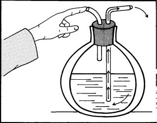
In the years from 1870 to 1900, the natural gases were liquefied and Dewar developed a vacuum flask to store cryogenic fluids. These advances permitted development of tissue-freezing techniques. In 1899, A. Campbell White of New York City described the use of liquid air to treat diverse types of skin lesions. Treatment was given by dipping a cotton swab into liquid air and applying the fluid quickly to the skin lesion, using repetitive application to freeze the entire lesion. White also suggested a wash bottle device that sprayed liquid air on skin lesions (Fig. 1).
In 1907, H. H. Whitehouse used freezing techniques for the treatment of a variety of disorders of the skin, including skin cancer. In the same year, Pusey reported similar varied uses of solidified carbon dioxide ( 78.5 8C), which was easier to handle and more easily obtained than the liquefied gases. For these reasons, carbonic snow remained in clinical use in succeeding years. In the 1930s, liquid oxygen ( 182.9 8C) had some use in the treatment of skin disease, but flammability and related safety considerations precluded general use.
In the 1930s and 1940s, the usefulness of solid carbon dioxide was increased by the development of new instru-
CRYOSURGERY 361
Figure 1. Dr. White’s ‘‘wash bottle’’ spray method of 1900, drawn as described in his article of that year. When the finger is placed over the air outlet, the pressure in the container produced by the boiling of liquid air in the container forces liquid air through the longer glass tube. The spray was then directed at the skin lesion.
ments, generally used to cool metal probes, sometimes with a mixture of solid carbon dioxide and acetone. These devices had little or no advantage over the use of the simple stick of solid carbon dioxide. Fluorinated hydrocarbons, commonly known as Freons, came into clinical use in the 1940s, but their freezing capacity was limited.
During these years, beginning in the 1940s with the extensive experiments of Temple Fay in Philadelphia with localized freezing of cancers by irrigation techniques, a number of reports dealt with the experimental production by local freezing of lesions in tissues, such as the liver or brain. In part, these experiments were made for purposes of physiological studies, but they required that an effort be made to develop instrumentation suitable for those purposes, and they showed the possibility of producing destruction of tissue by localized freezing, using diverse cryogenic agents.
In 1950, liquid nitrogen was introduced into dermatological practice by Allington, who described the use of cotton swabs dipped in this cryogen for the treatment of skin disease. As the availability of liquid nitrogen increased, cryosurgery gained in popularity. Nevertheless, cryosurgery remained a rather unimportant therapeutic modality because the freezing capability of cryogenic agents applied topically with cotton swabs was limited. Experimental studies showed that the depth of freezing with the swab techniques was 2 mm when liquid nitrogen was used as the cryogen, and carbon dioxide was even less effective. This depth of freezing was about the same as the thickness of normal skin, so the technique was suitable only for superficial lesions of the skin.
Cryosurgery as a therapeutic technique received a major stimulus through the development of cryosurgical apparatus by Cooper and Lee in 1961 (2). The apparatus used liquid nitrogen ( 196 8C) in a closed system, which permitted continuous and rapid extraction of heat from
362 CRYOSURGERY
tissues. The apparatus was originally designed to produce a cryogenic lesion in the brain for the treatment of Parkinsonism and other neuromuscular disorders. However, it was obvious that the apparatus had wider usefulness and, therefore, it was modified quickly and applied to the treatment of other types of diseases in diverse sites. In the following years, several types of cryosurgical apparatus using liquid nitrogen and other cryogenic agents were developed and found areas of usefulness in diverse benign and neoplastic conditions.
In the 1970s, some uses of cryosurgery were virtually abandoned, in part because of competition with other methods of local treatment, such as lasers and electrocoagulation. Other uses of cryosurgery became standard practice, especially in easily accessible areas, such as the skin and uterine cervix. In the 1980s, cryosurgical techniques for cardiac tachyarrhythmias were developed, but progress in other areas was slow.
In the 1990s, renewed interest in cryosurgery followed the development of intraoperative ultrasound, the improvement in cryosurgical apparatus, and the availability of percutaneous access techniques. The ultrasound image identified the site of the lesion, guided the placement of the probe into the lesion, and monitored the process of freezing. More types of cryosurgical apparatus were available, were well suited to percutaneous or endoscopic use, and permitted the simultaneous use of multiple probes. As a result, cryosurgical techniques have evolved into new applications, such as the treatment of visceral and other deep tumors. A recently written history of cryosurgery provides greater detail of its evolution, including the pertinent references (3).
EFFECT OF FREEZING ON TISSUE
All types of cells can be devitalized by freezing. The mechanisms of injury are related to crystallization of water, solute concentration in the cells, and irreversible changes in cell membranes. In the absence of cryoprotective agents, which are used when freezing is used to preserve cells, ice crystal formation produces damage that makes cell survival unlikely. Intracellular ice formation, which occurs when cells are frozen rapidly, is lethal. Extracellular ice formation, which occurs when cells are frozen slowly and water has sufficient time to leave the cell, is not considered as certainly destructive, but the cellular water loss does result in hypertonic damage to the cells. Under cryosurgical conditions, both mechanisms of injury are operative.
Damage from direct cellular injury is enhanced by the effect of freezing on the circulation. After freezing and thawing, the involved area becomes congested, and effective circulation through small vessels ceases within 30 min. With stagnation of the microcirculation, the hypoxic cells die and necrosis follows. These effects are well known from studies of frostbite, in which the relative importance of direct cellular injury and vascular stasis have long been debated. The full scope of the effects of freezing on tissue is described in recent reviews (4–6). Though direct cell injury is important, in cryosurgery, microcirculatory failure
clearly is a major factor in cell death: The loss of blood supply deprives all cells in frozen tissues of any possibility of survival. This results in a uniform necrosis of the tissue, except at the periphery of the previously frozen area. At the periphery of the cryogenic lesion, where the freezing temperature is not sufficiently cold to kill all of the cells, some cells survive and some cells linger between life and death for days and may die showing signs of apoptosis, that is, gene-regulated cell death (7,8).
The cryogenic lesion is characterized by sharply circumscribed necrosis. As thawing takes place, the previously frozen area becomes edematous and discolored due to congestion and perivascular hemorrhage. At the periphery of the dark red area, which closely corresponds to the margins of the previously frozen tissue, a narrow, bright red zone due to hyperemia appears. Further evidence of injury develops slowly, and the later tissue response depends on the severity of freezing. If only the tissue is subjected to mild superficial freezing, as might be done for benign disease of the skin, then the response will range from inflammatory reaction to superficial necrosis. The more extensive freezing required by large tumors is followed by greater destruction of tissue. Sharply demarcated necrosis becomes apparent in 2 days. The time required for slough of the necrotic tissue depends in part on its stroma. Cellular tissue sloughs quickly, but skin and other tissues with large quantities of fibrous stroma resist structural change and the necrotic tissue requires many days for separation. In the skin, the eschar requires two more weeks for separation, leaving a clean, granulating wound, which heals at a normal rate. The delay in healing that is characteristic of cryosurgical wounds is due to the time required for separation of the necrotic tissue. Healing is commonly favorable with rather little scarring. However, whenever full thickness of skin is lost, some scarring is inevitable. Hyperplastic scars are unusual. Depending on the severity of injury, the pigmentation of the treated area may be diminished or lost. Sometimes increased skin pigmentation at the periphery of the injured area is seen as a result of increased melanoblastic activity, but this is only temporary. In deep tissues, such as the viscera, the necrotic tissue is slowly absorbed over weeks or even longer, depending upon the volume of tissue frozen. Scarring is minimal.
Though tissues are devitalized by freezing, the matrix or structure of the tissue may be little changed, and this preservation of the framework is important in later repair. The resistance of the collagen fibers in the skin to damage by freezing is important to the reparative process (9). It is manifest in the favorable healing frequently seen in the treatment of skin disease and in the peripheral nerves after freezing. Though degeneration of axons and Schwann cells occurs, the perineurium is preserved, and this serves as a pathway for regrowth of axons, leading to eventual return of nerve function. Similar effects follow the freezing and repair of major blood vessels. Larger blood vessels, such as the aorta, femoral arteries, carotid artery, and portal vein, are devitalized by freezing in situ. With thawing, the previously frozen vessel is slightly dilated due to loss of tone, but the function as a blood conduit is unimpaired. The endothelium is lost, but the stroma of the vessel wall serves
as the matrix for repair, commonly with some intimal thickening (10).
The effect of freezing on bone is of particular pertinence. Bone devitalized in situ by freezing is slowly resorbed and simultaneously replaced with new bone, a lengthy healing process that may take many months, depending on the volume of bone, similar to that which occurs with autogenous bone grafts. During repair, the devitalized bone maintains form and continues function, though bones subjected to considerable stress (as the femur) are susceptible to fracture in the first month or two when bone resorption is maximal (11). This favorable reparative response has permitted extensive freezing of bone tumors in order to avoid excision.
APPARATUS
A wide variety of cryosurgical apparatus, using diverse cryogens, such as liquid nitrogen, nitrous oxide, argon, and carbon dioxide, is available. Various Freons, which were used for cryosurgery in past years, are no longer used because of environmental concerns. The types vary from electronically controlled automated apparatus with probe heaters to inexpensive hand-held devices that are little more than thermos bottles with controls for cryogen flow. The cooling is produced by change in the phase of the cryogen, that is, evaporation of a liquid or solid, or by expansion of compressed gas through a small orifice [Joule–Thomson (J–T) effect]. Thermoelectric cooling (Peltier effect), produced by passing direct current through dissimilar metal junctions (thermocouples), has not been useful in cryosurgery.
Currently, most apparatus use liquid nitrogen ( 195.8 8C), argon ( 185.9 8C), or nitrous oxide ( 89.5 8C). Carbon dioxide, which sublimes at 78.5 8C, though commonly used in past years, is little used in current times. Liquid nitrogen cools by change in phase, that is, changing from a liquid to a gas. Argon, nitrous oxide, and carbon dioxide are used as pressurized gases that cool by the J–T effect. The freezing capability of these cryogenic agents varies substantially, and this determines the choice of equipment for a particular disease. The cryogenic agents may be applied directly to the tissue, typically as a spray of liquid nitrogen, though nitrous oxide has been used in this way also. Freezing with liquid nitrogen applied via a cotton swab is another example of direct use. The cryogens may also be used in probes, which are a means of confining the cryogen in a closed system. At its tip, the metal probe has a heat-exchange surface that is applied to the tissue to be frozen. At this contact area, heat transfer to the probe results in tissue cooling. The freezing capacity varies with the size of the probe, the temperature of the probe, and the areas of the contact with the tissue. The heat removing capacity of probes varies from 10 to 100 W, depending on the features of probe construction and the cryogen that cools it.
The coldest cryogenic agent in clinical use is liquid nitrogen. It has the greatest freezing capability and is the best agent for destruction of large volumes of tissue, as is required in the treatment of cancer. Nevertheless, in
CRYOSURGERY 363
current practice, pressurized argon is commonly used for the treatment of neoplastic disease. The use of multiple probes simultaneously compensates for the somewhat lesser freezing capability of argon. The other cryogens are useful for less serious lesions, such as nonneoplastic or benign neoplastic disease, for which lesser degrees of freezing will suffice (Table 1).
Liquid nitrogen is a clear fluid that is odorless and nonflammable. Its boiling point is 195.8 8C at atmospheric pressure. The liquid will expand to 750 times its volume under normal atmospheric pressure. It must be stored in a double-walled, vacuum-insulated container with provision for pressure relief and liquid nitrogen withdrawal. The most popular containers for office or clinic use have a capacity of 15–35 L, which provides a holding time of 60–90 days and, depending on the rate of use, will require refilling every 4–6 weeks. Liquid nitrogen will evaporate at a rate of a few percentage points per day, depending on the quality of the container. Withdrawal devices, basically spigots, are used to transfer the liquid nitrogen from the storage container to the cryosurgical instrument.
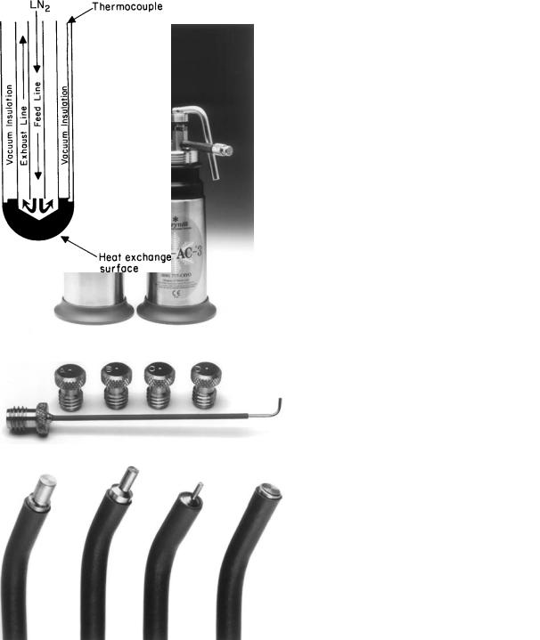
Hand-held cryosurgical apparatus, weighing 1 kg when empty, are commonly used, especially in dermatological practice (Fig. 2). They are basically small containers (thermos bottle construction) with storage capacities of 250–500 mL and with suitable on–off controls to initiate and control the spray of liquid nitrogen. Some devices allow pressure to build up in the container by means of a heat exchanger in the wall or top. Most have Luer lock fittings in the nozzle so that a variety of spray apertures, needles, or nozzles can be attached. These range in size from 24 to 15 gauge. The smaller aperture sizes are hindered by a tendency to become occluded by the development of frost in use, but this can be alleviated by bypass nozzle systems. Problems that must be solved in the construction of handheld devices include the design of a delivery tube that permits adequate heat exchange and prevents drip of liquid nitrogen from the delivery system nozzle. Most hand-held devices can be fitted with probes of diverse shapes and sizes, including those suitable for treatment of oral or gynecological diseases. The hand-held devices, relatively heavy when the container is filled with liquid nitrogen, are somewhat more difficult to use with a probe than with a spray because it is cumbersome to hold the weight steady in the hand while the probe is adherent to the tissue. Motion may cause fracture of the bond between probe and tissue, and ineffective freezing may result. With the spray technique, the small movement caused by the weight in the hand is not important because there is no direct bond to tissue.
The automated apparatus, cooled by liquid nitrogen, available from several companies, is almost always used with probes. The feed lines leading to the probes are insulated, usually by vacuum (Fig. 3). The control of the flow of liquid nitrogen is achieved by regulators. Most systems require pressurization, which is facilitated and speeded with an internal heating device. A heater in the probe tip speeds release from the tissue at the conclusion of freezing.
The automated apparatus first available in the early 1960s provided for control of the probe temperature in the
364 CRYOSURGERY
Figure 2. Modern handheld liquid nitrogen cryosurgical unit of the type commonly used in the treatment of skin diseases. The taller unit is 11 in. (280 mm) in height, has a capacity of 16 oz (500 mL), and weighs 30 oz (846 g) when filled. The shorter unit is 8.5 in. (215 mm) in height, has a capacity of 10 oz (300 mL), and weighs 24 oz (618 g) when filled. The devices may be fitted with a large selection of cryogen spray tips with apertures ranging from 0.4 to 1 mm (shown in Fig. 2b) and probes ranging in size from 1 mm to 4 mm in diameter at the tip. The probes allow precise control of freezing and may be used for cutaneous or mucosal lesions. (Photographs by courtesy of Brymill Cryogenic Systems, Ellington, CT 06029.)
Figure 3. Diagram of a typical probe cooled by liquid nitrogen. The liquid nitrogen passes from a reservoir in the console, down the vacuum-insulated feed line, to the heat exchange surface at the probe tip. The probe tip is cooled by a change in phase (liquid to gas), then the gas is returned to the console. Probes of many sizes and shapes are available. A thermocouple may be placed in the probe tip.
range of þ36 to 160 8C (2). The apparatus was soon modified to enable treatment of diverse tumors (12). In the 1990s, an advance in cryosurgical equipment technology featured the development of a technique to use liquid nitrogen super cooled to ca. 209 8C in new equipment. The super cooling was achieved by passing pressurized liquid nitrogen through a heat exchanger immersed in a liquid nitrogen chamber ( 209 8C) held under vacuum. The supercold liquid nitrogen was circulated to the probe, producing probe surface temperatures in the range of165 to 185 8C, depending on the diameter of the probe (13). Used with multiple probes, this apparatus had substantially greater tissue-freezing capability than the earlier technology. Nevertheless, this new apparatus did not improve to a significant extent on the probe cooling rate which was only 20 8C/min at 5 mm from the probe (14). The time required to reach a tissue temperature of 50 8C at a distance of 1 cm from the probe was 5–10 min (15). In comparison, the argon gas J–T apparatus cooled a probe much faster (16).
Recently, liquid nitrogen-cooled apparatus based on new technology equals the fast cooling rate of argon J-T apparatus and produces probe temperatures in the range of170 to 180 8C. This system uses a submersible nitrogen pump to generate the operating pressure to cool the probes with liquid nitrogen. Up to six vacuum-insulated probes may be cooled simultaneously (Cryo6. Erbe Co., Tubingen, Germany).
The cryosurgical apparatus that cool by the J–T effect are lightweight, portable, and quickly responsive in cooling or warming. Pressurized gas is passed through a small
nozzle and expands, cooling the probe. This type of apparatus, using diverse kinds of gases, has been available for many years, but the cryogens commonly used in the modern apparatus are argon and nitrous oxide.
Argon, a colorless, odorless gas that boils at 185 8C, has been used in J–T type apparatus since late in the 1960s. The gas in current devices is stored in steel cylinders that are pressurized at 3000 psi. In use, the probe temperature is ca. 130 8C at coldest. The cooling efficacy is pressure dependent. As the pressure in the cylinder falls, the cooling capacity diminishes. Argon permits the use of probes of very small diameter, as small as 17-gauge needles. In treatment, the use of such small probes requires that multiple probes be placed in the lesion. A larger probe, such as 3 mm in diameter, permits greater gas flow in the conduit, and will freeze a larger volume of tissue than the needle structures. Therefore, fewer probes may be used for the same volume of tissue. Since argon is a noble gas and is normally in the atmosphere, venting the gas from the apparatus into the operating area is not a safety concern.
Nitrous oxide ( 89.5 8C) is a colorless, nonflammable gas, commonly available in clinics and hospitals in the familiar ‘‘E’’ cylinders, which hold 2.72 kg (6 lb) of N2O at a pressure of 5.1 MPa (740 psig). The withdrawal of the nitrous oxide gas depletes the gas pressure in the cylinder, which affects the rate of freezing. The gas cylinder may be enclosed in a warming jacket to provide some heat in order to maintain gas pressure at an appropriate level. There is a safety consideration. Cryosurgical units using nitrous oxide that do not provide for venting of the exhaust to the outside air may expose personnel to some ill effects, such as impaired performance and cognition. Such units should be used only in well-ventilated rooms. Older devices, which may exhaust into the room 20–90 L of N2O/min, may be hazardous. Most new devices provide for gas scavenger systems to safely exhaust the nitrous oxide (Fig. 4).
In clinical use, the differences in cooling rate are important. Argon will cool a probe to 100 8C in 1 min, and to130 8C in 2 min. Nitrous oxide will cool a probe to ca.80 8C in the same time. In contrast, liquid nitrogen cools more slowly but will become colder, the probe temperature reaching 160 to 180 8C in 5 min, the final temperature depending on the engineering features of the apparatus (14– 16). However, the new type of liquid nitrogen apparatus with a design based on a reciprocating bellows pump will produce fast freezing of a probe, perhaps to 180 8C in < 1 min.
Sprays of cryogen can also be provided by nitrous oxide apparatus. If the fine droplets of liquid nitrous oxide are released on a surface, the droplets, instead of vaporizing to gas, recrystallize into solid nitrous oxide particles that lie on the surface or fly in all directions. To keep this partialpressure effect from occurring, the droplets must be surrounded by pure nitrous oxide gas, necessitating an inverted-cup shield around the applicator tip.
Carbon dioxide is a colorless gas that is used as a cryogen in solid and gaseous form. Solid carbon dioxide ( 75 8C) has been used for direct application to tissue for100 years. Carbon dioxide is also available as a compressed gas contained in E cylinders. In J–T apparatus, it provides probe temperatures of ca. 60 8C.
CRYOSURGERY 365
Figure 4. Modern cryosurgical device, cooled by nitrous oxide, used in cardiac cryosurgery for the treatment of atrial fibrillation and other arrhythmias. The console houses the primary gas supply in cylinders, the gas circuits, and the controlling electronics and software. The device cools the catheter probe by passing the pressurized gas through a restricting orifice at the probe tip. The probe is a steerable cryoablation catheter with leads to connect to the console for cryogenic gas flow, pressure monitoring at the catheter tip, and cardiac electrical signal recording (Fig. 3b). (Photographs by courtesy of CryoCor Inc., San Diego, CA 92121.)
366 CRYOSURGERY
Devices that cool by thermoelectric principles have limited freezing capability and can provide a probe tip temperature of ca. 20 to 30 8C at a low cooling rate. These devices have been satisfactory for ophthalmic use, but not for the treatment of tumors. The efficiency of these devices may be improved by combining with the technology of heat pipes, which then could provide probe temperatures of 50 to 60 8C (17). Nevertheless, the applicability of thermoelectric cooling to cryosurgery is limited.
TECHNIQUE
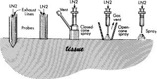
The two basic techniques for the use of cryosurgical apparatus are the direct application of the cryogen to the tissue, as in a spray of liquid nitrogen, or use of the cryogen in a closed system with probes. The choice of technique is in part a matter of personal preference and in part a matter of fitting the technique to the nature, size, and location of the disease. Probe techniques in general provide an easily controllable and a greater depth of freezing. The use of a probe is essential in freezing in less accessible areas of the body. Spray techniques, widely used in dermatological practice, permit easy application to accessible surfaces to achieve the superficial, and perhaps wide, freezing usually desired. Variants in these techniques blur the apparent sharp distinction between spray and probe techniques (Fig. 5).
Figure 5. Diagrammatic representation of differences and similarities in probe, closed-cone, open-cone, and spray techniques, commonly used in the treatment of skin diseases. The two probes shown on the left are closed systems in which the cryogen does not come into direct contact with the tissues. In the closed systems, the liquid nitrogen, after change of phase, is vented as a gas somewhere along the return line, usually in the console of the apparatus. In the open systems, the three devices on the right, the cryogen is sprayed directly on the tissues. Surface freezing techniques by pressing a probe against the tissue or by spraying cryogen are commonly used. However, the pointed probe shown on the left may be inserted into the tissue to achieve deeper freezing. This penetration of the tissue causes a wound, which may cause some problems. In the cone techniques, the dispersion of the spray is limited and the effect of confining the spray is to produce an open probe. In the cones, the heat-exchange surface is on the skin and the venting of the gas is at the side or top of the device. The open-spray technique is effective in treating wide areas with irregular outline, but care has to be taken to avoid dispersion of the spray to unwanted areas.
The Spray Technique
Spraying of the cryogen, usually liquid nitrogen, from the nozzle of an appropriate apparatus is an efficient method of producing rapid freezing of tissue. When used as a spray, the cryogen is used at its coldest temperature, and it produces superficial freezing rather quickly and easily over wide areas if required. The more superficial the lesion, the more suitable is the use of a spray, especially if the surface is irregular, as over a bony surface, or if the area of disease is extensive. The spray often is used intermittently, especially for small lesions. Steady spraying, as in an effort to freeze deeply in one site, causes the liquid nitrogen droplets to strike the tissue without vaporizing and run off the frozen tissues to freeze in undesired areas (unless spraylimiting devices are used), especially if excessive pressure is being used in the apparatus. Other methods of control are to reduce the pressure in the spraying device, if possible, or to use a smaller nozzle size, but reduced cryogen flow results in lessened capability of freezing the tissues. Problems with the dispersion of the spray are best corrected by the use of spray-confining devices such as hollow cones placed over the lesion so that the liquid nitrogen may be sprayed into the hollow device (Fig. 6). The use of the cone devices also has the effect of creating an open probe and is a useful technique of improving depth penetration of spray techniques, as required for the treatment of invasive skin cancers. Similar devices, such as funnels and hollow cylinders, have been used to confine liquid nitrogen as it is poured over bone tumors after removal by curettement.
The Probe Technique
The cryogen is circulated in a closed system, using metal probes for contact with the tissue to provide a heat sink or heat-transfer surface. Various sizes and shapes of probes, ranging from rod-like probes 1 cm in diameter to 18 gauge needle size and catheter shapes, to fit diverse anatomical sites and disease dimensions are available. The probes may have flat surfaces for contact with the tissue or may be pointed to allow insertion into the tissue. Slip-on metal end pieces, which fit over the end of the probe to modify the freezing surface, increase the versatility but also slow the freezing capability. The lines that feed the liquid nitrogen to the probe tip are vacuum insulated or insulated with appropriate materials, which increases the efficiency of the cryogen and provides for the safety of the user. The J–T types of apparatus have thin cryogen feed conduits that require no insulation.
In use, the physician selects one or more probes as appropriate to the disease. The manner of use is to apply the freezing surface of the cryoprobe to the lesion and allow the cryogen to flow. The cold probe acts as a heat sink and produces tissue freezing by removing heat from the tissue faster than blood supply and conduction restores it. Surface contact freezing is performed by pressing the freezing surface of the probe firmly on the tissue in order to ensure a good contact for heat exchange. This contact is improved by the use of water-soluble hospital lubricating jelly between the probe and the tissue. Greater depth of freezing may be achieved by increased pressure on the probe or by penetration of a sharp, pointed probe into the tumor. The
CRYOSURGERY 367
Figure 6. Canine liver being frozen with a large probe, 1 cm in diameter cooled with liquid nitrogen to 160 8C. The white frosted area is the tissue frozen after 3 min of contact. The depth of freezing is about the same as the lateral spread of frost from the side of the probe. With proper technique, the area of necrosis will closely approximate the visibly frozen area. (Reprinted with permission from J. Am. Med Assoc., 204, 566, 1968.)
penetration technique has the disadvantage that a wound is produced, and this may later bleed. Care must be taken to avoid motion of the probe during freezing because this might fracture the bond with the tissue and interfere with heat exchange. Heat exchange also depends on the gradient of temperature between the tissue and the probe, so the probe is always used as cold as possible when tissue destruction is sought. The larger the gradient, the faster the rate of freezing.
Freeze–Thaw Cycles
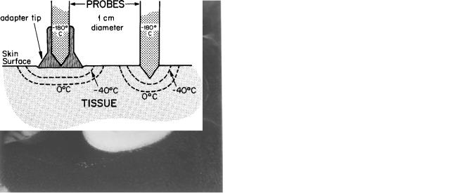
As freezing progresses, the tissue turns white (frosted in appearance) and hard, a change that begins at the area of contact with the cryogen and extends to incorporate an increased volume of tissue as time passes (Fig. 6). As treatment continues, in the case of lesions in easily accessible areas, such as the skin, the extent of freezing is judged by inspection and palpation. Estimation of depth of freezing may be difficult, but physicians experienced in the techniques can make reasonably accurate estimates of depth. With surface contact freezing by probes, the shape of the frozen volume of tissue is roughly hemispheric, so the depth of freezing can be judged to be about the same as the lateral spread of freezing from the probe. Significant modification in the shape of the frozen area may be achieved by the selection of different probe shapes (Fig. 7). The shape of the frozen area is also modified by the amount of pressure placed on the probe because increased pressure compresses the tissue and increases the depth of freezing. Another factor influencing the shape of the frozen area is the presence of major blood vessels, which provide a source of heat. In freezing with the spray techniques, a more superficial freezing may be expected and often is desired.
Figure 7. Diagram showing the effect of probe configuration on the shape of the frozen tissue. The probe on the right held with some pressure on the tissue causes indentation of the tissue, or minimal penetration, and produces a roughly hemispheric frozen zone. The same probe, fitted with a freezing tip adapter to form a wide, flat freezing surface, produces a wider but less deep frozen area. In each frozen zone, the 40 8C isotherm is shown to delineate its approximate location. The location of this isotherm varies slightly with the rate of cooling the tissue. Rapid cooling moves the isotherm slightly more toward the periphery.
However, if the spray is confined with a cone, the effect on the tissue is similar to probe freezing.
Freezing with the probe or spray continues until the desired volume of tissue, including a margin of apparently normal tissue, is treated or until the frozen area no longer enlarges. This is easy to observe in accessible tissues, such as the skin. In the use of a spray of liquid nitrogen, the nozzle of the device is moved about to distribute the cryogen evenly over the target, and this freedom of movement allows wide areas to be treated. On the other hand, probe freezing takes place from a selected site of application, and the rate of expansion of the frozen area slows as an equilibrium is established between the heat loss from the tissue via the probe, and heat brought to the area by the circulation of blood. For this reason, it is difficult to freeze tissue to a distance > 2–3 cm from the probe. This means that large lesions cannot be frozen completely in a single application of the probe. In these circumstances, the plan of treatment must include freezing from multiple sites with successive applications of the probe, which improves the width of the frozen area and also increases the depth of freezing in overlapped frozen areas to a slight extent. For large cancers, the simultaneous use of multiple probes is advantageous and time saving.
When used for the destruction of tissue, as for the treatment of tumors, cryosurgery must be performed in a manner that produces a predictable area of necrosis. The techniques to achieve this goal stress the rapid freezing of the tissue, slow thawing without assistance from warming devices, and immediate repetition of the freezing process in order to maximize destruction. Some modifications of these basic factors in technique are necessary for the diverse diseases in different parts of the body, especially if the intent is to destroy some cells while preserving others, but, in general, the cited basic technique provides the basis of effective therapy.
368 CRYOSURGERY
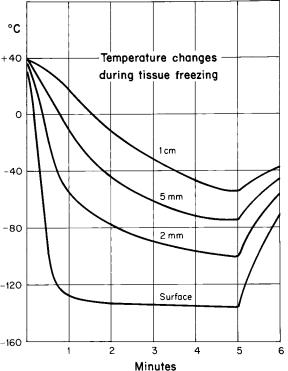
Rapid freezing promotes the formation of intracellular ice crystals, which are considered almost certainly lethal for cells. This occurs only in the tissue close to the freezing probe or in contact with the spray of cryogen. The rate of freezing of the tissue varies inversely with the distance from the site of application of the cryogen (Fig. 8). Close to the probe, temperature changes of the order of > 10 8C/min may be achieved, and this is considered rapid freezing. However, 2 cm from the probe, the cooling rate is slow, perhaps of the order of 2–5 8C/min. The possibility of tissue survival is enhanced with slow freezing rates, but fortunately even slow freezing rates have lethal potential because of cellular dehydration and related deleterious effects.
The thawing rate should be slow and unassisted because this increases the time that the tissues spend in a phase when recrystallization phenomena can add to the cell injury. Tissue often thaws in about the same time as was required for freezing, but this depends on the volume of frozen tissue. Large volumes of frozen tissue warm slowly. In some therapeutic applications, a probe heater is used to speed release from the tissues after freezing. This practice also slightly speeds thawing of the tissues, but this probably does not increase the chance of cell survival if the
Figure 8. Graph showing temperature changes recorded from thermocouples inserted in the tissue at diverse distances from a probe cooled by liquid nitrogen. The tracing identified as ‘‘surface’’ is from a thermocouple placed in the contact area between the probe surface and the tissue. The other tracings show the temperature changes at 2, 5, and 7 mm from the edge of the probe. The most rapid and deepest cooling of the tissue is at the point of contact. The greater the distance from the probe, the slower the cooling, the less the depth of freezing, and the shorter the duration of freezing.
technique is otherwise correct. Certainly, no warming solutions should be used to thaw the tissue, because it is well established that quick thawing of tissues reduces injury from freezing, whether in cryopreservation, cryosurgery, or frostbite.
Repetition of freezing maximizes damage by subjecting the tissues again to the same mechanism of thermal injury. To take full advantage of the lethal effect of repetitive freezing, it is necessary that the frozen tissues be completely thawed (> 0 8C) before the next freezing cycle because then the entire volume of the tissue will pass through recrystallization phases with their attendant injurious effects. The second freezing cycle is usually faster and more effective in cooling the tissue than the first cycle and achieves a slightly greater depth of freezing because the latent heat in the previously frozen tissue is reduced and because the microcirculation has begun to fail in the interval between freezings, reducing the heat supply. The lethal isotherm is then deeper in the frozen tissue. For this reason, it is advisable to wait a few minutes between freezing–thawing cycles.
The freezing of tissue in cryosurgery is a heat-transfer process, and the mechanics of extraction of heat from the tissue directly influence cryosurgical technique. The duration of each freezing–thawing cycle of treatment is dependent on factors that affect the rapidity of tissue freezing, such as efficiency of heat exchange, gradient of temperature, blood supply to the area, attainment of a desired temperature goal, and on the size of the lesion. The temperature of the cryogen is also important: the larger the gradient between cryogen and tissue, the faster the cooling. It is also a function of the contact area between cryogen and tissue: a wide area for heat exchange cools tissues faster.
Thermal modeling techniques have been used to predict the size of the frozen area that may be produced by a probe. The blood flow, the probe temperature, the area of contact with the tissue, and the duration of application are known. This work has contributed to the understanding of the functions and cooling capacity of cryosurgical equipment, has provided a method of estimating the effect on the tissues, and has shown direction in equipment design and in the planning of treatment. The modeling techniques have been useful in the several aspects of cryobiology, including the mechanisms of injury to cells. Nevertheless, before clinical use, it is necessary for the physician to practice with the cryoprobes or with a spray of cryogen in test materials in order to develop the ability to predict the size and shape of the frozen field as a function of time, temperature gradient, and heat exchange surfaces. The heat diffusion equations that must consider the frozen area and a moving solid–liquid interface as freezing progresses are complex and are best reviewed in source material (5,18).
MONITORING TECHNIQUE
Many cryosurgical procedures, especially those for benign diseases and small cancers of the skin, are performed using only observation and palpation of the frozen tissue to determine the progress of treatment and judge its adequacy. In
easily accessible sites, those physicians with considerable experience in cryosurgery can achieve satisfactory results without the use of monitoring techniques to guide therapy. However, the temperature of frozen tissue cannot be determined by its appearance: Frosted tissue looks the same at any freezing temperature. Equally important, the depth of freezing is difficult to judge in many situations, although the relationship between the depth of freezing and the lateral spread of freezing from a probe is an important clinical aid. The clinical evaluation, if not perfect, may produce an error in treatment that may be of critical importance in the treatment of malignant disease. Effective cryosurgery must yield predictable and certain necrosis.
To guide cryosurgical procedures and permit reasonable certainty of the death of tissues, methods of monitoring the freezing process have evolved. These methods include (1) the measurement of tissue temperature by thermocouples; (2) the measurement of electrical impedance on resistance in tissue; (3) the measurement of heat lost from the tissue by a heat flowmeter; (4) thermography and; (5) the imaging techniques, which are ultrasound, computerized tomography (CT); and magnetic resonance (MR).
Thermocouples
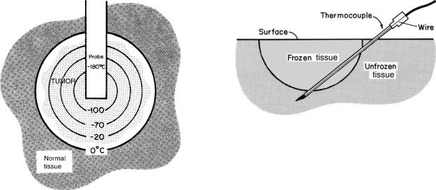
The most commonly used method of monitoring is by the insertion of needle-mounted thermocouples into the tissue at appropriate sites to measure tissue temperature (Fig. 9). Thermocouples are formed by the junction of two dissimilar conductors, commonly iron and constantan, or copper and constantan, in a closed electric circuit (Fig. 10). When the junction of the conductors is held at a temperature, an electromotive force (emf) proportional to the temperature difference will be generated. An instrument, such as a potentiometer or pyrometer, is used to measure this emf and provide a readout in terms of temperature.
Thermocouples perform several important functions in cryosurgical treatment. The insertion of thermocouples into the tissue in appropriate locations monitors the progress of the freezing treatment and ensures that temperatures destructive for tissues are attained. Thermocouples can also be used to ensure that tissues adjacent to a diseased area are not frozen and are hence safe from freezing injury. If used with a recorder, thermocouple measurements on the tracing provide written evidence of treatment. Thermocouples often are built into the probe tips, so that the temperature of the probe can be monitored. Though this provides useful information, confirming that the apparatus is working, probe temperature tells nothing about tissue temperature, except by estimations based on experience with the performance of the probe.
In cryosurgical treatment, especially for cancer, it is important to know that destructive temperatures are produced in the tissues. In the freezing–thawing cycles used in cryosurgery, tissue destruction is a multifaceted process involving the freezing rate, the tissue temperature, the duration of freezing, the thawing rate, and repetition of the freezing–thawing cycle. Separation of the relative destructive effects of these components is difficult, but the easiest to control and measure is the coldest temperature attained
CRYOSURGERY 369
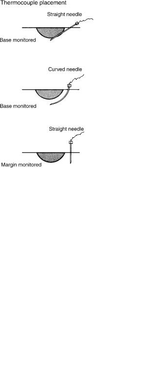
Figure 9. Diagram showing typical sites of thermocouple insertion. Three useful methods are shown. At the top is a thermocouple inserted at an angle from the side of the lesion through normal tissue. The thermocouple tip rests directly beneath the lesion and shows the temperature at the depth of the tissue. It probably is the mostcommonmethod of thermocouple usage. In thecenter isshowna curved needle used for base monitoring. This method avoids passing the shaft of thethermocouplethroughthefrozen tissue. Atthebottom is shown an alternative method of thermocouple use. The thermocouple is inserted at the border of the lesion. With this technique, the temperature registered at this thermocouple is interpreted as being the same temperature that would be measured at the border beneath the tumor. This method assumes that the depth of freezing is approximately the same as the lateral spread of freezing from the probe. The advantage of this technique (and the use of a curved thermocouple) is that the thermocouple shaft remains outside of the frozen area until the advancing ice front incorporates the tip. More than one thermocouple may be used.
Figure 10. Diagram of a basic thermocouple-millivoltmeter circuit consisting of a copper and a constantan wire welded together to form a measuring junction, the thermocouple. The free ends are connected to the millivoltmeter, which measures the emf associated with the temperature at the thermocouple. In the upper right of the illustration is shown the mounting of the thermocouple in a hypodermicneedle, which can beinsertedinthetissueatappropriate sites to measure temperature changes during freezing–thawing cycles.
370 CRYOSURGERY
Figure 11. Drawing demonstrating the gradients in temperature that exist in tissue being frozen with a probe at 180 8C. The incorporation of a tumor in a frozen mass is depicted. The gradients are substantial, ranging from 0 8C at the edge of the frozen volume to nearly 180 8C adjacent to the probe. Cell Death would not occur in the entire frozen area. If treatment were to cease at this stage, the tissue in the 0 to 20 8C range would probably survive and the tumor would grown again.
in the cryosurgical treatment. Hence, a lethal temperature goal is used in cryosurgery.
Early in cryosurgical experience, it was thought that15 or 20 8C was a proper lethal temperature goal. Substantial tissue damage results from a tissue temperature of20 8C, but this temperature is not safe for the treatment of malignant disease. In nonneoplastic disease, usually conservative freezing is wise and, therefore, tissue temperatures of the order of 20 to 30 8C are satisfactory. Temperatures of 40 8C are satisfactory for superficial skin cancers. The treatment of more aggressive cancers, as in the oral cavity, requires repetitive freezing to tissue temperatures of 50 8C or colder if cure is to be achieved (Fig. 11).
The accuracy of thermocouples is a matter of interest since treatment depends in part on this measurement. Some minor error is inherent in the temperature recording system, which consists of the thermocouple, the electrical leads, and the readout device. The readout device is commonly a potentiometer with a digital readout, which is sufficiently accurate for the purpose of cryosurgery. An important source of error is from conductance of heat along the thermocouple needle shaft. If the needle shaft passes through a frozen area, the reading from the tip may be falsely low (Fig. 12). Under other conditions, heat may be added to the temperature-measuring system from extraneous sources anywhere between the thermocouple needle tip and the recorder. However, with proper use, the error due to heat conductance is minimal and does not interfere with the thermocouple function of supplementing clinical judgment. Based on experimental data, temperatures in the range of 20 to 50 8C, produced in short freezing cycles, may be associated with an error of 2 8C due to heat
Figure 12. Diagram illustrating a mechanism for thermocouple error due to heat conduction. The thermocouple needle enters the tissue at an angle, just as commonly done in clinical practice. During freezing, the expanding ice front incorporates the needle shaft. The cooling of the shaft affects the thermocouple at the needle tip, and this may produce a falsely low reading. Also, the hub of the needle and the adjacent needle shaft in unfrozen tissue may be warmed by the tissue or the air, which may affect the reading in the opposite way. The magnitude of this possible error is a source of concern, but proper thermocouple use can avoid the error.
conductance (19). This differential is of little importance in cryosurgical techniques. The error due to heat conductance in thermocouples is less significant than the error produced by the positioning of the thermocouples in the tissue. A 1-mm variation in thermocouple placement in the tissue represents 10–15 8C difference in the temperature recorded in usual cryosurgical freeze–thaw cycles. Therefore, the important errors in thermocouple use are produced by the accuracy of placement rather than by heat conduction from extraneous sources.
Impedance–Resistance Measurements
Another method of quantification of freezing injury is a technique that measures the impedance or resistance changes to the passage of a small electric current through the tissues being frozen. Unfrozen tissue is a conductor of electricity because of the electrolyte content of the tissue fluid. During freezing, the formation of ice crystals in tissue and the removal of water from the tissue results in decreased electrical conductivity. When practically all of the extracellular water is crystallized, electrical impedance or resistance rises to the high levels. This change is interpreted as being associated with tissue death.
The techniques of impedance–resistance measurements require the insertion of needle electrodes in or about the tumor (Fig. 13). These conduct the small electric current from the line or battery-powered device to the tissue. The measurement is made between two electrodes. Both electrodes may be placed in the treated area, but it is preferable to measure between one electrode in the treated area and a distant reference electrode. The initial impedance–resistance in unfrozen tissue is of the order of 1–2 kV. When the entire measuring electrode is incorporated in frozen tissue, the impedance–resistance rises to megohm levels. This change seems to occur quickly, at least in comparison to the changes in temperature (Fig. 14).
CRYOSURGERY 371
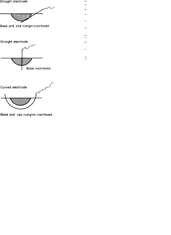
Figure 13. Diagram of electrical impedance–resistance monitoring system showing alternative methods of placement of the electrode. The entire electrode must be incorporated in the frozen tissue before the change in impedance–resistance rises to the megaohm level. Multiple electrodes may be used to increase the monitoring sites. The second electrode, which is placed in a more remote site to complete the electrical circuit, is not shown.
Impedance measurements were introduced into cryosurgical techniques by Le Pivert et al. (20), who measured electrical impedance in the tissue to the passage of a low frequency (1000 Hz) alternating current between electrodes. Impedance values of 0.5–1.0 MV were equated to40 8C and were associated with tissue destruction. This destructive effect on the tissue was reported to be independent of repetition of the freezing–thawing cycles.
Other investigators, using Le Pivert’s instrument in experiments on canine skin, found that an impedance of 1 MV corresponded to a tissue temperature of ca. 30 8C, but the range of temperatures about each impedance value was sufficiently great to cause concern about the possibility of tissue survival at the 1 MV value. Using a line powered 1000 Hz low current impedance meter, the relationship between tissue impedance and temperature and the subsequent development of tissue necrosis was investigated. An impedance of 10 MV was not always associated with tissue death, and the range of temperatures about any impedance value was considerable (Fig. 15). Comparison of the electrical characteristics of the tissue and the tissue temperature with the border of necrosis that was obvious in a few days showed that the tissue temperature was the more accurate predictor (21).
Figure 14. Graph illustrating simultaneous temperature and impedance changes. The temperature in the tissue was cooled to 22 8C at which time the cryosurgical apparatus was turned off. The tissue cooled further to 25 8C because the cold probe continued to function as a heat sink. Then the warming cycle began. The impedance rose steadily after the tissue cooled to 8 8C. When the impedance reached 10 MV 40 s later, the cryosurgical apparatus was turned off. The impedance continued to rise and the trace passed off the chart, returning 1 min later in the warming cycle. The temperature and impedance changes in thawing were more gradual in thawing than during freezing.
At this time, devices to measure tissue impedance– resistance during cryosurgical procedures are little used. The incorporation of an impedance electrode into the freezing surface of an endoscopic probe confirms that the probe is cooling properly, but yields no information about tissue temperature (22). Nevertheless, such a device would confirm fixation of the probe to the tissue. An impedancemeasuring device could be useful in the determination of depth of freezing. The use of multiple electrodes about the circumference of a tumor to monitor cryosurgery is a use that requires further testing.
Whatever method of quantification of cryogenic injury is used to supplement clinical judgment in cryosurgery, whether thermocouples or electrodes are inserted in the
Figure 15. Graph showing the relationship between impedance and temperature. The data for the graph are taken from mean values during the cooling cycle. The standard error of the mean is shown.
372 CRYOSURGERY
tissues, it is important to recognize that their placement in itself may be a source of error. It is equally difficult to determine the relationship of the electrode or thermocouple to the margin of the disease. Both thermocouple and impedance devices should be viewed as adjunctive to clinical judgment in therapy.
Heat Flowmeter
The heat lost from the tissue can be measured by means of a heat flowmeter (23). This is done by attaching a differential thermocouple to the probe tip. As heat passes from the tissue into the probe, the temperature electromotive force, proportional to the temperature differential, develops across the disk. This may be recorded so that total heat exchange is used as the monitor of the cryosurgical procedure. Though the technique was used in the treatment of patients, it has not proven useful generally.
Thermography
The use of thermography to monitor the progress of freezing tissue is best considered as a research method. The thermograms, recorded in monochrome or in color, define isotherms down to 40 8C. Bradley’s experiments with thermography, comparing the freezing characteristics of different freezing techniques, have shown the faster freezing capability of the spray devices in comparison with the probe techniques and have confirmed the importance of pressure on the tissue from the probe in modifying the shape of the frozen area. Though in vitro tissue testing permits evaluation of depth of freezing, clinical use provides only surface freezing evaluation in most circumstances (24,25).
Ultrasound
Ultrasound used during cryosurgery provides a real-time image of the frozen volume of tissue. Since ultrasound provides a more global view of the frozen tissue than do thermosensors, this imaging technique has come into wide use for monitoring the freeze–thaw cycle, especially for visceral tumors. Ultrasonography offers the possibility of matching the extent of the neoplasm with the volume of tissue frozen in treatment. Frozen tissue is hypoechoic, so the ultrasonic image is black. The edge of the frozen tissue is hyperechoic and appears as a bright line. As freezing continues and the volume of frozen tissue increases, the hyperechoic rim moves away from the probe, leaving the hypoechoic zone behind it. Therefore the process of tissue freezing can be observed during the cryosurgical procedure. Experience with ultrasound monitoring in hepatic and prostatic cryosurgery is now substantial. The correlation between the ultrasound image and the actual diameter of the frozen tissue is excellent, though it has become evident that ultrasound overestimates the volume of tissue frozen. Much of the frozen volume is obscured by distortions and reflections of the image. Sonography does not provide an image beyond the near edge of the ice because of complete posterior acoustic shadowing (26,27). Some compensation for this limitation can be obtained by viewing the
frozen volume from another angle. Three-dimensional (3D) ultrasound is in development (28).
The correlation between tissue temperature and the ultrasound image is of considerable importance because effective treatment depends on achieving an appropriate tissue temperature goal. The temperature of frozen tissue cannot be determined from its ultrasonic image. However, the hyperechoic rim of the image is 0 8C and the probe temperature is generally known, so inferences about the steep gradients of temperature in the tissue can be made. For example, using a probe at 160 8C on tissue for 5 min, the 20 8C isotherm is 70% of the distance from the probe to the periphery of the frozen tissue. The 40 8C isotherm, which is commonly used as a goal in tumor therapy, is60% of the distance from the probe to the frozen boundary. A rapid cooling rate moves the isotherm toward the periphery.
Computerized Tomography and Magnetic Resonance
Other investigational methods of monitoring are radiological. The absorption of X rays in biological tissue is proportional to the tissue density. Water, which is the major component of most biological tissues, changes density upon freezing and can be visualized. Computerized tomography (CT) can show the entire cross-section of the lesion. The development of the frozen volume of tissue can be seen on a series of CT images taken at frequent intervals. Magnetic resonance (MR) will provide a 3D image of the frozen volume of tissue. When used with thermal models, the temperature changes in the tissue can be quantified (29,30). Magnetic resonance requires the use of MR-compatible probes, which have been developed and used to a limited extent. Electrical impedance tomography (EIT) has been proposed as a method to provide real time imaging and provides a global image by introducing low amplitude alternating current (ac) into the body and thereby measurs the electrical potentials on the surface of the body. These potentials are analyzed to create a tomographic image (31). At present, the high cost of apparatus and logistical considerations will limit the usefulness of these imaging techniques in cryosurgery. However, it seems likely that image guidance will achieve greater applicability as monitoring techniques during the freezing process.
To summarize, clinical judgment, the prime factor in the control of the freezing of tissue, requires assistance from the monitoring techniques. Depending on the clinical circumstances, including the nature and the site of the disease, either tissue temperature measurement by thermosensors or ultrasound imaging is commonly used. The limitations of these techniques in providing thermal information during cryosurgery are well defined. To compensate for the limitations, wherever practical, the use of both thermosensors and imaging techniques are advised.
CLINICAL USES
Cryosurgery has established a firm place in medical practice for the treatment of diverse diseases in different parts of the body. Lesions in accessible sites, such as the skin or
oral cavity, may be treated with little need for anesthesia and without risk of hemorrhage. Treatment for many conditions can be given under local anesthesia in an office or outpatient clinic, avoiding the need for hospitalization, with its attendant inconvenience, cost, and risk. Cryosurgical treatment of viscera and other deep-seated disease is more difficult because of accessibility, but these techniques have become well developed in the past 15 years, benefiting from percutaneous technology and ultrasound imaging. Cryosurgical techniques deserve emphasis as an excellent choice of therapy on high surgical risk patients, especially those who have problems difficult to manage by other methods of treatment.
Skin Diseases
Cryosurgery is widely used in the treatment of skin diseases. Many non-neoplastic lesions, and a variety of benign tumors and cancers of the skin are successfully treated commonly with a hand-held device containing liquid nitrogen. Non-neoplastic skin lesions are easily treated by cryosurgery with little risk, but skin cancers require further comment.
Cryosurgery has become a standard technique of treatment for skin cancer in any location, and joins a variety of treatment methods, including surgical excision or radiotherapy as well as other special techniques. Most skin cancers are small, commonly not > 3 mm in depth and < 1 cm in surface diameter. These are easily treated by cryosurgery, and a cure rate in excess of 97% has been achieved. Certain types of skin cancer, called sclerosing or morphea type of basal cell carcinomas and tumors of the scalp, are difficult to cure by cryosurgery, perhaps because of difficulty in determining the extent of disease, or because of biologic behavior, or because of the richness of blood supply. These require aggressive freezing therapy in order to achieve a cure. In general, however, the histologic type of the cancer is not important, and either squamous cell or basal cell cancers can be treated. Melanomas are also easily destroyed by freezing, but most primary melanomas, with the possible exception of lentigo maligna, are better treated by excision in order to establish the diagnosis and to permit staging of the extent of the disease. Cryosurgery is well chosen for certain skin cancers in special situations. These include the following:
1.Multiple superficial small skin cancers, as commonly seen from excessive exposure to sunlight. A large number of cancers can be treated quickly and easily in comparison to multiple surgical excisions.
2.Cancers about the ears, nose, and eyes. The irregular contours of the head in these sites and the tightness of the skin over the underlying bone make treatment difficult at times. In these locations, cryosurgery offers ease of treatment and improved cosmetic results.
3.Cancers arising in irradiated skin. Such cancers require conservative management because new cancers will develop elsewhere in the damaged skin.
4.Cancers that persist after radiotherapy excision or other methods of treatment. These are often advanced cancers that present problems in management.
CRYOSURGERY 373
In these special situations, the results of cryosurgery compare favorably with surgical excision or radiation therapy.
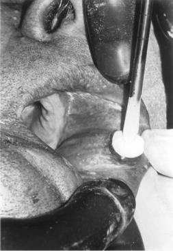
Oral Diseases
Benign and malignant tumors and precancerous conditions of the oral cavity are well suited to treatment by cryosurgery (Fig. 16). An important use is for the treatment of leukoplakia, which is the general descriptive term for white patches in the mouth and includes a number of different pathological conditions. Some of these, such as epithelial dysplasia and papillary epithelial hyperplasia, are precancerous. A biopsy, preliminary to definitive treatment, is necessary to differentiate between those lesions that are relatively innocuous, those that are precancerous, and those that are carcinoma in situ. All may be treated by cryosurgery. Such disease is superficial, and either nitrous oxide or liquid nitrogen apparatus may be used because the tissue needs to be frozen only to a depth of3 mm. The results are excellent, and most patients will remain free of disease. Persistent disease or recurrent disease usually results from failure to correct the etiologic factors, that is, continuation of the use of alcohol and tobacco or continued irritation of the oral mucosa for other reasons. In dysplastic disease or carcinoma in situ, 10% of patients will require additional treatment over a 2-year period.
Oral Cancer
Malignant tumors of the oral cavity are aggressive cancers that are difficult to cure by conventional treatment, that is,
Figure 16. Photograph showing the treatment of a benign blood vessel tumor (hemangioma) by freezing in situ. A cryoprobe, 5 mm in diameter, is applied. The frozen zone is extending over the lesion. Freezing will continue until the entire tumor is encompassed. No thermocouple monitoring is necessary with this benign lesion.
374 CRYOSURGERY
surgical excision and/or irradiation. In general, the 5-year survival rate is only 30% of all patients who acquire the disease. Cryosurgery can be used in special circumstances, especially for high surgical risk patients whose extensive associated disease would make general anesthesia and extensive operation prohibitive in risk. Cryosurgical techniques are also suitable when the cancer is on or adjacent to bone because the underlying bone limits the depth of penetration of a tumor and facilitates its destruction by freezing. In selected patients, the survival rate achieved by cryosurgical treatment appears comparable to that provided by surgical excision. However, the result can be achieved at a lessened cost to the patient in terms of operative mortality and postoperative functional disability. Nevertheless, cryosurgery is seldom used for oral cancer in current surgical practice.
The further away from direct vision, as is possible in the oral cavity, the more difficult is the application of cryosurgery. Cryosurgery has been used for cancers in the pharynx, larynx, bronchi, and esophagus with treatment given via endoscopy. Vision is limited, so accurate and extensive freezing is difficult. In the pharynx and larynx, occasional successes in attempts at cure have been achieved, but current experience provides little reason to choose cryosurgery in preference to radiotherapy or excision. Experience in the trachea and esophagus is limited to a few patients treated for palliation of symptoms. Obstruction can be relieved and tumor size kept under control by cryosurgery repeated every few months, but invasive growth continues. The presence of necrotic tissue in the larynx is a threat to the airway. In general, palliation of incurable cancer is provided better by radiotherapy or chemotherapy.
Bronchial Tumors
Considerable experience has matured in recent years with the cryosurgical treatment of bronchial tumors, including cancers, which produce symptoms by obstruction of the air passages. The treatment is via endoscopy, freezing the tumor with nitrous oxide-cooled probes, which then opens the airway and relieves the symptoms. With the removal of the obstructing tumor, irradiation of the remaining tumor may be used more safely (32).
Nose and Throat Diseases
Cryosurgery has been used for a wide variety of diseases of the nose and throat, including mucosal dysplasia, tonsillitis, nasal polyps, and rhinitis. However, generally other methods of treatment are chosen for these conditions. Special probes and techniques are necessary, and commonly the treatment can be done under local anesthesia. In chronic nasal airway obstruction due to hypertrophy of the nasal turbinates, freezing of the excess nasal tissue is followed by slough of the hypertrophic tissues, which improves the nasal airway and decreases the secretions. The treatment should be conservative so that normal tissue is not unnecessarily frozen. Healing occurs over a 2- or 3-week period and results in a reduced amount of turbinate tissue with a normal appearing mucosa and relief of symptoms.
Cryosurgery may be used for tonsillectomy. Cryosurgery is well chosen for adult patients who have blood dyscrasias or who are high surgical risks, because cryosurgery may be performed with little or no blood loss and low risk of postoperative bleeding. Nitrous oxide or liquid nitrogen apparatus may be used. The chance that tonsillar remnants may remain after freezing, which may lead to persistent symptoms, means that careful attention must be given to technique to ensure that all tonsillar tissue is frozen. Repetitive treatments may be used to eliminate tonsillar remnants.
Gynecological Diseases
Cryosurgical treatment has become common in gynecological practice in recent years. A wide variety of inflammatory and neoplastic diseases of the vulva, vagina, and cervix may be treated by freezing in situ. Since tissue diagnosis is important, it must be recognized that advances in colposcopy have improved the diagnostic ability of the physician to differentiate between inflammatory and neoplastic diseases.
Chronic cervicitis, which is inflammatory disease of the uterine cervix, is a principal use for cryosurgery. Careful evaluation of the extent of the disease must exclude the possibility of invasive carcinoma. Cryosurgery is effective treatment for dysplastic disease and carcinoma in situ. The differentiation between inflammatory disease and premalignant or malignant disease must be made by pap smear or biopsy before treatment is begun. The results of therapy are excellent, but if the disease was carcinoma in situ, persistent disease must be expected in 9% of patients.
Cryosurgical techniques are seldom used in the treatment of invasive malignant disease of the vulva, vagina, and cervix. Though it has been used for cancer in some potentially curable patients who are not candidates for one reason or another for conventional therapy, sometimes with excellent results, too few patients have been treated to permit evaluation of its ability to cure cancer in these sites. More commonly, cryosurgery has been used to relieve pain due to cancer after other methods of treatment have failed. Under these conditions, cryosurgery will diminish the size of the cancer, reduce malodorous discharges, control bleeding, and ameliorate pain.
Proctological Diseases
Cryosurgery has been used to treat a variety of proctological disorders, ranging from relatively minor conditions such as anal fissures and hemorrhoids to large, incurable cancers. However, anal fissures and hemorrhoids may be treated successfully by a variety of different surgical methods. For example, injection of sclerosing agents into small to moderate sized hemorrhoids is a commonly used effective therapeutic method. These alternative techniques are good therapeutic methods, so cryosurgery has little place in the treatment of non-neoplastic proctological diseases.
Cancers of the anus and rectum may be treated by cryosurgery in selected patients who are at high surgical risk for conventional surgical excision. Best suited for treatment are carcinomas that are exophytic rather than ulcerated and infiltrating. In the rectum, the ideal cancer
for cryosurgery is within reach of the examining finger and on the posterior and lateral walls where freezing is safer. Similar criteria have been used for selection of patients for therapy by electrocoagulation. It is best to reserve cryosurgery for patients who are difficult problems in management by excisional surgery.
Diseases of the Prostate Gland
Cryosurgical treatment of prostatic disease was introduced into clinical practice in the mid-1960s, had extensive trial in the 1970s, and then fell into disuse in the 1980s. However, after intraoperative ultrasound became available and when improved cryosurgical equipment was developed, then cryosurgery for prostatic cancer became a viable alternative to excisional surgery. The technique requires the passage of multiple thin probes, as few as six, as many as twenty, through the skin of the perineum into the prostate gland. The probes are placed in an appropriate spatial relationship to include the entire gland during the freezing. Then the probes are cooled, using pressurized argon in a J–T apparatus or using liquid nitrogen. The process of freezing is monitored by ultrasound imaging and commonly also by placement of thermosensors in the peripheral areas of the prostate (33). The cryosurgical treatment is suitable for most stages of prostatic cancer, including those patients who have persistent disease after irradiation. The long-term beneficial results are similar to those of excisional surgery.
The effect of prostatic cryosurgery on metastatic disease is controversial. Regression of metastatic deposits of prostatic cancer following repeated freezing of the prostate has been attributed to the release of tumor antigens from the frozen tissue and the subsequent development of prostatic tumor-specific antibodies. In patients with bone metastases, relief of bone pain often is achieved for several months, but radiological evidence of reduction of metastatic tumor is uncommon. In experimental animals bearing tumors, cryosurgery has been shown to produce an immune response, manifest in a lower incidence and a slower growth of recurrent tumor. Though objective evidence of benefit from a humoral or a cell-mediated immunological response in men following prostatic cryosurgery is lacking, considerable interest in the possibility of benefit by this mechanism is evident.
Visceral Tumors
In addition to the prostate gland, tumors in diverse organs have been treated by cryosurgery. In general, the tumor requires operative exposure, which can be acquired by minimally invasive percutaneous techniques in some cases. The freezing is done with ultrasound monitoring whenever feasible.
Tumors of the liver, either primary in the liver or metastatic from cancers in other organs, have received a wide clinical trial of cryosurgery. Conventional surgery of liver tumors is associated with two major problems, which are the technical difficulty of the operation, chiefly related to hemorrhage, and the fact that the majority of hepatic tumors are not suitable for surgical excision. With treatment by cryosurgery, prolonged survival may be achieved
CRYOSURGERY 375
in 25% of patients, tumor recurs in the liver in 25%, and extra-hepatic disease becomes manifest in 50% of patients. Recurrence in the cryo-treated site occurs in 20% of patients (34). When one considers the fact that the patients selected for cryosurgery are those considered inoperable or unsuited for excisional surgery, the increased survival and chance of cure represents good results.
The application of cryosurgical techniques to kidney tumors is in clinical trial. The successful use of ultrasound to monitor the freezing process in tumors of the prostate and the liver has encouraged use in tumors of the kidney, especially when conservation of kidney function is critical to the patient. At the present time, in patients with marginal kidney functional reserve, partial nephrectomy is an option in therapy. In a similar manner, cryosurgical ablation offers the possibility of conserving renal tissue. The techniques and tissue effects of renal cryosurgery are similar to those in other viscera, such as the liver. Early clinical results indicate that cryosurgery is useful in the treatment of small tumors of the kidney in patients with associated disease (35).
Bone Tumors
Cryosurgical techniques have achieved a small place in the management of bone tumors. Cryosurgery is a conservative method of management and is better suited to the management of benign bone tumors rather than malignant tumors. The best technique requires that cryosurgery be used in combination with curettage, removing most of the soft tumor by scraping with a curette. This forms a cavity in the bone, and cryosurgery is used to freeze the cavity, destroying any residual tumor. In bone tumors, the freezing is performed with diverse techniques, including use of probes or by spraying or pouring liquid nitrogen into the bone cavity. When the cryosurgical treatment is finished, the bone cavity is filled with bone grafts or acrylic cement for added support. Cryosurgery is not yet used widely for bone tumors, but the results achieved by a few surgeons have been outstanding and have shown that amputation of selected long bones can be avoided (36).
Rhythm Disorders of the Heart
In recent years, cryosurgical techniques have been adapted to the treatment of arrhythmias of the heart, which is particularly useful for disabling abnormally fast heart rates that begin in the atrium. To prevent the ventricle from following the fast atrial rate, cryosurgical ablation of the atrioventricular node (AV node) is used to produce heart block. Then a pacemaker is implanted to maintain the cardiac rate. Similar techniques can be used to inactivate irritable foci in the ventricle or accessory pathways of conduction that produce dangerous fast heart rhythms. Cardiac cryosurgery is commonly done with catheter cryoprobes, passed into the heart from a peripheral blood vessel. The control of the freezing depends on electrophysiological monitoring that confirms that the desired freezing has been produced (37). Recent research related to cardiac disease has been in the direction of using warmer freezing temperatures to elicit a specific tissue response. The object is to stimulate angiogenesis as treatment of myocardial ischemia or to inhibit myogenesis as needed to
376 CRYOSURGERY
control the smooth muscle response to the injury caused by balloon angioplasty (38). To achieve these therapeutic goals may require the use of adjunctive chemotherapy.
COMPARISON WITH OTHER METHODS OF TREATMENT
The advantages of cryosurgery are most evident when used to treat an accessible lesion without excising any tissue. When used in this way, the technique is quick, relatively painless, and associated with little or no blood loss. Since usually no tissue is excised, there is no opportunity for tumor cell implantation in the open wound. Conservation of tissue, especially bone, is possible. The lethal effect of freezing extends into bone and destroys any tumor cells that may be present, while the devitalized matrix remains as a structural basis for later repair. Most patients have little discomfort after surgery because of the desensitizing effect of cold on sensory nerves. Nerve function, lost as a consequence of freezing, commonly returns after several months. Wound healing is surprisingly good, but is delayed by the need to await necrosis and sloughing of devitalized tissue. Soft tissue wounds heal in a month. The healing of soft tissue is favorable, and extensive scarring is rare. Bone repair may require a year or longer for completion.
The principal disadvantages are that an entire specimen is not available for histologic examination and healing is slower than with excision and closure. The lack of a specimen is a circumstance shared by other methods of treatment, such as electrocoagulation and radiotherapy. In fact, only complete surgical excision yields the specimen for study as a whole. Slow healing is not entirely disadvantageous, since it provides time to study the open wound and is associated with favorable healing.
The chief limitation with cryosurgery, especially when dealing with cancer, is the difficulty in freezing sufficient tissue. The best presently available apparatus is barely adequate for large cancers because tissues are poor conductors of heat and are provided with a source of heat that limits the extension of freezing. The amount of tissue that can be frozen in a single application of a probe is small in comparison with the size of many cancers. Depth of freezing beyond 2 cm is difficult to achieve. Multiple applications of the probe, or the use of multiple probes and repetition of freezing are the methods of compensating for the difficulties of freezing sufficient tissue.
As a form of producing local necrosis of tissue, cryosurgery must be compared with the other methods of producing similar effects, including conservative local excision and electrocoagulation. Freezing in situ requires less anesthesia than electrocoagulation, and the freezing process is more easily controlled and yields a necrotic lesion of more predictable size. The scar after healing is less with cryosurgery than with electrosurgery. Conservative local excision is applicable to many cancers, but has greater risk of bleeding and infection than cryosurgery. Cryosurgery is better for lesions that rest on underlying bone because the extensive destruction can be achieved without excising bone and producing undesirable and unnecessary postoperative disability. In some advanced skin cancers that are difficult problems in management,
the results with Moh’s technique of serial excision and immediate histologic monitoring are strongly competitive with cryosurgery.
Laser therapy is competitive with other methods of tissue destruction. The advantages of laser therapy closely resemble those of cryosurgery from the standpoint of healing, speed of treatment of multiple lesions, and usefulness in many diseases. The limitations of laser therapy are related to the expense of the equipment, considerations of safety in use, and the difficulty in determining the needed depth of treatment.
Radiation therapy may also be used for selected problems in management of tumors and has advantages in difficult areas, such as about the eyelids, the nose, and the anus. The results in these sites indicate that radiotherapy is a legitimate alternative choice in many patients; nevertheless, cryosurgery can achieve the same results, yet faster. Radiotherapy is not a good choice for lesions with bone invasion.
In comparison to many other tools used by the surgeon, cryosurgical apparatus is simple to use and the techniques of cryosurgery are easily learned. Nevertheless, as with any other technique that physicians use to cure disease, the key to success in therapy is in the selection of patients suitable to cryosurgical treatment and in the proper use of the technique. Considerable attention must be placed on technique that ensures good heat transfer from the tissues and an appropriate amount of destruction by freezing. Physicians who use cryosurgery for cancer should be familiar with alternate accepted methods of treatment so that an appropriate choice of therapy can be made and a change to a different method of therapy will be made when appropriate. Under these conditions, in the hands of an experienced physician, cryosurgery can be used to solve many difficult problems in cancer therapy and will yield surprisingly good results in carefully selected patients (39).
FUTURE DIRECTIONS
Cryosurgery has achieved a modest stature in medical, dental, and veterinarian practice. In the past 15 years, considerable progress has been made in cryosurgical techniques as a result of improved technology. With the availability of percutaneous and endoscopic probes, visceral and other deep lesions can be treated. Nevertheless, the available apparatus, even with the use of multiple probes, still has limited freezing capability. Improvements in equipment are needed. Current efforts are focused also on improvements in the imaging techniques, which will facilitate the control of the tissue freezing process.
The current direction of research explores cryosurgical techniques that are combined with other methods of treatment, such as radiotherapy and cancer chemotherapy, for the treatment of invasive cancers (40). Rather little experience has been reported concerning combinations of cryosurgery with radiotherapy, but the methods are complementary, with the effects of radiotherapy longer lasting in contrast to the quick destruction by freezing in situ. In advanced cancers, cryosurgery has also been used in combination with the systemic or local
administration of cancer chemotherapeutic drugs. This use is based on the hypothesis that cells that are injured, but not necessarily killed by freezing in situ, might be more susceptible to complete destruction by an antineoplastic agent.
Current research shows continued interest in the specific immunological response against antigens of frozen tissue. The practical clinical benefit is in the possibility that freezing a cancer in its primary site would produce an immunological response that would destroy cancer in distant sites to which it had spread. Some reports suggest that such benefit has been achieved, especially in the treatment of advanced cancer of the prostate gland. Furthermore, a specific immunological benefit has been shown by several groups of investigators working with experimental tumor systems in animals. Unfortunately, evidence of clinical benefit remains unclear. Therefore, one can conclude only that the potential use of specific immunotherapy is an attractive feature of cryosurgery that requires further investigation.
Differences in the sensitivity to cold injury of the several types of cells are also of importance to the future development of cryosurgery (38). The cells of bone, osteocytes, are very susceptible to freezing and die at minimal subfreezing temperatures. In skin, the pigment-bearing cells, melanocytes, are highly sensitive to cold injury, and selective destruction of melanocytes can be achieved at temperatures of 4 to 20 8C in single short exposures. At this temperature range, the damage to other epidermal cells is minimal. Squamous cells of skin resist freezing injury at temperatures as cold as 20 8C. The importance of these differences is in the possibility that selective destruction of cells may be possible and some therapeutic advantage may be gained. The recent demonstration of the occurrence of apoptosis in the periphery of the cryogenic lesion points the way to molecular-based optimization of therapeutic freezing techniques, whether in the direction of selective destruction, partial preservation, or more complete destruction of cells. This appears to be the most potentially rewarding direction of cryosurgical research.
BIBLIOGRAPHY
1.Henderson A. Cold: Man’s assiduous remedy. Med Ann DC 1971;40:583–588.
2.Cooper I, Lee A. Cryostatic congelation: A system for producing a limited controlled region of cooling and freezing of biologic tissues. J Nerv Dis 1961;133:259–263.
3.Gage AA. History of cryosurgery. Sem Surg Oncol 1998;14: 99–109.
4.Gage AA, Baust JG. Mechanisms of tissue injury in cryosurgery. Cryobiology 1998;37:171–186.
5.Rubinsky B. Cryosurgery. Annu Rev Biomed Eng 2000; 2:157– 187.
6.Hoffmann NE, Bischof JC. The cryobiology of cryosurgical injury. Urology 2002;60:40–49.
7.Clarke DM, Hollister WB, Baust JG, VanBuskirk RG. Cryosurgical modeling: sequence of freezing and cytotoxic agent application affects cell death. Mol Urol 1999;3:25–31.
8.Yang WL, Addona T, Nair DG, Qi L, Ravikumar TS. Apoptosis induced by cryo-injury in human colorectal cancer cells is associated with mitochondrial dysfunction. Int J Cancer 2003;103:360–369.
CRYOSURGERY 377
9.Li AK, Ehrlich HP, Trelstad RL, Koroly MJ, Schattenkerk ME, Malt RA. Differences in healing of skin wounds caused by burn and freeze injuries. Ann Surg 1980;191:224–248.
10.Gage AA, Fazekas G, Riley EE. Freezing injury to large blood vessels in dogs. Surgery 1967;61:748–754.
11.Gage AA, Greene GW, Neiders ME, Emmings FG. Freezing bone without excision—an experimental study of bone cell destruction and manner of regrowth. JAMA 1966;196:770–774.
12.Gage AA. Cryosurgery in the treatment of cancer. Surg Gynecol Obstet 1992;174:73–92.
13.Baust JG, Gage AA, Ma H, Zhang CM. Minimally invasive cryosurgery—technological advances. Cryobiology 1997;34: 373–384.
14.Saliken JC, Cohen J, Miller R, Rothert M. Laboratory evaluation of ice formation around a 3-mm Accuprobe. Cryobiology 1995;32:285–295.
15.Popken F, Seifert JK, Englemann R, Dutkowski P, Nassir F, Junginger T. Comparison of iceball diameter and temperature distribution achieved with 3-mm Accuprobe cryoprobes in porcine and human liver tissue and human colorectal liver metastases in vitro. Cryobiology 2000;40:302–310.
16.Hewitt PM, Zhao J, Akhter J, Morris DL. A comparative laboratory study of liquid nitrogen and argon gas cryosurgery systems. Cryobiology 1997;35:303–308.
17.Hamilton A, Hu J. An electronic cryoprobe for cryosurgery using heat pipes and thermoelectric coolers. J Med Eng Technol 1993;17:104–109.
18.Diller KR. Engineering-based contributions in cryobiology. Cryobiology 1997;34:304–314.
19.Gage A, Caruana J, Garamy G. A comparison of instrument methods of monitoring freezing in cryosurgery. J Dermatol Surg Oncol 1983;9:209–214.
20.Le Pivert P, Binder P, Ougier T. Measurement of intra tissue bio-electrical low frequency impedance: A new method to predict preoperatively the destructive effect of cryosurgery. Cryobiology, 1977;14:245–250.
21.Gage AA, Augustynowicz S, Montes M, Caruana J, Whalen D. Tissue impedance and temperature measurements in relation to necrosis in experimental cryosurgery. Cryobiology, 1985;22: 282–288.
22.Homasson JP, Thiery JP, Angebault M, Outracht L, Maiwand
O.The operation and efficacy of cryosurgical nitrous oxidedriven cryoprobe. Cryobiology 1994;31:290–304.
23.Harly S, Aastrup J, Elbrand O. Heat exchange in cryosurgery of Meniere’s disease: Experimental and clinical studies. Cryobiology 1977;14:609–613.
24.Bradley P. Thermography as an aid to cryosurgery. Acta Thermographica 1977;2:83–90.
25.Pogrel MA, Yen CK, Taylor R. A study of infrared thermographic assessment of liquid nitrogen cryotherapy. Oral Surg Oral Med Oral Path 1996;81:396–401.
26.Brewer WH, Austin RS, Capps GW, Neifeld JP. Intraoperative monitoring and postoperative imaging of hepatic cryosurgery. Sem Surg Oncol 1998;14:129–155.
27.Saliken JC, Donnelly BJ, Rewcastle JC. The evolution and state of modern technology for prostate cryosurgery. Urology 2002;60:26–33.
28.Chin JL, Downey DB, Mulligan M, Fenster A. Threedimensional transrectal ultrasound guided cryoablation for localized prostate cancer in nonsurgical candidates: A feasibility study and report of early results. J Urol 1998;159: 910–914.
29.Gilbert JC, Rubinsky B, Wong ST, Brennan KM, Pease GR, Leung PP. Temperature determination in the frozen region during cryosurgery of rabbit liver using MR image analysis. Magn Reson Imaging 1997;15:657–667.
30.Mala T, Edwin B, Tillung T, Kristian HP, Soreide O, Gladhaug
I.Percutaneous cryoablation of colorectal liver metastases