

7.Johns HE, MacKay JA. A collimating device for 60CO teletherapy units. J Fac Radiol, London 1953-1954;5:239.
8.Attix FH. Introduction to Radiological Physics and Radiation Dosimetry. New York: John Wiley and Sons Inc.; 1986.
9.Greening JR. Fundamentals of Radiation Dosimetry. Medical Physics Handbook 6. Bristol, England: Adam Hilger; 1981.
10.Johns HE, Cunningham JR. The Physics of Radiology. 4th ed. Springfield, (IL): Charles C. Thomas; 1983.
11.ICRU Report 33. Radiation Quantities and Units. Bethesda, (MD): International Commission on Radiation Units and Measurements; 1980.
12.Bentel GC. Radiation Therapy Planning. 2nd ed. New York: McGraw-Hill; 1996.
13.Khan FH. The Physics of Radiation Therapy. 3rd ed. Philadelphia: Lippincott Williams and Wilkins; 2003.
14.Johns HE, Cunningham JR. A precision cobalt 60 unit for fixed field and rotation therapy. Am J Roentgenol 1959;81:4.
15.Cunningham JR, Ash CL, Johns HE. A double headed cobalt 60 teletherapy unit. Am J Roentgenol 1964;92:202.
16.Leung PM, Rider WD, Webb HP, Aget H, Johns HE. Cobalt-60 therapy unit for large field irradiation. Int J Radiat Oncol Biol Phys 1981;7:705.
17.Rogers DWO, Bielajew AF, Ewart GM. Co beam contamination from the source capsule (Abstr.). Med Phys 1984;11:401.
18.American Association of Physicists in Medicine (AAPM), Task Group 51, A protocol for the determination of absorbed dose from high energy photon and electron beams.Med Phys1983;120:741.
19.A Code of Practice for Absorbed Dose Determination in Photon and Electron Beams. Vienna: International Atomic Energy Agency (IAEA); 1987.
20.Cunningham JR, Woo M, Rogers DWO. The dependence of mass energy absorption coefficient ratios on beam size and depth in a phantom. Med Phys 1986;13:496.
21.Karzmark CJ, Deubert A, Loevinger R. Tissue-phantom ratios-an aid to treatment planning. Br J Radiol 1965;38:158.
22.Cunningham JR, Shrivastava PN, Wilkinson JM. Program IRREG–Calculation of dose from irregularly shaped radiation beams. Comp Prog Biomed 1972;2:192.
23.Takahashi S. Conformation radiotherapy-rotation techniques as applied to radiography and radiotherapy of cancer. Acta Radiol 1965;242 (Suppl): 1.
24.Rawlinson JA, Cunningham JR. An Examination of Synchronous Shielding in 60-Co Rotation Dose Distributions. Radiology. 1972;102:667.
25.Battista JJ. Cobalt-60 Radiation Therapy: Fifty Years Review and More. London: Ontario; October 27th 2001.
26.Glasgow GP. Cobalt-60 Teletherapy. Chapt. 10. In: Van Dyk J, editor. The Modern Technology of Radiation Oncology. Madison (WI): Medical Physics Publishing; 1999.
See also PHANTOM MATERIALS IN RADIOLOGY; RADIATION DOSIMETRY FOR ONCOLOGY; RADIOTHERAPY TREATMENT PLANNING, OPTIMIZATION OF; X-RAY THERAPY EQUIPMENT, LOW AND MEDIUM ENERGY.
COCHLEAR PROSTHESES
FRANCIS A. SPELMAN
University of Washington
Seattle, Washington
INTRODUCTION
Cochlear prostheses (also called cochlear implants) bypass acoustic processing of sound by the cochlea and convert
COCHLEAR PROSTHESES |
133 |
acoustic signals into electrical currents. These currents are delivered via intracochlear electrodes, which directly stimulate the auditory nerve fibers that connect the cochlea to the central nervous system. Cochlear prostheses convert auditory signals into minute electrical currents that stimulate auditory nerve cells via electrodes placed near viable nerve cells. Cochlear implants differ profoundly from acoustic hearing aids. They stimulate the cells of the auditory nerve directly, bypassing the hair cells of the organ of Corti. Acoustic aids increase the mechanical signals that are delivered to the hair cells, aiding their depolarization and the delivery of signals to the auditory nerve. Since the introduction of commercial implants nearly 30 years ago, cochlear prostheses have become one of bioengineering’s prominent success stories: > 60,000 people use cochlear implants worldwide. The devices provide patients with a means to overcome deafness. Their success is such that, since the time that the article was written about cochlear implants in the first edition of this Encyclopedia, the cochlear implant has been recommended for people who are severely deaf, rather than reserving the implant for the profoundly deaf (1). Cochlear prostheses provide the standard treatment for people who are profoundly deaf.
In addition to cochlear prostheses, some prostheses are implanted surgically in the central nervous system as auditory brainstem implants, in the cochlear nucleus, or as mid-brain implants in the inferior colliculus.
This article is an update of the article Cochlear Prosthesis in the 1st ed. of this Encyclopedia (2).
CANDIDATES FOR IMPLANTS
Hearing loss can occur in either one or both ears. The common classifications of hearing impairment are mild (21–40 dB), moderate–severe (61–70 dB), severe (71–81 dB), and profound (90þ dB) (3). Here dB (20 log10[P2/P1]) is the sound pressure, P2, referenced to normal hearing thresholds, P1, usually measured at 500, 1000, and 2000 Hz. It refers to the increase of sound pressure that must be used for a subject to reach hearing threshold. Blanchfield et al. number the severely to profoundly deaf between 464,000 and 738,000, all of whom are candidates for cochlear implants (4).
Some prostheses are implanted surgically in the cochlear nucleus. The numbers of patients receiving those devices are much smaller than those who receive implants in the cochlea, 300 people (5). The candidates come primarily from subjects with neurofibromatosis (6–8). The morbidity and mortality with central implants is small, and the success is reasonable. The subjects do not do as well as those with the cochlear prostheses described below, but are able to decode speech (6). The emerging field of central auditory implants will not be covered further in this article because the numbers of users are relatively small at this time.
THE AUDITORY SYSTEM
A complete description of the functioning of the peripheral auditory system is beyond the scope of this article. However, to understand the operation of the prosthesis, one
134 COCHLEAR PROSTHESES
Figure 1. A sketch of the peripheral auditory system, external ear, ear canal, eardrum, middle ear, and inner ear (2).
must know a little about the anatomy and physiology of the peripheral auditory system, which consists of the external ear, the middle ear, and the inner ear (9). Figure 1 shows the auditory system in a simplified form. Sound impinges on the external ear and is guided by way of the ear canal to the tympanic membrane (eardrum). The tympanic membrane vibrates with a relatively large displacement and low pressure. The ossicles (bones) of the middle ear act as an acoustic impedance transformer to change the vibration to relatively small displacement and high pressure at the oval window. The cochlea, the spiral-shaped organ of the inner ear, contains the cells that convert mechanical motion into the electrochemical signals that are recognized by the nervous system (9,10). Several sites on the World Wide Web provide animations of the operations of the components of the auditory system. One such site may be found at, http://www.neurophys.wisc.edu/animations. Other
animations and data are maintained in a ‘‘virtual library’’ that has been assembled by the Association for Research in Otolaryngology at its web site http://www.aro.org. Geisler refers to both sites in his work, From Sound to Synapse (9).
THE AUDITORY PERIPHERY
The peripheral auditory system (Fig. 1) consists of the external ear, the middle ear and the inner ear (9). The external ear guides acoustic waves through the external auditory meatus to the tympanic membrane, which vibrates in response to air moving in the ear canal. The middle ear acts as a mechanical transformer, a system of levers and pistons, to match the air-driven tympanic membrane to the fluid-filled inner ear, the cochlea (9).
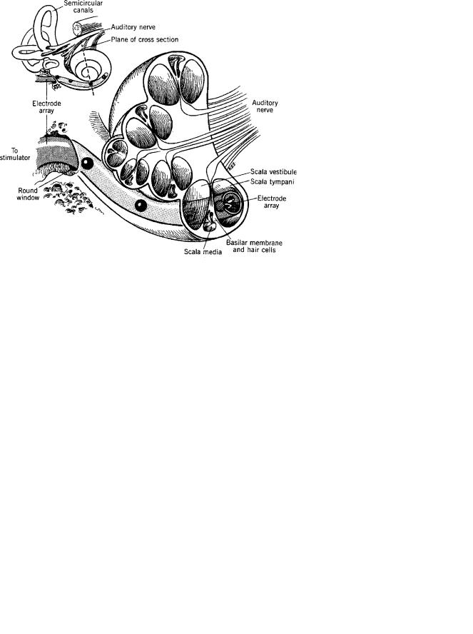
Figure 2 shows a cutaway view of the inner ear and its three chambers or scalae, that is, the scala vestibuli and the scala tympani, which communicate via the helicotrema, an opening at the apical end of the cochlea, and the scala media, which is isolated from the other two scalae by membranes (9,10). The stapes (stirrup) of the middle ear drives the fluids of the scala vestibuli and in doing so deflects the membranes of the scala media (10). One of these membranes, the basilar membrane, bears the hair cells, the motion-sensitive cells that excite the VIII cranial nerve (9,10).
The inner ear acts as a transduction and signal processing mechanism. Auditory information is decomposed into its fundamental frequencies by the frequency-sensitive basilar membrane. Amplitude, phase, and frequency information is carried by the cells of the auditory (VIII cranial) nerve. Simplistically, sounds are decomposed into their spectral peaks (11).
Figure 2. Cutaway view of the cochlea of the inner ear, showing the three chambers or scalae of the ear, and an artist’s conception of a cochlear electrode array inserted into the scala tympani of the cochlea (2).
Each of the 30,000-odd fibers of the auditory nerve has an auditory threshold function that is sensitive to a small range of frequencies. All threshold minima lie within 10– 15 dB; fibers have dynamic ranges that can be as much as 30–40 dB at their characteristic frequencies (9,12). The rate at which a single peripheral fiber fires is a monotonically increasing function of the acoustic stimulus at its characteristic frequency. The dynamic range of a fiber depends on a number of factors, including its threshold and its spontaneous firing rate, the latter of which can be as large as 100 spikes/s (9).
The responses of auditory nerve fibers are nonlinear. At low intensities, the responses of single nerve fibers mimic the frequency spectra of the complex sounds that stimulate the ears of experimental animals (13). At higher intensities, the spectra produced by the responding fibers are dominated by the low frequency component of the speech sound (its first formant) and the distortion products of that frequency (13). Recent evidence provides strong support for nonlinear system to preserve speech sounds at low and high intensity, in quiet and in noise (9).
In summary, the auditory system has a number of features that enable it to decode sound: (1) specific cells are excited at threshold by specific acoustic frequencies; (2) increasing intensity of an acoustic signal causes an increasing spread of influence from cells for which it is the best frequency, to cells that respond at threshold to other frequencies; (3) the intensity of a particular signal appears to be coded both by the rate at which cells fire and by the numbers of cells excited by a particular stimulus; (4) nonlinear properties of the auditory system cause the suppression of one cell’s response to one frequency by stimulation with another frequency, by saturation of rate and by the production of distortion products in the system’s response to high intensity excitation; and (5) frequency information contained in complex stimuli is preserved in the temporal responses of auditory neurons.
HISTORY OF COCHLEAR PROSTHESES
The first report of electrical stimulation of the ear is attributed to Volta, in a paper read to the British Royal Society in July of 1800 (14,15). He reported that his approach, using perhaps 50 V excitation, was uncomfortable, sounding like the boiling of fluid. He did not repeat the study. More recently, Djuorno and Eyries (16) reported the first attempt to excite the auditory nerve directly with electricity. Later, Doyle et al. reported results with electrical stimulation of the auditory nerve (17). Simmons performed an experiment a year later in which he went
COCHLEAR PROSTHESES |
135 |
further, stimulating the VIII auditory nerve and the inferior colliculus of a human patient, showing that it was possible for the subject to distinguish frequencies well below 900 Hz, but not >1000 Hz (18). Simmons demonstrated that both peripheral and central stimulation of the auditory system was possible. In 1964, the House Ear Institute began an extensive series of surgeries to implant cochlear prostheses, reporting on their long-term effects in 1973 (19). The first experiments on multichannel cochlear prostheses were initiated by Simmons et al. in 1979 (20). Their results were promising, and now multichannel implants are the standard of the industry.
Since the first experiments, cochlear prostheses have been built and applied worldwide, receiving approval from governmental agencies and remarkable success in > 60,000 patients. Indeed, cochlear prostheses are considered the standard treatment for profoundly and severely deaf adults. Three commercial firms, Cochlear Corp. (Sydney, Australia), Advanced Bionics Corporation (Valencia, CA; recently purchased by Boston Scientific Corporation), and Med-El Corporation (Innsbruck, Austria) produce cochlear implants successfully. The early cochlear implants were single-channel devices (21), but all of the cochlear prostheses that are implanted today are multichannel devices (22).
THEORY OF OPERATION
The cochlear implant operates on the premise that, if the hair cells of the auditory system are damaged, they can be bypassed and that neurons can be driven directly with very small electrical signals. Figure 3 shows a greatly simplified block diagram of a cochlear implant. Acoustic signals are transduced by a microphone, whose small electric signal is amplified. An external processor decomposes the electrical analogue of the acoustic signal. In the processors that are produced today, processing is digital, with the processor analyzing the instantaneous frequency content of the acoustic signal in the frequency domain. The signals are sent across the skin via a radio frequency link (in the VHF band) that transmits both information and power from the outside of the subject to the inside. These transcutaneous signals are shown with bidirectional paths. Data can be transferred in both directions, providing information to therapists about the condition of the electrodes and the state of the auditory system of the patient. The data flowing to and from the external signal processor are serial bit streams.
The transcutaneous bit streams can have rapid rates: consider that the sampling rate of the audio signal can
|
|
|
Transcutaneous |
|
|
|
|
|
|
Signals |
|
|
|
Acoustic |
Microphone |
External |
Internal Signal |
Controlled |
Currents |
|
and |
Digital Signal |
Processor/ |
Current |
to |
||
Signal |
||||||
Amplifier |
Processor |
Decoder |
Sources |
Electrodes |
||
|
Figure 3. Simplified block diagram of a cochlear implant. Four blocks are shown, a microphone and amplifier, external digital signal processor; internal signal processor/decoder; and, controlled current sources (see text; after Ref. 23).
136 COCHLEAR PROSTHESES
exceed 20,000 samples/s, and that updates of information delivered to the internal processor may present data at 5000 or more data points per second. The data must include the electrode(s) that are being driven, the amplitude and the pulse width of the current pulse that is applied. Rates can exceed 80,000 pulses/s (25). The internal signal pro- cessor–decoder decodes the incoming bit stream. It distributes drive signals to the current sources, selecting specific sources to drive, and setting the amplitude and duration of the control signals.
The current sources drive the electrodes of the cochlear implant’s electrode array. Those electrodes are placed in the scala tympani of the cochlea, and direct the current drive signals to the neurons of the auditory nerve, the VIII cranial nerve. The electrodes of the array are placed in proximity to the neurons, in order to reduce the threshold currents necessary to excite the cells, and to reduce current spread within the inner ear (26).
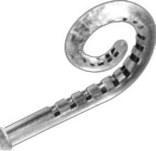
Figure 4 shows one of the cochlear electrode arrays that is produced commercially by Cochlear Corporation (Sydney, Australia). There are 24 contacts in all, two of which are placed outside of the cochlea, leaving 22 contacts that may be driven to excite neurons of the auditory nerve. The contacts may be driven as monopoles (single internal current sources, referenced to an return contact external to the inner ear), as dipoles (pairs of internal current sources) or as combinations of three or more contacts. The contacts are placed along the inside of the spiraling, silicone carrier. The carrier is shaped to fit snugly against the modiolus of the scala tympani. The contacts can be driven singly or in combinations, for example, as dipoles or as multiple sources.
The three manufacturers of cochlear implants use scala tympani arrays that are similar to the array shown in Fig. 4. However, other approaches to stimulate the auditory nerve cells are possible. Normann and his colleagues have tested monolithic electrode arrays that are designed to penetrate the auditory nerve directly (27). Like others before him, Normann realized that bringing electrode contacts near the neurons will reduce thresholds and
Figure 4. Picture of the Nucleus 24 Contour electrode array, showing 24 contacts and a shape that is designed to appose the modiolar wall of the scala tympani (24).
limit the spread of excitation (28–30). The concept has not been tested chronically in human subjects.
The concept of an information channel is critical to the understanding of the cochlear prosthesis. A channel may drive current to one electrode, but it often distributes drive to two or more electrodes. Field shaping and steering techniques suggest the use of multiple electrodes for each channel (31–33). Indeed, demonstrations by Bierer and Middlebrooks (33,34) showed that the quadrupolar configurations (called tripolar in the Bierer paper) produce more focused stimuli than either monopolar or bipolar excitations. Recent experiments in cats have upheld the finding, showing that multipolar stimulation allows two triads of electrodes to be driven simultaneously without significant crosstalk (35). It is clear that a channel may involve several electrodes driven simultaneously, and cannot be defined as the information conveyed by a single contact on an electrode array. While one contact may be driven at a time, bipolar stimulus configurations are common and multipolar configurations may emerge soon.
Signal processing techniques have changed dramatically since the time that the first version of this article was written. The number of available electrodes has more than doubled. In common to most processors is a bank of filters, analogue or, commonly, digital. The filtered signals are decomposed into time-varying envelope signals that are compressed and delivered as either amplitude modulated pulses or width modulated pulses. The pulses are delivered with a variety of strategies.
Continuous interleaved stimulation (CIS) is a technique by which a single electrode is stimulated at a time in order to eliminate field interactions between and among channels when electrodes are driven as monopoles (31). The electrodes receive signals from specific filters. The signals are converted to symmetrical, rectangular, biphasic current pulses whose amplitudes may be proportional to the envelope of the filter signal and whose width is invariant. Conversely, amplitude can be held constant and width can be varied. More recently, Advanced Bionics Corporation has used a processor whose repetition rate can be 5800 pulses/s per channel, to develop rapid updates of channels in the CIS paradigm. In a recent processor, HiRes, stimulation rates can be as much as 5800 pps when two widely spaced channels are driven simultaneously, and drops by one-half when the two channels are driven sequentially (36).
The n-of-m strategy employs a larger number of filters, m, than there are electrodes, n (37). Depending on which filters contain the maximum acoustic energy, pulses are delivered to appropriate electrodes. The cochlea is organized tonotopically along the basilar membrane. Hence, each electrode’s field excites a specific group of characteristic frequencies in perceptual space. Those filters that exhibit the maximum energy determine the electrodes that will be driven by a given temporal sample of the acoustic signal. Biphasic, symmetrical, rectangular pulses are delivered to specific electrodes, n, at particular sample times. Because of the field interactions between electrodes no more than two channels are driven during a given sample.
Other techniques include simultaneous analog stimulation (SAS), in which widely separated electrodes
are driven simultaneously to increase the rate at which information is transferred to the auditory nerve. The field interactions are reduced by driving electrodes that are separated by several millimeters in the inner ear (38). Simultaneous analog stimulation is a special case of the ‘‘filters with compression’’ technique described by Eddington >20 years ago (39). Today, fewer electrodes are driven simultaneously, but they are updated more rapidly (40). Thus, SAS is a variation of both Eddington’s filters and CIS. Eddington described a means by which electrodes were assigned the compressed analog outputs of filters. Those analogue signals were delivered continuously to the electrodes.
A potentially exciting new technique of stimulation takes advantage of the stochastic behavior of auditory neurons. If a stimulator provides high rate conditioning pulses to its electrode array, it is possible to simulate the stochastic firing frequencies of the cells of the auditory nerve (41). This approach has been tested in small numbers of European patients with what appears to be dramatic success, particularly with auditory signals in noise (Rubinstein, personal communication; see below).
Despite the richness of the processing techniques that have been employed, there are still hurdles to be overcome. The number of true, simultaneous channels is too small. It should be at least 16; there is often a mismatch between the frequency assigned to an electrode and its position in the cochlea; the signals that are delivered to the neurons do not contain fine temporal information; the phase information between channels is not preserved; and, there may be neurons missing, causing some electrodes and critical frequencies to be missing as well (42). Future implants may be able to address some of the concerns that are raised here.
EVALUATION OF COCHLEAR PROSTHESES
When human subjects first used cochlear implants, the numbers of subjects were small and tests were not standardized. As the devices improved, standard tests were developed and used across the centers at which implantation was being done (15). The tests include materials that are open and closed set. The test subjects do not review open set materials prior to the test, whereas closed set materials are reviewed before testing takes place. Subjects participate in word tests and sentence tests. In the former, single words are presented while in the latter sentences are presented and the subjects can deduce parts of the sentence logically.
In addition to providing word and sentence tests, consonant (C) and vowel (V) discrimination tests are included in the test batteries. In these tests, nonsense utterances, CVCs or VCVs, are presented and the subject must identify the appropriate vowel or consonant.
Open word tests are difficult while sentence tests are relatively easy. For example, implant users have steadily increased their comprehension of sentences from much <10% with early single channel devices to 80% or above with today’s multichannel devices (22). Many implant users are able to converse on the telephone, a significant result, since they cannot rely on the cues presented by lip
COCHLEAR PROSTHESES |
137 |
reading in that situation. Still, word comprehension from open-word sets remains relatively low, between 40 and 50%, and most users dislike listening to music (43). Clearly, the context that comes from sentence structure and content is important to comprehension, and the complex spectral content of music makes it difficult.
Cochlear implants are a great bioengineering success. Wilson used an aviation metaphor recently, likening the cochlear implant to a DC-3, a reliable workhorse of an aircraft without the sophistication of a twenty-first century transport airplane (43). The implant has advanced from the single-channel stage of the Wright flyer, but has yet to reach its pinnacle.
THE BENEFITS AND RISKS OF IMPLANTATION
Cochlear implants provide clear benefits to their users. For example, hearing-impaired children learn language more rapidly with cochlear implants than they do with hearing aids (44). Adults do well and benefit from their implants, particularly when they are dealing with speech in quiet. However, for patients to achieve the greatest benefits from the device, their prostheses should be adjusted individually for the minimum and maximum stimulation levels for each electrodes in the array, the stimulation rate, and the speech processing strategy (45). Skinner suggests that for best results the parameters should be adjusted for the maximum dynamic range: from quiet sounds to maximum sounds that are ‘‘. . .not too loud. . .’’ (45).
A recent survey of patients from Toronto, Ontario, Canada, was taken of 42 early deafened adult users. Of the 30 who responded, >96% said that they were satisfied with the implant, >93% would undergo the procedure again, and 90% said that they would recommend the implant to another person in the same situation (46). The subjects were encouraged by family and peer support and bolstered by having a positive attitude before, during and after the process of implantation and therapy.
There are risks associated with the surgery, but they are quite small. Cunningham et al. (47) reviewed the cases of 462 adults and 271 children in a private tertiary care center for the years 1993–2002. They found that the overall incidence of infection postoperatively was 4.1%. Major infectious complications occurred in 3.0% of the cases; those complications required surgical intervention (47). Bacterial meningitis was found in 26 of 4264 children receiving cochlear implants in the United States (48). That was found to be associated with a particular electrode array that used a positioner to place it near the modiolar wall. The array was subsequently withdrawn from the market (http://www.fda.gov/cdrh/safety/cochlear.html), and there have been no other reports of the occurrence of meningitis. Cunningham et al. (47) recommended that children undergo vaccination before implantation to prevent bacterial infections.
THE COST OF IMPLANTATION
A recent article cited the cost of cochlear implant treatment as > $40,000.00, of which $20,000.00 is the approximate

138 COCHLEAR PROSTHESES
cost of the device itself (49). Despite the high cost of the device and the surgery, the cochlear prosthesis is beneficial when compared with the long-term costs of other medical device procedures (50,51). Garber et al. (49) asked why the cochlear implant has limited access despite its success and the likely market, and surveyed 25 of 231 practices and 96 of 213 hospitals to try to learn what caused the limits of availability. They concluded that both the practitioners and hospitals lose money when they provide cochlear implants, limiting access to the devices. The cochlear implant is approved in the United States for Medicare, Medicaid, and insurance reimbursement.
THE FUTURE OF COCHLEAR PROSTHESES
In a recent review, Wilson et al. (52) suggested that the future held combined acoustic and electrical stimulation, bilateral implants, new electrode designs and closer mimicking of processing in the normal cochlea. This article discusses electrode designs, combined acoustic, and bilateral stimulation and the closer mimicking of processing in the normal cochlea.
HIGH DENSITY ELECTRODE ARRAYS
Electrode arrays have remained much the same for more than a decade. They are built manually on substrates of silicone, using Pt–Ir (90–10%) alloyed electrodes. The group of Dr. Kensall Wise at the University of Michigan has proposed the use of high density arrays that are made on silicon substrates using IrO contacts (54,55). If such arrays can be built for human use, they will reduce the cost of building electrode arrays while increasing the specificity of excitation of cells. Another approach to the problem is to build electrode arrays on multilayered polymer substrates (Fig. 5). Sample arrays have been used to demonstrate the use of high density arrays in animal studies, with clear
Figure 5. Photograph of a 12-site sample array made by Advanced Cochlear Systems (Snoqualmie, WA) to insert into the scala tympani of a cat (53). The width of each gold electrode contact is 100 mm
independence of channels driven in the first turn of the scala tympani (Snyder, Corbett, Bonham, Rebscher, and Johnson, personal communication).
The goal driving the development of these high density arrays is to increase the specificity of stimulation and to allow several independent groups of cells to be driven simultaneously (32,56). The work of Jolly (32) and Bierer and Middlebrooks (33,57) suggested that this might be the case. More recent work has confirmed the earlier results and extended them (35,34). The benefit of focused multipolar stimulation and of simultaneous excitation of several independent groups of neurons is not without cost. More driven electrodes require greater current consumption. Current consumption is increased with focusing, since focused stimuli require more applied current to reach the same potential fields in conducting media (58). It is likely that high density electrode arrays will be a part of cochlear implants, but there are engineering challenges to be met before it will happen.
COMBINED ACOUSTIC AND ELECTRICAL STIMULATION
Preliminary studies of combined electrical and acoustic stimulation have been done successfully in both Europe and the United States (52,59,60). The subjects come from the substantial population of people who preserve some hearing for frequencies <1 kHz, but who are severely impaired for frequencies > 1 kHz. Two questions arise immediately. (1) Can low frequency hearing be preserved after an electrode array has been placed in the high frequency regions of the inner ear? (2) Can acoustic and electrical stimuli be applied simultaneously and successfully?
The likelihood of success is great, particularly if patients have short electrode arrays implanted, avoiding damage to the delicate structures of the inner ear. That concern is critical in the case of the hybrid stimulation scheme, since low frequency information will come via the normal, albeit amplified, acoustic pathway. Two manufacturers, Cochlear Corporation (Sydney, Australia) (60) and Med-El (Innsbruck, Austria) (59) have produced electrode arrays for the purpose and have tested them in clinical settings. The Med-El array has an implanted length of 31.5 mm (59), while the Cochlear Corporation array’s length is 10 mm in its latest version (60). Both have had extensive laboratory tests and have been used clinically. Clinical tests confirm the initial hypothosis: when patients suffer primarily from high frequency hearing loss, the use of hybrid stimulation is likely to provide great benefit, and may well increase the numbers of people who can have near-normal hearing (52). Electrical and acoustic stimuli can be combined by implanting one ear with a cochlear prosthesis and using a hearing aid in the contralateral ear. This approach has had some reports of success and is currently under study in research laboratories.
NORMAL PROCESSING: CONDITIONING PULSES
In the 1990s, investigators began to consider the issue of the stochastic behavior of neurons (61) and that high rate
conditioning stimuli might improve the behavior of cells in the auditory nerve, decreasing thresholds and increasing dynamic ranges (52). They proposed to use electric currents with 5 kHz pulse trains of brief pulses, biphasic rectangular pulses of 40 ms duration for each phase (62). Rubinstein and various colleagues pursued the idea further, suggesting that high rate stimuli might mimic stochastic resonance in neurons and improve signal processing in cochlear implants (60). Computer models validated the concept, as did initial tests in a human subject (63). An extensive neurophysiological study confirmed the idea in experimental animals (62).
Rubinstein and Frijns did preliminary tests for the use of high rate, low amplitude conditioning pulses in the processors of some human subjects, reporting success in the majority of their subjects (Rubinstein, personal communication). The concept is certainly a logical and promising idea; whether it will provide a dramatic improvement to cochlear implants is something that will be learned from further experiments in human subjects.
NORMAL PROCESSING: FINE STRUCTURE
Present cochlear implants impose low pass filter functions on the acoustic signals that they decode. Signals are filtered, and their envelopes detected, with a concomitant loss of fine structure. Fine structure is defined as information spanning frequencies from 500 to 10 kHz (22). Speech can be well understood in quiet environments. The users of present-day cochlear implants rarely enjoy music. Some of that may be improved by increasing the fine structure of the signals delivered to the ear via the cochlear implant. The Hilbert transform provides a potential approach to providing both amplitude information and fine structure (22,64,65). Smith et al. (63) determined that the envelope of the transform was important for speech perception, while the fine structure determines localization and pitch.
Processors that employ Hilbert transforms have yet to be produced in quantity. Although prototypes exist, they have not made their way into cochlear implants (64). The development of implants that can reproduce the fine structure of signals is likely to improve cochlear prostheses.
BILATERAL IMPLANTS
Binaural hearing is critical to sound localization and the extraction of auditory signals in noise. In addition, binaural implants may allow listeners to employ the ‘‘head shadow’’ benefit to hear a specific voice in the face of sounds produced by a competing crowd of people (52). Wilson notes promising results from several centers at which patients have received bilateral implants (52). He reports improvements in speech comprehension, as well as the results of several careful psychophysical studies that were focused on the balance between the prostheses that were implanted. Wilson and his colleagues concluded that bilateral implants are likely to provide clear benefits. While users are tolerant of some timing and amplitude mismatches, the careful matching of stimulus sites, that is, electrode locations, may be necessary for success (52). Another issue to
COCHLEAR PROSTHESES |
139 |
consider is the cost of bilateral implantation. Bilateral implantation incurs the cost of two cochlear prostheses and two surgeries. Does the benefit accrued by the patient double? That remains to be seen at the time of this writing.
CONCLUSION
Cochlear prostheses are a clear bioengineering success story. More than 60,000 patients have benefited worldwide. Many users can talk on the telephone and communicate effectively without visual aids, like lipreading. The design of the cochlear prosthesis is likely to improve, even as the number of implantees grows rapidly, indeed, at double-digit rates. With that rich background and rapid growth, there are opportunities for bioengineers to produce even better cochlear prostheses.
ACKNOWLEDGMENTS
This work was sponsored by grants R43DC000531 and R43DC04614 of the National Institutes of Health.
BIBLIOGRAPHY
1.NIH NIH Consensus Statement: cochlear Implants in Adults and Children. Bethesda, MD, National Institutes of Health; 1995.
2.Webster JG. Encyclopedia of medical devices and instrumentation. New York: John Wiley & Sons; 1988.
3.Blanchfield BB, Feldman JJ, et al. The severely to profoundly hearing impaired population in the United States: Prevalence and demographics. Policy Anal Brief H Ser 1999; 1 (October): 1–4.
4.Blanchfield BB, Feldman JJ, et al. The severely to profoundly hearing-impaired population in the United States: prevalence estimates and demographics. J Am Acad Audiol 2001;12(4): 183–189.
5.Kuchta J. Neuroprosthetic hearing with auditory brainstem implants. Biomed Tech (Berlin) 2004;49(4):83–87.
6.Otto SR, Brackmann DE, et al. Multichannel auditory brainstem implant: update on performance in 61 patients. J Neurosurg 2002;96(6):1063–1071.
7.Schwartz MS, Otto SR, et al. Use of a multichannel auditory brainstem implant for neurofibromatosis type 2. Stereotact Funct Neurosurg 2003;81(1–4):110–114.
8.Kanowitz SJ, Shapiro WH, et al. Auditory brainstem implantation in patients with neurofibromatosis type 2. Laryngoscope 2004;114(12):2135–2146.
9.Geisler CD. From Sound to Synapse: Physiology of the Mammalian Ear. New York: Oxford University Press; 1998.
10.Dallos P, Popper AN, Fay RR, editors. The Cochlea. Springer Handbook of Auditory Research. New York: Springer; 1996.
11.Sachs MB, Young ED. Encoding of steady-state vowels in the auditory nerve: representation in terms of discharge rate. J Acoust Soc Am 1979;667(2):470–479.
12.Liberman MC. Auditory-nerve response from cats raised in a low-noise chamber. J Acoust Soc Am 1978;63(2):442–455.
13.Sachs MB, Young ED. Effects of nonlinearities on speech encoding in the auditory nerve. J Acoust Soc Am 1980; 68:858.
14.Volta A. On the electricity excited by mere contact of conducting substances of different kinds. R Soc Philos Trans 1800;90: 403–431.
140 COCHLEAR PROSTHESES
15.Clark GM. Cochlear Implants: Fundamentals and Applications. New York: Springer-Verlag; 2003.
16.Djourno A, Eyries C. Prothese Autitive par excitation electrique a distance du nerf sensoriel a l’aide d’un bobinage inclus a demcure. Presse Med 1957;35:14–17.
17.Doyle JB, Doyle HD, et al. Electrical stimulation in eighth nerve deafness. Bull LosAngeles Neurol Soc 1963;18:148.
18.Simmons FB, Mongson CJ, et al. Electrical stimulation of the acoustic nerve and inferior colliculus in man. Arch Otolaryngol Head Neck Surg 1964;79:559.
19.House WF, Urban J. Long term results of electrode implantation and electronic stimulation of the cochlea in man. Ann Otolaryngol Rhinol Laryngol 1973;82:504.
20.Simmons FB, Mathews RG, et al. A functioning multichannel auditory nerve stimulator. A preliminary report on two human volunteers. Acta Otolaryngol 1979;87(3–4):170–175.
21.House WF, Berliner KI. Cochlear Implants: from idea to clinical practice. In: Cooper H, editors. Volume 1, Cochlear Implants: A Practical Guide. San Diego, CA: Singular Publishing Group, Inc.; 1991. p 9–33.
22.Zeng F-G. Trends in cochlear implants. Trends Amplif 2004;8(1):1–34.
23.Spelman F. Cochlear Prostheses. In: Ratner BD, Hoffman AS, Schoen FJ, Lemons JE, editors. Volume 1, Biomaterial Science: An Introduction to Materials in Medicine. Amsterdam: The Netherlands Elsevier Academic Press; 2004. p 658–669.
24.Anonymous. Nucleus 24 Contour, Cochlear Americas. 2004.
25.Kessler DK. The CLARION Multi-Strategy Cochlear Implant. Ann Otol Rhinol Laryngol Suppl 1999; 177(Apr.):8-16.
26.Jolly CN, Gsto¨ttner W, et al. Principles and outcome in perimodiolar positioning. Ann Otolaryngol Rhinol Laryngol Suppl 2000;185(12):20–23.
27.Hillman T, Badi AN, et al. Cochlear nerve stimulation with a 3-dimensional penetrating electrode array. Otolaryngol Neurotol 2003;24(5):764–768.
28.Simmons FB. Electrical Stimulation of the Auditory Nerve in Man. Arch Otolaryngol 1966;84 (July, 1966):24–76.
29.White MW, Merzenich MM, et al. Multichannel cochlear implants: Channel interactions and Processor design. Arch Otolaryngol 1984;110:493–501.
30.Arts HA, Jones DA, et al. Prosthetic stimulation of the auditory system with intraneural electrodes. Ann Otolaryngol Rhinol Laryngol Suppl 2003;191:20–25.
31.Wilson BS, Finley CC, et al. Better speech recognition with cochlear implants. Nature (London) (July 18, 1991); 352:236– 238.
32.Jolly CN, Spelman FA, et al. Quadrupolar stimulation for cochlear prostheses: Modeling and experimental data. IEEE Trans Biomed Eng 1996;43(8):857–865.
33.Bierer JA, Middlebrooks JC. Cortical responses to cochlear implant stimulation: Channel interactions. J Assoc Res Otolaryngol. 2004;5(1):32–48.
34.Snyder RL, Bierer JA, et al. Topographic spread of inferior colliculus activation in response to acoustic and intracochlear electric stimulation. J Assoc Res Otolaryngol 2004;5(3): 305–322.
35.Bonham B, Snyder RL, et al. The neurophysiological effects of simulated auditory prosthesis stimulation: channel interaction, current steering and channel morphing. San Francisco, CA: University of California at San Francisco; 2004.
36.Anonymous. New methodology for fitting cochlear implants. Valencia, CA: Advanced Bionics Corporation. 1–5; 2003.
37.McDermott HJ, McKay CM, et al. A new portable sound processor for the University of Melbourne/Nucleus Limited multielectrode cochlear implant. J Acoust Soc Am 1992;91: 3367–3371.
38.Anonymous. Clarion S-Series. Sylmar, CA, Advanced Bionics, Inc.; 1997.
39.Eddington DK. Speech discrimination in deaf subjects with cochlear implants. J Acoust Soc Am 1980;68:885–891.
40.Anonymous. PULSARci Cochlear Implant, Med-El; 2004.
41.Rubinstein JT, Hong R. Signal coding in cochlear implants: Exploiting stochastic effects of electrical stimulation. Ann Otol Rhinol Laryngol 2003;112(9, Part 2):14–19.
42.Moore BCJ. Coding of sounds in the auditory system and its relevance to signal processing and coding in cochlear implants. Otolaryngol Neurotol 2003;24(2):243–254.
43.Wilson BS. The History of Cochlear Implants. Neural Interfaces Workshop, Hyatt Regency Bethesda Hotel, Bethesda, MD: NIDCD, National Institutes of Health; 2004.
44.Skinner MW. Cochlear implants in children: What direction should future research take? 2001 Conference on Implantable Auditory Prostheses, Pacific Grove CA; 2001.
45.Skinner MW. Optimizing cochlear implant speech performance. Ann Otolaryngol Rhinol Laryngol Suppl 2003;191: 4–13.
46.Chee GH, Goldring JE, et al. Benefits of cochlear implantation in early-deafened adults: the Toronto experience. J Otol 2004;33(1):26–31.
47.Cunningham CD, 3rd, Slattery WH, 3rd, et al. Postoperative infection in cochlear implant patients. Otolaryngol Head Neck Surg 2004;131(1):109–114.
48.Reefhuis J, Honein MA, et al. Risk of bacterial meningitis in children with cochlear implants. N Engl J Med 2003;349(5): 435–445.
49.Garber S, Ridgely MS, et al. Payment under public and private insurance and access to cochlear implants. Arch Otolaryngol Head Neck Surg 2002;128(10):1145–1152.
50.Cheng AK, Niparko JK. Cost-utility of the cochlear implant in adults. Arch Otolaryngol Head Neck Surg 1999;125(11): 1214–1218.
51.Niparko JK, Kirk KI, et al. Cochlear Implants: Principles and Practices. Baltimore MA: Lippincott Williams & Wilkins; 2000.
52.Wilson BS, Lawson DT. Ann Rev Biomed Eng 2003;5:207–249.
53.Corbett SS, III, Johnson T, Rebscher S, Carson M, Ketterl J, Snyder R. unpublished results.
54.Weiland JD, Anderson DJ. Chronic neural stimulation with thin-film, iridium oxide electrodes. IEEE Trans Biomed Eng 2000;47(7):911–918.
55.Weiland JD, Anderson DJ, et al. In vitro electrical properties for iridium oxide versus titanium nitride stimulating electrodes. IEEE Trans Biomed Eng 2003;49(12): 1574-1579.
56.Clopton BM, Spelman FA. Technology and the future of cochlear implants. Ann Otolaryngol Rhinol Laryngol Suppl 2003;191:26–32.
57.Bierer JA, Litvak L, et al. Effects of electrode configuration on psychophysical measures of channel interaction in cochlear implant subjects. Soc Neurosci 2003.
58.Spelman FA, Pfingst BE, et al. The effects of electrode configuration on potential fields in the electrically-stimulated cochlea: models and measurements. Ann Otol Rhinol Laryngol 1995;104(Suppl. 166):131–136.
59.Adunka O, Kiefer J, et al. Development and evaluation of an improved cochlear implant electrode design for electric acoustic stimulation. Laryngoscope 2004;114(7):1237– 1241.
60.Gantz BJ, Turner C. Combining acoustic and electrical speech processing: Iowa/Nucleus hybrid implant. Acta Otolaryngol 2004;124(4):344–347.
61.Rubinstein JT, Abbas PJ, et al. Stochastic Resonance: Can it be exploited by speech processors? Conference on Implantable Auditory Prostheses, Pacific Grove, CA; 1997.