

Molecular and Cellular Signaling - Martin Beckerman
.pdf370 15. Apoptosis
FIGURE 15.5. Central components of the mitochondrial PTPC: Depicted in the figure are the central elements—the ANT located in the inner membrane and the VDAC situated in the outer membrane. They form a pore through which Cyto c, Smac/DIABLO, and a number of different effector molecules can diffuse through and into the cytoplasm. Also shown are a variety of key Bcl-2 family members. A proapoptotic Bcl-2 protein, for example, Bax, is depicted bound to the ANT/VDAC, having displaced an antiapoptotic Bcl-2 protein, while several other Bax/Baks form a pore.
of the intermembrane space and the matrix. It is composed of voltagedependent anion channels (VDACs) located in the outer mitochondrial membrane and the adenosine nucleotide translocator (ANT) situated in the inner membrane. The PTPC also includes the peripheral benzodiazepine receptors, creatine kinases, hexokinase II, and cyclophilin D.
The mitochondrial PTPC is the main target of Bcl-2 signaling and is the main site of their proand antiapoptotic actions. If a molecule can cross a membrane by means of simple passive diffusion, driven only by a concentration gradient, then that membrane is permeable to that molecule. In apoptosis, the Bcl-2 proteins regulate the permeability of the mitochondrial membrane to the apoptosis-promoting molecules. Bcl-2 proteins act as sensors of stress signals and as regulators of mitochondrial membrane permeability. They regulate membrane permeability by binding to and altering the pores formed by the ANT and VDAC, and by oligomerizing and forming pores by themselves.
BH3-only proteins stimulate the release from mitochondria of Cytochrome c and other apoptosis-promoting factors. The BH3-only protein tBid, for example, promotes the permeability-increasing activities of Bax and Bak proteins, stimulates the remodeling of the mitochondrial cristae, and triggers the release of Cytochrome c. Members of the anti-apoptotic branch of the family inhibit apoptosis by sequestering BH3-only proteins thereby preventing their stimulation of Bax and Bad. There are two kinds of BH3 protein actions. Bid-like BH3 proteins facilitate the oligomeriza-

15.10 Mitochondria Release Cytochrome c |
371 |
tion of Bax and Bak, while Bad-like BH3 proteins bind Bcl-2s and displace Bid-like peptides from them.
15.10Mitochondria Release Cytochrome c in Response to Oxidative Stresses
Oxidative phosphorylation is carried out in mitochondria. One of the key agents in this process is Cytochrome c, an evolutionary ancient electron carrier. It is a small protein, consisting of 104 amino acid residues and a covalently attached heme group. Cytochrome c is part of the machinery that transfers electrons through several protein complexes located in the inner mitochondrial membrane. It carries electrons from the cytochrome reductase complex to the cytochrome oxidase complex. The electron transport activity leads to the pumping of protons from the matrix side to the cytoplasmic side of the inner mitochondrial membrane. The biochemical process, oxidative phosphorylation, generates ATP by utilizing the energy released during the electron transfer from NADH or FADH2 to O2.
Recall that atoms in stable molecules are tied together by bonds consisting of pairs of electrons, one with spin up and the other with spin down. When bonds are broken the molecules may end up with one or more unpaired electrons. Molecules possessing unpaired electrons are referred to as free radicals. The presence of an unpaired electron renders the molecules highly reactive. Because of the presence of an electron the molecule has a propensity to “steal” electrons from other molecules, in many cases breaking bonds to acquire the electrons. Chain reactions can be produced in the cell, and free radicals are highly dangerous. Most commonly encountered free radicals in the cell involve oxygen and these are called reactive oxygen species (ROS).
Mitochondria are the major cellular source of reactive oxygen species. In mitochondria, a small fraction, perhaps 2 to 5%, of the molecular oxygen being reduced by the respiratory electron transport chain is converted to superoxide (O2-) and then to hydrogen peroxide (H2O2) or to the hydroxyl radical (OH-). The ROS cause cellular (oxidative) stresses because they can oxidize DNA, proteins, and lipids. The presence of ROS influences cellular physiology in many ways and triggers protective reactions involving the upregulation of antioxidants and detoxifying enzymes. Antioxidants are free radical scavengers. They supply electrons to the free radicals, allowing them to form bonds and converting them nonreactive forms.
Cytochrome c is loosely bound to the inner mitochondrial membrane by cardiophilin and other anionic lipids. When ROS levels rise it disrupts the binding of Cytochrome c thereby making it easier to release. Once
Cytochrome c is released it stimulates assembly of the apoptosome leading to activation of Caspase 9 and Caspase 3. Thus, oxidative stress conditions arising when ROS activity levels in mitochondria become excessive and are no longer controlled by antioxidant scavengers are signaled by Cytochrome

372 15. Apoptosis
c release from the mitochondria. The Cytochrome c molecules act as local signaling molecules that convey stress messages from the mitochondria to the apoptosome.
15.11Mitochondria Release Apoptosis-Promoting Agents
Several different kinds of molecules are sent out through the PTPC, as indicated in Table 15.4. Cytochrome c and deoxyadenosine triphosphate (dATP) convey signals that are required for assembly of the apoptosome and the activation of Caspase 9. Two other regulators of the apoptosome, Smac/DIABLO and Omi/HtrA2 are also released from the mitochondria. A number of apoptotic effectors are released in addition to the activators and regulators of apoptosome and Caspase 9 functions. These include Apoptosis inducing factor (AIF) and Endonuclease G, which target nuclear chromatin and degrades it.
Chromatin condensation and fragmentation of nuclear DNA is an important part of apoptosis. A number of proteins fragment DNA and target chromatin in response to proapoptotic signals. One of these, the CAD DNase, was discussed in Section 15.7. Two apoptotic enzymes are released by mitochondria that target chromatin and fragment DNA. One of the DNA chopping proteins, or DNases, is Endonuclease G. This enzyme is released from mitochondria in response to stimulation by proapoptotic proteins. Once it is released into the cytosol it translocates to the nucleus where it fragments DNA into nucleosomal-sized fragments. In the first nuclear steps of apoptosis, DNA repair is halted and chromatin condensation occurs, which turns off DNA transcription. Another apoptosis promoting agent sent out from the mitochondria is Apoptosis-inducing factor. This protein is released from mitochondria at an early stage in the apoptosis process. It is responsible for peripheral chromatin condensation and for large-scale chromatin fragmentation.
TABLE 15.4. Agents released by mitochondria: Abbre- viations—Second mitochondrial activator of caspases (Smac); direct IAP binding protein with low pI (DIABLO).
Agent |
Action |
Cytochrome c |
Required for assembly of the |
|
apoptosome |
dATP |
Required for assembly of the |
|
apoptosome |
Smac/DIABLO |
Proapoptotic regulator |
Omi/HtrA2 |
Proapoptotic regulator |
AIF |
Early acting nuclear factor |
Endonuclease G |
Later acting nuclear factor |
|
|
15.12 Role of Apoptosome in (Mitochondrial Pathway to) Apoptosis |
373 |
15.12Role of Apoptosome in (Mitochondrial Pathway to) Apoptosis
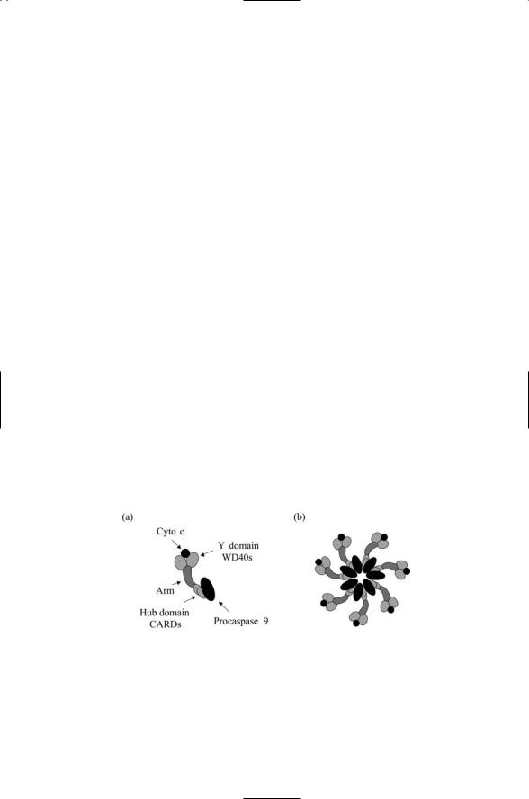
The apoptosome is the main control point in the mitochondrial pathway to apoptosis. When released from the mitochondria, Cytochrome c interacts with a 130-kDa adapter protein called Apaf-1 located in the cytoplasm just outside the mitochondria. This adapter contains three domains (Figure
15.6a). It has an N-terminal caspase recruitment domain (CARD), a central domain that binds dADP/ATP, and a C-terminal domain containing a series of WD-40 repeats. When Cytochrome c binds to the WD-40 repeats to override the autoinhibition, and deoxyadenosine triphosphate (dATP) hydrolysis occurs, Apaf-1 is able to form oligomers leading to the formation of a 1-MDa apoptosome.
The apoptosome is a sevenfold symmetric platform. It is wheel-shaped with seven spokes radiating out from a central hub (Figure 15.6b), and functions to activate Procaspase 9 when these zymogens are incorporated into it. When fully saturated, seven Procaspase 9 molecules are bound to the seven Apaf-1 CARD domains and seven Cytochrome c molecules are bound to the Y domains. The formation of an apoptosome containing activated Caspase 9 molecules triggers the apoptosis process by interacting with and activating Caspase 3.
The apoptosome is a major control point where proapoptotic agents released by the mitochondria converge and where Caspase 9 and Caspase 3 interact and activate one another. At the apoptosome, Caspase 9 processes and activates Caspase 3. A feedback loop in which processed Caspase 3 cleaves and activates Caspase 9 amplifies the production of activated caspases. Several other proteins are present, and form complexes with the caspases. Among these are caspase inhibitors and caspase counterinhibitors.
FIGURE 15.6. Apaf-1 and the apoptosome: (a) Apaf-1 molecule bound to Cytochrome c and Procaspase 9—Shown are a pair of WD-40 repeats in the C- terminal Y domain that bind Procaspase 9, and a pair of CARD domains in the N- terminal that bind Cyto c. A flexible arm containing a CED4 homology motif that binds dATP/ADP connects the N- and C-terminal regions to one another. (b) Fully assembled apoptosome illustrating how the individual Apaf-1 molecules associate into a sevenfold symmetric platform.

374 15. Apoptosis
15.13Inhibitors of Apoptosis Proteins Regulate Caspase Activity
Inhibitor of apoptosis proteins (IAPs) bind to and inhibit the activities of caspases once they have been converted by proteolysis from zymogens to caspase monomers and homodimers. The presence of these regulators (and their counterregulators) establishes a second tier of control over the catalytic activities of caspases. The defining feature of the IAPs is the presence of one or more BIR (baculoviral IAP repeat) domains (Figure 15.7). These are found in the eight mammalian IAPs discovered to date. A second prominent feature of the IAPs is the presence of a RING domain in the C- terminal of some but not all IAPs.
Caspases must form homodimers to become catalytically active. The homodimerization partner supplies a sequence crucial for catalysis, a sequence that that monomeric form of the caspases lacks. Crystal structure studies of Caspase 9 in complex with the BIR3 domain of an X-linked AIP (XIAP) protein show how the IAP proteins inhibit the catalytic activities of the caspases. The BIR3 domain binds to the homodimerization domain of monomeric caspase, thereby preventing formation of caspase homodimers. This mechanism differs from that used by the IAPs to inhibit effector caspases such as Caspase 3 and Caspase 7. Rather than preventing formation of the homodimers, XIAP uses a sequence just forward of its BIR2 domain to block the active site of the effector caspases.
Inhibitor of apoptosis proteins possess a ubiquitin ligase activity. As shown in Figure 15.7, IAPs such as XIAP and c-IAP1 and c-IAP2 possess a RING finger domain in their C-terminus. This motif is often encountered in E3 ubiquitin ligases. In the case of IAPs bearing this motif, there seem
FIGURE 15.7. Domain organization of the inhibitor of apoptosis proteins (IAP):
(a) Structure of XIAP. (b) Structure of c-IAP1 and c-IAP2. ILP-2 and Livin resemble XIAP except that they only have one BIR domain (BIR3 in the case of ILP-2 and BIR2 in the case of Livin). The other three mammalian AIP proteins—NAIP, Survivin, and Apollon/Bruce—have from one to three BIR domains but lack a RING finger domain. Abbreviations: X-linked IAP (XIAP); IAP-like protein (ILP); neuronal inhibitory apoptosis protein (NAIP); baculoviral IAP repeat (BIR); caspase recruitment domain (CARD).

15.15 Feedback Loops Coordinate Actions at Various Control Points |
375 |
to be two choices of substrates. In response to proapoptotic signals, the IAPs trigger their own ubiquitination. Alternatively, in the absence of proapoptotic signals, the IAPs act in an antiapoptoptic capacity by promoting the ubiquitination of activated Caspases 3 and 7.
15.14 Smac/DIABLO and Omi/HtrA2 Regulate IAPs
Smac/DIABLO is an IAP counterregulator. It is released from mitochondria in response to proapoptosis signals, and once released disrupts the ability of IAPs to inhibit the caspases. The IAP family member XIAP is one of its main targets. In the absence of Smac/DIABLO, XIAP binds to the small subunit of Caspase 9. It does not bind to Caspase 9 in its procaspase form, but only to the cleaved and assembled Caspase 9 monomer. Smac/ DIABLO disrupts the ability of XIAP to inhibit Caspase 9 by binding to its BIR3 domain. A sequence of four residues in the N-terminal recognizes and binds a surface groove in the BIR3 domain.
The protein Omi/HtrA2 is another IAP counterregulator released by mitochondria in response to proapoptotic signaling. Like Smac/DIABLO it promotes apoptosis by antagonizing the IAPs ability to bind to and inhibit activation of Caspases 3, 7, and 9. Upon release from the mitochondria Omi/HtrA2 forms complexes with XIAP and disrupts the latter’s caspaseinhibiting functions. Although Omi/HtrA2 binds to the IAP in a manner resembling that of Smac/DIABLO, its manner of action is different. Unlike Smac/DIABLO, Omi/HtrA2 is a serine protease and may act in a proteolytic manner to degrade and thus limit the inhibitory activities of the IAPs.
15.15Feedback Loops Coordinate Actions at Various Control Points
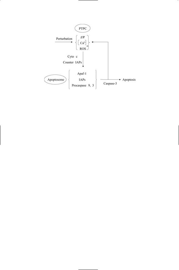
The apoptosome and the mitochondrial PTPC are major control points for apoptosis. These control points communicate with each other and with the DISC in order to arrive at live or die decisions. The communication between PTPC and apoptosome is summarized in Figure 15.8.In the figure,a stress-gen- erating perturbation causes the loss of inner mitochondrial transmembrane potential Ym, produces ROS, and/or elevates the free calcium concentration [Ca2+]m in the mitochondrial matrix. In response, the mitochondrial membranes become more permeable to Cytochrome c and other proapoptotic agents. These agents leave the mitochondria. Some
(cytochrome c and dATP) trigger formation of the apoptosome and others
(Smac/DIABLO and Omi/HtrA2) activate the caspases by binding and inhibiting the IAPs. Caspase 3 activated at the apoptosome diffuses to and enters the mitochondria. It then cleaves a number of components of the mitochondrial electron transport chain. These feedback operations trigger the
376 15. Apoptosis
FIGURE 15.8. Mitochondrial PTPC—Apoptosome circuit: Perturbations of the mitochondrial permeability transition pore complex (PTPC) stimulates the release of apoptosis-promoting factors such as Cytochrome c and counter inhibitor of apoptosis proteins (IAPs). These act at the apoptosome to activate Caspases 3 and 9.
stepped-up production of ROS and efflux of Cytochrome c, which then acts at the apoptosome to amplify the amount of activated Caspase 9 and Caspase 3.
Another feedback loop connects events at the apoptosome with those occurring at the DISC. The connecting link is Caspase 6. Caspase 3 cleaves and activates Caspase 6, which then migrates to the DISC where it stimulates Caspase 8, thereby amplifying the strength of the apoptotic signal at the cell surface as was discussed in Section 15.7. The BH3-only protein Bid completes the circuit by connecting actions taking place at the DISC with those occurring at the PTPC.
Apoptosis is controlled by a plethora of positive feedback loops that ensure that once the appropriate thresholds are passed there will be a firm commitment to apoptosis. The presence of thresholds ensures that random excursions and perturbations do not unnecessarily commit the cell to apoptosis when it ought not to. An equally important ensemble of negative feedback loops generates the threshold dependences.
15.16Cells Can Produce Several Different Kinds of Calcium Signals
Calcium is an important signaling intermediary, or second messenger, and was introduced as such in Chapter 8. Calcium can also function as a first messenger. Calcium is well suited to function as a signaling molecule. It is able to accommodate from 4 to 12 oxygen atoms in their primary coordination sphere. This property allows the calcium ions to contact multiple

15.17 Excessive [Ca2+] in Mitochondria Can Trigger Apoptosis |
377 |
partners and trigger large conformational changes when binding a protein. In addition, calcium does not diffuse very far because of the presence of a large number of calcium buffers in the cytosol.
The normal intracellular calcium ion concentration is about 100 nM. This level is several orders of magnitude lower than the calcium ion concentrations in the extracellular spaces, which is roughly 2 mM. To maintain the intracellular calcium concentrations at the resting levels, the Na+/Ca2+ ion exchanger and pumps such as the plasma membrane calcium ATPase
(PMCA), remove calcium from the cell. In addition, the sarco-endoplasmic reticulum calcium ATPase (SERCA) embedded in the SR/ER ships calcium from the cytosol into the intracellular stores. Because of the presence of cellular machinery keeping calcium levels low, transient local increases in calcium concentration can be produced easily and serve as a signal.
Lipid second messengers relay signals from the plasma membrane to the intracellular stores. These molecules trigger the release of calcium when they bind inositol (1,4,5) triphosphate receptors (InsP3Rs). A second kind of calcium release channel, the ryanodine receptor (RyR), releases calcium from the intracellular stores, as well. Several different kinds of calcium signals can be produced. One kind of calcium signal, the calcium “wave,” is a global signal that propagates over large distances across the cytosol. The propagation of these waves over large intracellular distances is made possible by positive feedback in which calcium released from stores in one locale diffuses to and triggers the release of calcium from nearby stores that sets off further releases, thereby generating a wave of calcium. Calcium can be released from stores in a more localized manner. The local releases of calcium from intracellular stores are called “puffs” when produced by InsP3Rs and “sparks” when facilitated by RyRs.
15.17Excessive [Ca2+] in Mitochondria Can Trigger Apoptosis
Calcium signals are sent to the mitochondria under normal conditions and also under abnormal conditions. Calcium signals are sent to the mitochondria in response to increased signaling at the plasma membrane in order to spur increases in metabolic activity to support the elevated signaling load. This coupling of metabolism and calcium is made possible by the presence in the mitochondrial matrix of a number of calcium sensitive metabolic enzymes. Increased calcium signaling to the mitochondria also occurs under abnormal conditions of cytosolic calcium overload (i.e., disruptions in calcium homeostasis) and ER stresses. In these situations, the changes in mitochondrial physiology drive the cell towards apoptosis.
Calcium ions are cycled between the SR/ER and mitochondria. The membranes of these organelles are in close proximity to one another and

378 15. Apoptosis
highly local and directional signaling similar to that occurring at chemical synapses takes place. One of the early events taking place in stressgenerated apoptosis is an increased permeability of the PTPC to calcium entry. This event leads to increases in permeability to cytochrome c, resulting in its increased efflux from the mitochondria.
Combinations of proapoptotic conditions such as oxidative stresses and calcium signaling between the ER and mitochondria can initiate apoptotic responses under condition where one or the other condition by itself cannot. The cooperation between the two is mediated by a positive feedback loop in which ROS generated in the mitochondria promote increased Ca2+ release from the ER. The calcium accumulates in the mitochondria and triggers further increases in ROS production. This process generates the progressive depolarization of the inner mitochondrial membrane and increases membrane permeation leading to cell death.
The PTPC is not the only control point where Bcl-2 family proteins exert their regulatory influences. They also act at the endoplasmic reticulum where they influence the release of Cytochrome c occurs through their modulation of calcium signaling. They help maintain homeostatic control over movement of calcium into and between the two organelles (the ER and the mitochondria), and mobilize calcium release. Proapoptotic Bax and Bak proteins localize to both the endoplasmic reticulum and mitochondria where they control calcium trafficking between the two organelles. In the ER, Bax and Bak help initiate the apoptosis process by contributing to Caspase 12 activation, and in the mitochondria they help activate Caspase 7.
15.18p53 Promotes Cell Death in Response to Irreparable DNA Damage
In response to DNA damage signals, the p53 protein will either halt the cell cycle to allow for repairs or promote apoptosis if the damage is irreparable. The signaling proteins that convey messages to p53 do so through phosphorylations and acetylations of specific residues. The specific mix of phosphorylations and acetylations determines in large measure the response that will be made by p53. For example, phosphorylation of p53 on Ser46 stimulates transcription of apoptosis-promoting genes, but does not stimulate transcription of cell cycle genes. A second factor that guides the p53 response is the mix of cofactors present at the promoter sites. As is the case for other transcription factors, the cofactors help determine which genes get transcribed and which ones do not. In the case of p53 two other p53 family members—p63 and p73—work together with p53 to transcribe proapoptosis genes.
The p53 protein can shift the balance of proand antiapoptosis factors in at least two distinct ways. First, in its role as a transcription factor, the p53 proteins can step up the transcription of proapoptotic genes. As
15.19 Anti-Cancer Drugs Target the Cell’s Apoptosis Machinery |
379 |
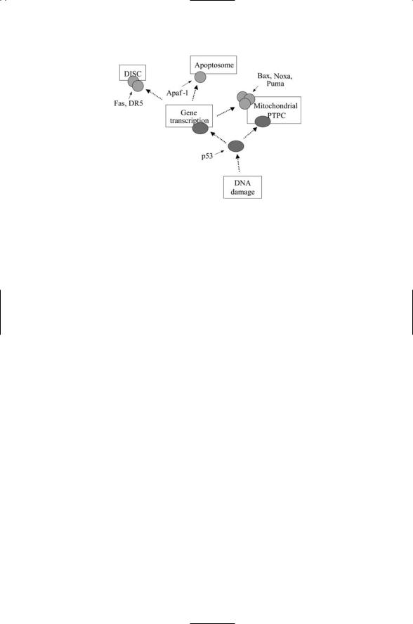
FIGURE 15.9. Regulation of apoptosis by p53: DNA damage signals are conveyed to p53 in the form of posttranslational modifications of specific residues such as phosphorylation on Ser46. In response, p53 (along with p63 and p73, not shown) stimulates transcription of apoptosis-promoting proteins acting at the DISC, PTPC, and apoptosome. It also diffuses to the mitochondria where it interacts with antiapoptotic Bcl-2 family members to increase membrane permeability.
depicted in Figure 15.9, p53 can increase the numbers of propapoptotic proteins. These include death ligands and receptors at the DISC, the Apaf-1 scaffold proteins at the PTPC, and proapoptotic multidomain Bax, and BH3-only proteins, at the apoptosome. Second, p53 can translocate to the mitochondria where it can interact with the antiapoptotic Bcl-xL and Bcl-2 proteins to increase the efflux of Cytochrome c from the mitochondria. Recall from the last chapter that some of the most lethal mutations to p53 occur in its DNA binding domain. This domain contains the binding site for attachment to the Bcl-2 proteins so these mutations not only destroy p53’s ability to bind DNA in the nucleus but also negate its ability to interact with the Bcl-xL proteins at the mitochondria.
15.19Anti-Cancer Drugs Target the Cell’s Apoptosis Machinery
The machinery involved in regulating apoptosis is a major target of anticancer therapies. The goal of these therapeutic strategies is to induce apoptosis in the malignant tissue while leaving healthy tissue alone. Components of the key control loci—DISC, apoptosome, and PTPC—are the primary targets of many of these strategies. One set of strategies revolve about causing apoptosis-inducing damage to DNA and other cellular components, while another set of approaches focuses on causing oxidative damage in the mitochondria in a way that stimulates the release of proapoptotic factors.