

Molecular and Cellular Signaling - Martin Beckerman
.pdfProblems 245
FIGURE 2 FOR PROBLEM 10.1. Receptor-ligand bond dissociation data:The most probable unbinding forces are plotted against the logarithm of the loading rate. (a) The data fall along a single line. (b) There is a sharp break in the data, producing two linear regimes.
stant loading forces are applied to the bonds; that is, forces F of the form F = rt, where r is the loading rate and t is the time. Under these conditions the most probable unbinding force F* is related to the rate and to the characteristic length by
F * = |
kBT |
ln |
r |
|
. |
(10.4) |
|
|
|
|
|
||||
|
x |
koff (0) |
kBT |
|
|
|
|
|
|
|
x |
|
|
|
|
|
|
|
|
|
|
|
|
When the most probable unbinding forces of receptor-ligand pairs are plotted against the logarithm of the loading rate the data typically fall along a straight line. Two commonly encountered situations are depicted below. In (a), the data are distributed about a single straight line. What quantity does the slope of this line represent, and how does it relate to an energy landscape? In (b), there is a sharp break in the straight line so that there are two slopes. Interpret these results within an energy landscape picture, i.e., what does the energy landscape look like? Recall that typical thermal energies are on the order of 0.6 kcal/mol. For a distance x of 0.5 Åwhat is the corresponding force, expressed in Newtons?
References on the Theory of Slip and Catch Bonds
Bell GI [1978]. Models for specific adhesion of cells to cells. Science, 200: 618–627. Dembo M, et al. [1988]. The reaction-limited kinetics of membrane-to-surface adhe-
sion and detachment. Proc. R. Soc. Lond. B, 234: 55–83.

11
Signaling in the Endocrine System
The endocrine system consists of a number of glands containing specialized secreting cells that release signaling molecules into the bloodstream, other bodily fluids,and extracellular spaces.Glands of the endocrine system include the adrenal gland, the hypothalamus, and the parathyroid, pineal, pituitary, and thyroid glands. Hormone-secreting cells are found in many locations in the body. The thymus produces a variety of hormones that regulate lymphocyte growth, maturation and homeostasis. The pancreas contains groups of hormone-secreting cells organized into pancreatic islets (the Islets of Langerhans) that secrete insulin, glucagon, and somatostatin. Hormonesecreting epithelial cells are strategically situated in the gastrointestinal tract, gonads, kidneys and placenta, where they direct the mitogenic activities of cells and tissues that wear down and require constant replenishment, and angiogenetic activities that produce new blood vessels.
Cells of the endocrine system produce several different kinds of hor- mones—peptide and polypeptide hormones, steroids synthesized from cholesterol, hormones derived from amino acids, and hormones made from fatty acids. Some hormones are lipophilic while others are water soluble. Lipophilic hormones such as steroids, retinoids, and thyroids are small, relatively long-lived hormones able to pass through the plasma membrane and enter the cell where they bind nuclear receptors.Water soluble hormones are fairly short lived and bind to receptors embedded in the plasma membrane of the target cells. Two kinds of transmembrane receptors bind nonlipophilic hormones: Polypeptide hormones that promote growth and wound-healing are bound by receptor tyrosine kinases; all others are bound by G protein-coupled receptors, which will be discussed in the next chapter.
Polypeptide hormones are small compact molecules, ranging in size from 6 to 45 kDa. Epidermal growth factor (EGF) and nerve growth factor (NGF) were the first polypeptide growth factors to be discovered. These and other growth factors bind to receptor tyrosine kinases expressed by the appropriate recipient cells. Receptor tyrosine kinases are single chain proteins that pass through the plasma membrane once. They, along with nonreceptor tyrosine kinases, are exclusively found in metazoans. Several
247

248 11. Signaling in the Endocrine System
different classes of proteins help transduce polypeptide hormone signals into the cell. These elements include, besides receptor tyrosine kinases and the aforementioned nonreceptor tyrosine kinases, a variety of highly modular adapters, and several families of GTPases. Each of these classes of signal transducers will be examined in this chapter.
11.1 Five Modes of Cell-to-Cell Signaling
In the last chapter, signaling between cells was characterized as being either contact-mediated or short range or long range. This catalog of signaling modes can be generalized to encompass cytokine signals in the immune system, long range hormone signals in the endocrine system, and neurotransmitters and neuromodulators in the nervous system. The expanded ensemble of signaling modes contains five entries—endocrine, paracrine, autocrine, juxtracrine, and synaptic. In endocrine signaling, the sending and receiving cells can be far apart. As noted in the introductory remarks, the signals are conveyed from sending glands to distant points in the body by bodily fluids.
Paracrine signaling refers to signaling in which molecules are secreted directly into the extracellular spaces by an originating cell, and these molecules travel no more than a few cell diameters to reach a target cell. The signaling molecules are often immobilized by elements of the extracellular matrix and bind to receptors on the surface of their cellular targets. This mode of signaling is utilized by hormone-releasing epithelial cells, by cytokine-releasing endothelial cells and fibroblasts, by chemotropic factor-releasing cells that direct growth cones and migrating cells, and by morphogen-releasing cells in organizing centers that direct cell fate during development (to be discussed in Chapter 13).
Some hormones that are secreted act back on the cells releasing them.
In this autocrine mode of signaling, positive feedback serves to amplify the initial weak signals. This mode is encountered in the immune system (see, for example, signaling by IL-2 shown in Figure 9.1) and also in the endocrine system as part of growth factor signaling. It is made use of during development, and if not properly regulated leads to uncontrolled growth and proliferation in a variety of cancers.
Cell-to-cell (juxtacrine) signaling is frequently conveyed by direct contact between a receptor on one cell and a cell surface-bound ligand, or counterreceptor, on an adjacent cell. The nondiffusible signaling molecules participating in juxtacrine signaling may pass through the plasma membranes or may be tethered to the outer leaflet of the plasma membranes by GPI anchors. This mode of signaling includes situations where cell surface receptors bind ligands that are components of the extracellular matrix.
Synapses are junctions formed to promote sustained signaling between cells. Signaling through chemical synapses is the preeminent form of sig-

11.2 Role of Growth Factors in Angiogenesis |
249 |
naling between nerve cells. This synaptic signaling involves the diffusion of signaling molecules (neurotransmitters) across a synaptic cleft between the membranes of a pair of preand postsynaptic nerve cells. The membrane and submembraneous regions of synapses are specialized structures highly enriched in many different kinds of signaling molecules. Like synaptic signaling between neurons, immune system T cells use synaptic signaling when contacting antigen-presenting cells.
11.2 Role of Growth Factors in Angiogenesis
Several families of growth factors coordinate and control the formation of new blood vessels during angiogenesis. Whenever new tissue is made additional blood vessels must be created to supply oxygen and nutrients and remove waste products. The process of vascular development takes place in several stages. In the earlier stages, called vasculogenesis, precursor vascular endothelial cells migrate, differentiate, proliferate and assemble into an initial set of vascular connections of uniform size. In the later stages, termed angiogenesis, the initial latticework of tubules is refined through further differentiation, sprouting and branching to form a mature vascular system. The fully developed network contains large arteries that branch into progessively smaller blood vessels terminating in capillaries, and it contains a return system of progressively larger venous structures.
The endothelial and smooth muscle cells that form the lining and sheathing of blood vessels coordinate their activities by sending and receiving a variety of chemical signals. Some of the signaling molecules are vascular endothelial cell-specific; others such as platelet-derived growth factor (PDGF) and basic fibroblast growth factor (bFGF) are not. Members of the vascular endothelial growth factor (VEGF) family are prominent among the vascular specific growth factors. They are required for vasculogenesis and also play a role in angiogenesis. A second family of growth factors, the angiopoietins, binds to endothelial cell line-specific Tie receptors. They work together with endothelial-specific ephrins and with the VEGFs to coordinate and control angiogenesis. The VEGFs and angiopoietins operate through a paracrine mechanism to signal and recruit nearby cells. The ephrins remain attached to the plasma membrane of their originating cell and signal bidirectionally through a juxtacrine mechanism.
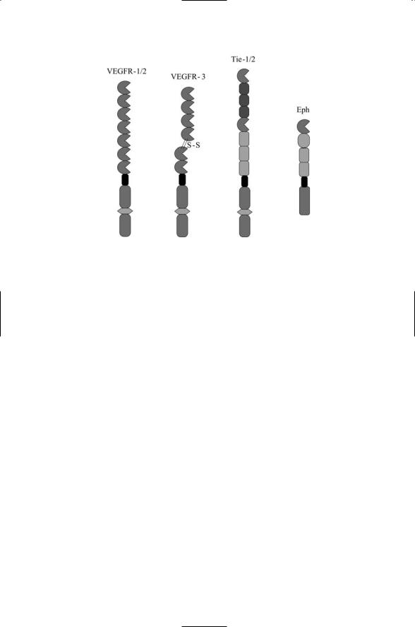
As shown in Figure 11.1, the VEGF, Tie, and Eph receptors involved in vascular development and repair are mosaic proteins. Their extracellular regions are composed of multiple domains arranged in a linear fashion.They differ from the receptors discussed in the last chapter by the presence of a tyrosine kinase domain in their cytoplasmic segment. The angiopoietins and ephrins promote the remodeling and branching, vascular maturation, and the attachment to surrounding support cells and extracellular matrix. For example, Ang1 promotes vascular maturation and stabilization while Ang2
250 11. Signaling in the Endocrine System
FIGURE 11.1. Angiogenesis receptors: The VEGFR-1/2 proteins contain 7 Ig-like domains followed by a transmembrane segment and a cytoplasmic kinase domain split by an insert. The VEGFR-3 chain differs slightly from VEGFR-1/2 in that the set of tandem Ig domains is shortened by one and contains a disulfide bridge. Tie receptors 1 and 2 contain an Ig-like domain followed by 3 EGF-like repeats and then another Ig domain followed by 3 FNIII repeats, a transmembrane segment, and the split kinase domain. Eph receptors contain an Ig domain, a cysteine-rich region, plus 2 FNIII repeats, a transmembrane segment, and a cytoplasmic kinase domain.
maintains the vascular system in a plastic state. The ephrins were discussed earlier in connection with growth cone navigation. The vascular-specific
Ephrin-B2 ligand and EphB4 receptor function in a roughly analogous manner in angiogenesis. They are differentially expressed in endothelial cells. The Ephrin B2 ligand functions as an arterial marker, and the EphB4 receptor operates as a venous marker.These signaling proteins help delineate the boundary between blood vessels that become arteries and those that become veins.
11.3 Role of EGF Family in Wound Healing
Growth factors of the EGF family coordinate and control wound healing and the replacement of cells in tissues that undergo rapid turnover. Wound healing is a complex process. Even in the case of a simple skin cut it requires the participation of agents of the nervous system to generate a pain signal, and elements of the immune system to generate an inflammatory and immune response. New tissue must be grown to replace the damaged material. The old damaged vasculature must be taken down and new vascula-

11.4 Neurotrophins Control Neuron Growth, Differentiation, & Survival |
251 |
ture installed, and the underlying connective tissue must be remodeled and restored.
In a skin cut, gloss keratinocytes (skin cells) migrate into the region of injury, and upon arrival proliferate and mature. Signaling by members of the EGF family of ligands and their receptors is essential for the proper activities of keratinocytes during wound healing. Several EGF family ligands are involved in repairing a skin wound. Among these are TGFa, HB-EGF, AR, and betacellulin. These ligands are synthesized by keratinocytes, along with their EGF receptors and supply proliferation and migratory signals, through an autocrine mechanism. In response to these signals the keratinocytes increase their production of integrins. Binding by these integrins to the ECM mediates substratum adherence by the keratinocytes and triggers the expression of collagesase-1. This enzyme degrades the ECM, a step required for remodeling and repair of damaged tissue. Continued autocrine signals maintain the expression of collagenase-1 during migration and repair.
Besides the skin, wound repair occurs in the gastronintestinal tract, in muscle tissue following injury, and at sites of chronic inflammation, to name just a few common examples. Primary sources of epithelial growth factors include the submaxillary salivary gland, the duodenal Brunner’s gland, epithelial cells in the mammary gland, and the kidneys. Growth factors are released into bodily fluids—saliva, serum, milk, and urine—and travel to sites of injury and growth. Epithelial cells at many places in the body are continuously renewed and growth factors are released locally near these sites as well. In these situations the growth factors operate in autocrine and paracrine manners rather than through an endocrine mechanism.
11.4Neurotrophins Control Neuron Growth, Differentiation, and Survival
Programmed cell death is an important process in the developing nervous system. It is used to remove cells that are extraneous, cells that were transiently produced to help in development but are no longer needed, and cells that failed to wire up properly to other cells. Neurotrophins are trophic factors, chemicals that stimulate growth and development. Neurotrophins such as NGF regulate the decision to survive or die, promote axonal and dendritic outgrowth, branching and remodeling, and help control synapse formation and plasticity. Other examples of trophic factors are ciliary neurotrophic factor (CNTF), glial-derived neurotrophic factor (GDNF),
PDGF, and bFGF.
The neurotrophin family of ligands and receptors contains four ligands and two receptors. The four ligands are NGF, brain derived neurotrophic factor (BDNF), neurotrophic factor 4 (NT4), and NT3. These ligands bind to two kinds of neurotrophin receptors, namely, p75NTR receptors and Trk receptors. As shown in Figure 11.2, the p75NTR receptor contains a cyto-

252 11. Signaling in the Endocrine System
FIGURE 11.2. Neurotrophin receptors: The p75NTR receptor has four cysteine-rich repeats (CR1–CR4), a transmembrane segment, and a cytoplasic death domain. The Trk receptors have a cysteine segment (C1), 3 leucine-rich repeats (LRRs), a second cysteine segment (C2), 2 immunoglobulin-like domains (Ig1, Ig2), an insert transmembrane segment, and a cytoplasmic kinase domain. All of the neurotrophins can bind p75NTR. NGF binds the TrkA receptor; BDNF and NT4 bind the TrkB receptor, and NT3 binds the TrkC receptor.
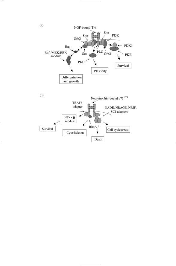
plasmic death domain, while the Trk receptors possess a cytoplasmic tyrosine kinase domain. The two receptors operate using different sets of adapter proteins to convey a variety of messages into the cell as indicated in Figure 11.3. The Trk receptors utilize Shc and Grb2 adapters, while p75 employs TRAFs and a set of four or more adapters (Figure 11.3b) to convey death and arrest messages. In general, p75NTR receptors convey apoptotic messages and Trk receptors convey survival messages, but as illustrated in Figure 11.3 the full picture is far richer. These and other growth factor receptors work together along with each other and with integrins and cadherins to convey messages to most or all control points in the cell. These include regulatory sites for gene expression (to promote growth and differentiation), sites of focal contact between cells and ECM (to control movement, adhesion, cell shape, and outgrowth), and mitochondrial control sites for apoptosis (to regulate death versus survival decisions).
11.5Role of Receptor Tyrosine Kinases in Signal Transduction
Receptor tyrosine kinases transduce signals from polypeptide growth factors into the cell. Except for receptors such as p75, polypeptide growth factors bind to members of the receptor tyrosine kinases (RTKs), singlepass transmembrane receptors possessing an intrinsic tyrosine kinase activity. These receptors contain three functional regions: an N-terminal
11.5 Role of Receptor Tyrosine Kinases in Signal Transduction |
253 |
FIGURE 11.3. Ligand-bound Trk and p75 neurotrophin receptors, and associated adapters: (a) NGF-bound Trk receptor. Second messengers and associated signaling intermediates involved in activating the PKB and PKC have been omitted to keep the figure from becoming too cluttered. (b) Neurotrophin-bound p75 receptor. Abbreviations: p75NTR-associated cell death executor (NADE), neurotrophin receptor-interacting melanoma-associated antigen (MAGE) homolog (NRAGE), neurotrophin receptor-interacting factor (NRIF), Schwann cell factor-1 (SC1).
extracellular region that contains one or more ligand-binding sites; a transmembrane segment; and a C-terminal cytosplasmic region that contains a catalytic domain and several phosphorylation sites.
The RTKs that bind polypeptide growth factors are highly modular linear arrays of domains. Among the domains present in the extracellular regions of these glycoproteins are Ig domains, fibronectin type III domains, cys- teine-rich domains and the EGF ligand-binding domains. A juxtamembrane region is situated just inside the transmembrane helix region and, as mentioned above, there is a tyrosine kinase domain near the C-terminal region. Docking sites are opened when (auto)phosphorylation of tyrosine residues in the activation loop of the kinase domain turns on that domain’s catalytic

254 11. Signaling in the Endocrine System
activity resulting in a subsequent phosphorylation of tyrosine residues. When this happens, proteins containing phosphotyrosine recognition modules are able to dock at these sites.
Like other single pass transmembrane receptors, ligand binding in the extracellular N-terminal region does not perturb the electrostatic environment enough to stabilize a conformation sufficiently different in its C- terminal cytoplasmic region to serve as a signal. Instead ligand binding promotes the formation of stable receptor dimers and oligomers. It is the bringing together of two or more cytoplasmic domains that initiates signaling inside the cell. In the case of RTKs, the close proximity of the two cytoplasmic kinase domains and accompanying phosplorylation sites enable cross or autophosphorylation of tyrosine residues.
The transmission of a signal into the cell interior by a receptor tyrosine kinase following ligand binding occurs in two stages. The first step is the autophosphorylation, or cross phosphorylation, of tyrosine residues in the activation loop of the catalytic domain. The bringing together of the cytoplasmic portions of the RTK triggers this step with the result that the catalytic activity of the kinase domain is turned on. The kinase domain then catalyzes the phosphorylation of one or more tyrosine residues in the cytoplasmic region outside of the catalytic domain. This second step makes available docking sites for signal proteins. Key to this second function is the presence of protein-protein recognition modules, compact domains that recognize short peptide sequences containing phosphorylated tyrosine, serine, and threonine residues.
Receptor dimers and oligomers can be formed in response to ligand binding in more than one way. Ligand-mediated dimerization triggers human growth hormone (hGH) signaling. As discussed in Chapter 9, a single ligand first binds to one receptor and then attaches to the second receptor to form a bridge that holds together a 1 : 2 ligand-receptor complex. A different strategy is observed in EGF-induced dimerization. In this case, the extracellular portions of the two receptors form the bridge resulting in a 2 : 2 complex. In more detail, EGF-EGFR binding triggers conformational changes that expose a loop on each receptor molecule. These loops bind to each other to form the bridge. As noted earlier, the ErbB2 receptor does not require a ligand for its activation. This happens because the bridging loop on this receptor is constitutively in the open for bridging conformation. The formation of a bridge by a pair of loops in contact with a ligand dimer is depicted in Figure 11.3.
11.6Phosphoprotein Recognition Modules Utilized Widely in Signaling Pathways
Signaling proteins diffuse from one location to another, are activated and turned off by binding and posttranslational modifications, and are recruited, assembled, and disassembled into signaling complexes at cellular control

11.6 Phosphoprotein Recognition Modules |
255 |
points. The arrangements of these events into signaling pathways so that a sequence of signaling steps can occur in the correct order at the right time in the right place is made possible through the use of modular signaling domains. Some of these domains supply a sort of glue that enables one protein to bind another, and for that operation to be followed by yet another binding operation leading to the recruitment of proteins and formation of signaling complexes. Other domains are specifically designed to recognize the presence of posttranslational modifications such as the addition of phosphoryl groups. To see the utility of this kind of modular domain, consider the receptor tyrosine kinases just discussed. The result of ligand binding is the presence of one or more phosphorylated tyrosine residues near the cytoplasmic C-terminus of the receptor. If this event cannot be recognized and responded to, signaling ends.
Src homology-2 (SH2) and phosphotyrosine binding (PTB) domains recognize short peptide sequences containing phosphotyrosine residues. The SH2 domain is the prototypic recognition module. It consists of approximately 100 amino acid residues, and is found on proteins that assemble and disassemble in the vicinity of the plasma membrane in response to autoand cross-phosphorylation of tyrosine residues in the cytoplasmic portion of transmembrane receptor molecules. It is the first discovered and largest family of modules that recognize phosphorylated tyrosines. SH2 domains recognize phosphorylated tyrosines and a specific flanking sequence of three to six amino acid residues located immediately C-terminal to pY. Phosphotyrosine binding (PTB) domains also recognize short peptide sequences containing phosphotyrosine, but, in contrast to SH2 domains, amino acid residues N-terminal and not C-terminal to pY belong to the recognition sequence. PTB domains recognize turn-loop structures and, in particular, hydrophobic amino acid residues five to eight residues located N-terminal to pY.
14-3-3 proteins are small, 28 to 30 kDa in size. There are at least seven isoforms of the mammalian 14-3-3 proteins; they are abundant and are expressed in all eukaryotic cells. Unlike most of the other domains discussed in this section, 14-3-3 domains are not embedded in larger proteins but exist as independent units that form homodimers. These proteins bind with high affinity to peptide sequences containing phosphoserine residues followed by a proline two positions towards the C-terminal. As dimers they are able to bind to signaling molecules such as Raf-1 containing tandem repeats of phosphoserine motifs. The 14-3-3 proteins localize their binding partners within the cytoplasmic compartment and keep them sequestered from the nucleus and membranes. These proteins operate in the growth and apoptosis pathways and in cell cycle control.
Forkhead-associated (FHA) domains are conserved sequences of 55 to 75 amino acid residues. Whereas SH2 and PTB domains recognize sequences containing phosphorylated tryosines, FHA domains can recognize peptide sequences containing phosphorylated threonines, phosphorylated serines, and phosphorylated tyrosines with a pronounced affinity for