

Molecular and Cellular Signaling - Martin Beckerman
.pdf360 15. Apoptosis
the cell surface as apoptotic bodies containing fragments of DNA and other macromolecules.
Malfunctions in the cellular machinery that controls apoptosis are encountered in many disease situations. Excessive apoptotic cell death occurs in Alzheimer’s disease, Parkinson’s disease, Huntington’s disease, and ALS (Lou Gehrig’s disease). Too little cell death is a hallmark of cancer. In B-cell leukemia, for example, key regulators of the decision circuitry that determines whether a cell survives or dies are overexpressed.
Apoptosis is suppressed and because population control is lost leukemia develops.
15.1Caspases and Bcl-2 Proteins Are Key Mediators of Apoptosis
Apoptosis is largely carried out by caspases, proteolytic enzymes that catalyze the cleavage of specific molecules and groups of molecules in response to activating signals. Caspases target critical repair, splicing, and replication components, they cut up membranes and cytoskeleton regulators, and they destroy cellular DNA. They also stimulate the expression of markers on the cell surface that tag the cell for orderly destruction and engulfment by neighboring cells. This orderly disassembly of a cell occurs in a way that prevents damage due to leakage.
Bcl-2 proteins are a second group of proteins intimately involved in apoptosis. They function as sensors and regulators of the apoptosis program. They were first identified in B-cell lymphomas and, since then, mutated forms have been found in a variety of cancers. These proteins are characterized by the presence of one or more Bcl-2 homology (BH) domains, the ability of some to form pores in internal membranes, and their propensity to either promote or inhibit the release of apoptotic signals and agents from the internal membranes.
Apoptosis can be initiated by death signals sent into the cell from other cells and by stress signals generated within the cell. Signals sent by other cells instructing a cell to undergo apoptosis are received by death receptors belonging to the tumor necrosis factor (TNF) family. When a death ligand binds the death receptors, a death inducing signaling complex is formed that initiates the apoptosis process. Death signals are also triggered by cellular stresses detected internally in organelles such as the endoplasmic reticulum, Golgi, nucleus, and mitochondria. Apoptosis signaling is sent in response to conditions such as irrevocable DNA damage in the nucleus, unfolded protein stresses in the ER, and oxidative stresses in the mitochondria. Two loci, one within the mitochondria and the other just outside that organelle, serve as the main control points for the launching of apoptotic responses.
15.2 Caspases Are Proteolytic Enzymes Synthesized as Inactive Zymogens |
361 |
15.2Caspases Are Proteolytic Enzymes Synthesized as Inactive Zymogens
The activity of enzymes that chop up and digest molecules is tightly controlled in the cell. One common strategy for controlling proteolytic enzymes, or proteases, is to synthesize them in an inactive form that requires further processing for their activation. One common kind of inactive form is a zymogen, a proenzyme containing a prodomain that must be removed in order to create an active form of the enzyme. Zymogens are converted to catalytically active forms by their proteolytic cleavage into two or three pieces followed by assembly of the catalytic subunits into complexes. Examples of proteases synthesized as zymogens include digestive enzymes that reside in the stomach (pepsin) and pancreas (trypsin), and also include blood-clotting enzymes (thrombin).
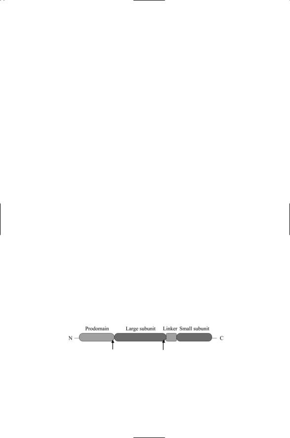
Caspases are cysteine aspartate-specific proteases. They break peptide bonds after Asp residues, i.e., at Asp-X sites, and possess a highly conserved cysteine residue in their catalytic site. Caspases are synthesized as zymogens. They contain a prodomain in the amino terminal region that regulates the proenzyme, followed by a large domain, approximately 20 kDa in size, and then a small domain, about 10 kDa in size. The proenzyme is activated by proteolytic cleavage at two Asp-X sites, one situated at the end of the prodomain and the other separating the large and small domains (Figure 15.1).
The tetramer is the functional (active) form of the caspase. It is constructed in several stages. In the first stage, two zymogens associate to form a zymogen homodimer. Adjacent large and small subunits (left hand pair and right hand pair) are part of the same polypeptide chain with the small units placed on the inside and the large subunits on the outside. In the next stage, each of the polypeptide chains making up the caspase zymogen dimer is cleaved at the Asp-X sites. These cuts induce conformational changes in the subunits that are part of the caspase activation process because they bring the caspases closer to conformation supporting catalysis. The similarity in conformations can be seen in Figure 15.2 where zymogen and caspase homodimers are compared side-by-side. Differences in the crucial loops L1
FIGURE 15.1. Caspase domain structure: The N-terminal prodomain is followed by a large subunit, a linker, and a small subunit. The location of the two Asp-X cleavage sites is indicated in the figure by arrows. Following cleavage, two large and two small subunits associate to form a caspase tetramer with the two small subunits inside and two large subunits on the outside.

362 15. Apoptosis
FIGURE 15.2. Structure of caspase-7 homodimers as determined by means of X-ray diffraction measurements: (a) Procaspase-7 zymogen homodimer, and (b) Caspase- 7 homodimer. The figures were prepared using Protein Explorer using atomic coordinates deposited in the Brookhaven Protein Data Bank under accession codes 1K88 (a) and 1K86 (b).
to L4 that determine the catalytic cleft are clearly visible in the left and right hand panels of the figure. The caspases are still not fully activated in the more open caspase form, but a final set of changes occurring during substrate binding renders the caspases fully active.
15.3Caspases Are Initiators and Executioners of Apoptosis Programs
More than a dozen different kinds of caspases have been identified in mammals. Those that have been found in humans have been placed in one of three groups in Table 15.1. Caspases belonging to Group I are associated with inflammatory responses. These caspases were first named for inter- leukin-1b converting enzyme (ICE), and then renamed caspases 1, 4, and 5. These enzymes process pro-inflammatory cytokines. The remaining two groups of caspases are specifically associated with apoptosis, either as initiators that convey signals through their proteolytic actions or as effectors that degrade cellular components. Group II caspases are effectors. They proteolytically degrade a variety of cellular components and are thus the executioners of the apoptosis program.
Group III caspases are apoptosis initiators. They act upstream of the effectors and activate them in response to proapoptotic signals and events. The four caspases appearing as Group III caspases all have large prodomains in which there is either a DED (death effector domain) or a CARD (capsase recruitment domain) protein–protein interaction domain.

15.4 There Are Three Kinds of Bcl-2 Proteins |
363 |
TABLE 15.1. Mammalian caspases and their roles in the cell: Group III initiator caspases act upstream of the Group II effector caspases. The executioners have small prodomains and require assistance of the initiators for their activation. Four element consensus sequences that the caspases recognize and cleave are presented in column 4. The most conserved residue in the consensus sequence is the Asp (D) residue proximal to the cleavage site while the residue in the fourth position mostly determines the substrate specificity.
|
|
Consensus |
|
Group |
Caspase |
sequence |
Prodomain |
|
|
|
|
I: ICE |
1 |
(WL)EHD |
Large, CARD |
|
4 |
(WL)EHD |
Large, CARD |
|
5 |
(WL)EHD |
Large |
II: Effectors |
3 |
DExD |
Small |
|
6 |
(ILV)ExD |
Small |
|
7 |
DExD |
Small |
III: Initiators |
2 |
DExD |
Large, CARD |
|
8 |
(ILV)ExD |
Large, DED |
|
9 |
(ILV)ExD |
Large, CARD |
|
10 |
(ILV)ExD |
Large, DED |
|
|
|
|
These regulatory sequences target the procaspases either to adapters bound to death receptors at the cell surface or to adapters positioned near mitochondria. At these locations the initiator caspases are positioned to respond to proapoptotic signals. Caspases 8 and 10 contain a pair of DEDs. Caspase 2 and 9 contain CARDs. The effector (Group II) caspases do not have a large prodomain in their N-terminus but instead possess a small N- terminal peptide. One other initiator caspase has been found—Caspase 12, a murine (mouse) caspase that is localized to the endoplasmic reticulum.
15.4 There Are Three Kinds of Bcl-2 Proteins
Bcl-2 proteins are central regulators of caspase activity and of the cell decision concerning whether or not to undergo apoptosis. Bcl-2 proteins can be grouped into three subfamilies according to their domain structure and their proor antiapoptotic activities (Table 15.2). The defining characteristic of the Bcl-2 proteins is the presence of one or more BH domains. The Bcl-2 and Bax subfamilies contain multiple BH domains while the Bad subfamily only possesses a BH3 domain.
There are four kinds of BH domains, designated as BH1 through BH4.
A typical Bcl-2 subfamily member contains at least three of the BH domains, namely, BH1, BH2, and BH3. In addition, it has a hydrophobic tail that anchors the protein to the outer membrane of mitochondria, the endoplasmic reticular membrane, and the outer nuclear envelope. Members of the Bcl-2 subfamily inhibit apoptosis by restricting membrane permeabil-

364 15. Apoptosis
TABLE 15.2. Bcl-2 family of apoptosis regulators: Listed are the numbers of amino acid residues in the proteins.
Bcl-2 subfamily: |
|
Bax subfamily: |
|
Bad subfamily: |
|
Antiapoptotic |
size (aa) |
Proapoptotic |
size (aa) |
Proapoptotic |
size (aa) |
|
|
|
|
|
|
A1 |
172 |
Bak |
211 |
Bad |
197 |
Bcl-2 |
239 |
Bax |
192 |
Bid |
195 |
Bcl-xL |
233 |
Bcl-xS |
170 |
Bik |
160 |
Bcl-w |
193 |
Bok |
213 |
Bim |
196 |
Boo |
191 |
|
|
Blk |
150 |
Mcl-1 |
350 |
|
|
Bmf |
186 |
|
|
|
|
Hrk |
91 |
|
|
|
|
Noxa |
103 |
|
|
|
|
Puma |
193 |
|
|
|
|
|
|
ity through interactions with mitochondrial membrane components and by binding to and sequestering members of the proapoptotic Bax group.
The proapoptotic Bax subfamily consists of proteins that possess a BH3 domain, a hydrophobic transmembrane tail, and at least one other BH domain. Some have a BH4 domain; others have BH1 and BH2 domains and perhaps a BH4 domain. Their 3-D structure is similar to pore-forming bacterial toxins. The Bax proteins interact with the proteins embedded in the outer mitochondrial membrane to increase membrane permeability, and they can form pores by themselves in membranes when they oligomerize.
The proand antiapoptotic, multi-BH domain proteins have electrostatic and structural properties that enable them to not only operate in the cytoplasm but also insert into the membrane to make pores. Their polypeptide chains are organized into sets of eight a-helices. There are three layers of two a-helices and a pair of short capping helices at one end of the chain. The organizaztion of the protein and the correspondence between helices and BH domains is presented in Figure 15.3. Several structural features support pore forming. The structure is fairly flexible and so can rearrange itself with little energy penalty. Two of the helices—a5 and a6—are able to span the membrane. There are several disordered, flexible regions, and there are three cavities. Charge-wise, the bottom of the protein is lined with basic (positively charged) residues that complement the acidic (negatively charged) membrane surface, and there is a pronounced hydrophobic cleft surrounded by basic residues. The picture that emerges from examinations of these structures is that of a5 and a6 along with the corresponding a5 and a6 helices from dimerization partners forming a pore, with a2 to a4 forming a binding groove, and the C-terminus forming an anchor.
The Bad subfamily is referred to as the BH3-only family. Some members of this group have a transmembrane anchor while others do not. When activated by proapoptotic stimuli, these proteins translocate to the mitochondria and stimulate apoptotic responses. BH3-only proteins function as cellular sentinels. In unstressed cells Bid, Bim, and Bmf are immobilized in

15.5 How Caspases Are Activated 365
FIGURE 15.3. Structure of the antiapoptotic protein Bcl-xL: Shown in part (a) of the figure is a ribbon diagram of the portions of the protein whose structure could be determined through X-ray crystallography (i.e., highly disordered regions are not included in the model). Gray-scale shadings highlight the four BH regions. The correspondence between BH regions and the eight a-helices is presented in part (b) of the figure. The figure was generated using Protein Explorer from Brookhaven Protein Data Bank entry 1AF3.
the cytoplasm. Bid is a sensor of death signals sent into the cell through the death receptors, and is localized in the vicinity of the death receptors. Bim is sequestered at microtubule associated myosin V motors where it awaits activation by cytokine and other stress signals. Bmf is also immobilized at myosin V motors where it responds to loss of cell attachment (anoikis) signals. Two other BH3-only proteins, Bik and Blk, function in the endoplasmic reticulum as sensors of cellular stress. The remaining BH3-only proteins are regulated at the transcriptional level. Noxa and Puma are transcribed in a p53-dependent manner and may be regarded as DNA damage sensors, while Hrk and Bim are upregulated in response to growth factor deprivation and cytokine withdrawal.
15.5 How Caspases Are Activated
Caspases are activated by external suicide instructions and internal stress signals. Cells receive death instructions from other cells. These messages are conveyed by cell-to-cell messengers called death ligands. The messages are received by death receptors embedded in the plasma membrane. The death messages are transduced into the cell interior through a multiprotein signaling complex formed by the activated death receptors. This complex is

366 15. Apoptosis
called the death-inducing signaling complex, or DISC. The DISC is the control point for external signal activation of initiator Caspases 8 and 10 and signal to the BH3-only sensor protein Bid.
The counterpart to DISC for internal stress signals is a signaling complex called the apoptosome. This multiprotein signaling complex is formed just outside the mitochondria in response to internal stress signals. Initiator caspase 9 is activated at this control point in response to the stress-induced release of proapoptotic factors from the mitochondria. The release of the mitochondrial factors is triggered by activity at the mitochondrial control point called the permeability transition pore complex, or PTPC, where multidomain Bcl-2 proteins are localized and BH3-only sensor proteins converge when activated.
Several families of positive and negative regulators control the caspase machinery. The regulatory proteins ensure that apoptosis is not triggered inappropriately in response to random perturbations and aberrant signals. External and internal signals are integrated together, and both contribute to the live or die decision. If strong signals are sent into the cell instructing it to undergo apoptosis, and strong stress signals are also present within the cell, the decision is fairly simple—caspases will be activated and the cell will die. If, as is normally the case, neither external nor internal death signals are present the cell will live. All other situations are more complex. Cellular context comes into play through adjustments in the expression levels of the positive and negative regulators that determine the set, balance, or commitment point for apoptosis. The remainder of the chapter is devoted to exploring how the apoptosis control system works.
15.6Cell-to-Cell Signals Stimulate Formation of the DISC
The death receptors that transduce the death messages into the cell belong to the tumor necrosis factor (TNF) superfamily. The TNF superfamily in humans includes 19 Type II ligands, single-pass transmembrane proteins with cytoplasmic N-terminals, and at least 29 receptors, mostly Type I (extracellular N-terminals). Recall from Chapter 9 that these receptors are widely expressed in the immune system where they respond to variety of growth, proliferation, and death signals. Their extracellular region is characterized by the presence of from 2 to 5 repeats of a cysteine-rich motif containing a number of disulfide bridges. Their cytoplasmic portions contain docking sites for several different kinds of adapters that mediate the recruitment of key signaling elements to the receptors.
The death receptor family includes the TNF-a, Fas/Apo-1, and TNFrelated apoptosis-inducing ligand (TRAIL) receptors (Table 15.3). The receptors and ligands operate as homotrimeric proteins. Signaling begins when a trio of ligands binds to a trio of receptors. This event triggers the

15.7 Death Signals Are Conveyed by the Caspase 8 Pathway |
367 |
TABLE 15.3. Members of the TNF family of receptors containing death domains in their cytoplasmic region: Abbreviations—Death receptor (DR); TNF-related apoptosis inducing ligand (TRAIL).
Death receptor |
Alternative name(s) |
Fas |
Apo-1, CD95 |
TNF R1 |
CD120a |
DR3 |
Apo-3 |
TRAIL R1 |
Apo-2, DR4 |
TRAIL R2 |
DR5 |
DR6 |
|
|
|
recruitment of the adapters to the cytoplasmic portion of the receptors leading to the assembly of a DISC. In forming a DISC, the TNF-associated death domain (DD) proteins are first recruited to the cytoplasmic portion of the TNF receptors and bind by means of their death domains. Proteins with death effector domains (DEDs) bind next, and other binding events follow. In this manner, a DISC is formed with FADD, TRAF2, TRADD adapters serving as a starting point for three pathways—Caspase 8, NF-kB, and MAP kinase, respectively.
15.7Death Signals Are Conveyed by the Caspase 8 Pathway
The Caspase 8 pathway (Figure 15.4) begins when the Fas-associated death domain (FADD) protein is recruited to the nascent DISC. Zymogens with large prodomains such as Caspase 2, Caspase 8, and Caspase 10 are brought into close proximity with one another at the FADDs and as a result can form zymogen dimers and act on themselves to remove their prodomains. Several caspase molecules can enter, become activated, and leave, one after the other. Once activated these enzymes make contact with and activate the effector caspases such as Caspase-3.
One of the Caspase-3 substrates is the caspase-activated deoxyribonuclease (CAD) protein and its inhibitor ICAD. The CAD and ICAD proteins are the catalytic and regulatory subunits of a protein referred to as the DNA fragmentation factor, or DFF. The ICAD subunit remains bound to the CAD subunit in the absence of Caspase 3 activities and inhibits its enzymatic actions. Caspase 3 cleaves the ICAD thereby freeing the CAD
DNase and allowing it to move into the nucleus where it cleaves chromatin.
The BH3-only protein, Bid, is sited at the DISC. It functions as a sensor and as part of the circuitry that integrates externally and internally generated signals. As indicated in Figure 15.4, Caspase 8 cleaves the 22-kDa Bid sensor protein to create the 15-kDa tBid. Once formed, the tBids translo-

368 15. Apoptosis
FIGURE 15.4. Signaling through the DISC located at the plasma membrane: Depicted are the main positive-regulating signaling elements. Homotrimeric TNF ligands bind homotrimeric TNF receptors. In response, several adapter molecules are recruited to the cytoplasmic portion of the receptors. The FADD adapter mediates recruitment and activation of the Caspase 8 pathway. The TRAF2 and RIP proteins mediate signaling and activation of the IKKs, which disinhibit NF-kB from the IKKs. The TRADDs also signal to the JNK MAP kinase cascade.
cate to the mitochondrial PTPC where they promote the activities of proapoptotic Bcl-2 proteins. If the overall mix of BH proteins at the PTPC favors apoptosis, proapoptotic factors are released from the mitochondria leading to stimulation of the apoptosome and the sequential activation of
Caspase 3 and then Caspase 6, which further stimulates Caspase 8 activities in situations where the externally driven stimulation is weak.
15.8How Proand Antiapoptotic Signals Are Relayed
Proand antiapoptotic signals are relayed to the nucleus by NF-kB proteins and MAP kinases. The two other pathways activated by death ligand relay signals via NF-kB proteins and MAP kinase modules as is usual for other members of the TNF superfamily. These pathways were discussed earlier in
Chapter 9. Downstream signaling proteins establish contact with receptor interacting proteins (RIPs) and tumor necrosis factor receptor associated factor (TRAFs) that are recruited into the DISC. These pathways promote the expression of both proand antiapoptotic genes. The NF-kB module usually, but not always, acts to promote survival by raising the threshold for

15.9 Bcl-2 Proteins Regulate Mitochondrial Membrane Permeability |
369 |
apoptosis. The IKKs are the key point of convergence of a variety of regulatory signals triggered by cellular stresses. The IKKs are activated when recruited to and phosphorylated at the DISC. They, in turn, phosphorylate the IkBs, resulting in the activation of NF-kB. In their prosurvival mode, the NF-kB dimers translocate to the nucleus where they stimulate transcription of negative regulators of not only DISC signaling but also of mitochondrial proapoptotic signaling elements. As a consequence, the balance between proand antiapoptotic factors is shifted in favor of the antiapoptotic ones and apoptosis is prevented.
Recall from Chapter 9 that MAP signaling pathways convey stress (JNK and p38) and growth (ERK) signals from the plasma membrane to the nucleus where they influence transcription of a different sets of target genes. As shown in Figures 9.4 and 15.4, the JNK pathway begins in the DISC, where the TRADDs recruit and activate MEKK1, the first of the kinases in the MAP kinase cascade. The last kinase in the cascade is JNK. Once activated this kinase translocates to the nucleus where it phosphorylates members of the AP-1 family of transcription factors. A similar set of signaling steps occurs in the p38 pathway.
Transcription factors such as AP-1 family members and NF-kB reflect cellular conditions and prior signaling events in their transcriptional activities. Depending on the specific mix of coactivators and corepressors present, subunit composition, and the set of residues that have been phosphorylated (and acetylated), these transcription factors will either promote or inhibit apoptosis. For instance, the c-Jun transcription factor, an AP-1 family member activated at the end of the MAP kinase cascade, usually functions as a transcription activator, but can also function as transcription repressors when associated with corepressors. In NF-kB signaling, flexibility of response is provided by variations in subunit composition. Depending on Rel subunit composition, NF-kB will either promote apoptosis by expressing TRAIL receptors or inhibit apoptosis by expressing antiapoptotic survival factors.
15.9Bcl-2 Proteins Regulate Mitochondrial Membrane Permeability
Mitochondria occupy a central place in internal stress-induced apoptosis. As noted earlier in the chapter, the PTPC located in the mitochondria serves as a key control point for internal stress responses. The PTPC, or alternatively, the permeability transition pore (PTP), is formed at points of contact between the inner and outer mitochondrial membranes. These complexes are a conduit for the passage of agents such as Cytochrome c and Smac/DIABLO that trigger apoptosome assembly and activation of Caspase 9 (Figure 15.5). The PTPC encompasses the crucial inner membrane (IM) and outer membrane (OM) proteins along with key constituents