

Molecular and Cellular Signaling - Martin Beckerman
.pdf14.7 Translocated and Fused Genes Are Present in Leukemias |
339 |
mimicking the effect of active Wnt signaling. Similarly, certain mutations to b-catenin render that molecule insensitive to APC/GSK regulation leading to the same result. Mutations in APC or b-catenin are encountered in many colorectal tumors.
One of the main endpoints of signaling through APC/GSK/b-catenin is the transcription machinery in the nucleus. Upon translocation to the nucleus, b-catenin binds to the transcription factor Tcf-4/LEF to form a dimeric complex. Thus, the program of gene expression is altered when b- catenin is not properly regulated. A second end point of signaling through APC is the cytoskeleton. During cell division APC interacts with proteins in the ends of microtubules that form the mitotic spindles. APC contributes to the linking of microtubules to kinetochores, attachment sites on the chromosomes, and it contributes to signaling that the correct attachments are made. Cells containing mutated forms of APC exhibit chromosome instability, that is, they have incorrect numbers of chromosomes.
The Wnt pathway is not the only developmental pathway whose components can contribute to cancer. Aberrant signaling in the Hedgehog and TGFb pathways can promote cancer as well. Mutated forms of Patched and Smoothened, the two receptors that operate in the Hedgehog pathway, are encountered in basal cell carcinoma, the most common human cancer. Recall from the last chapter that Patched normally binds and sequesters Hh, and thus restricts its growth-promoting effects. It is therefore a tumor suppressor, while Smoothened, which transduces growth signals into the cell, acts as a growth stimulator. Gli transcription factors acting downstream from the aforementioned receptors in the Hedgehog pathway convey the growth signals to the nucleus. Raised levels of Gli activity are found in these cancers as a result of Gli mutations and aberrant receptor behavior.
As noted in the last chapter, Smads, acting as downstream signal transducers in the TGFb pathway, can promote or inhibit growth. In their capacity as growth inhibitors they inhibit G1 cyclin-dependent kinases Cdk4 and Cdk2 that act during the G1 phase of the cell cycle. (These kinases will be discussed later in the chapter.) The Smads stimulate production of p15Ink4b, a protein that binds to and inhibits these cdks, and prevents assembly of Cyclin D-cdk4 and Cyclin E-Cdk2 complexes. Mutations in components of the TGFb pathway—ligands, receptor, Smads (Smad2 or Smad4) and cofactors—are present in the majority of colon and pancreatic cancers.
14.7Translocated and Fused Genes Are Present in Leukemias
Member of the Bcl-2 family of proteins are central regulators of apoptosis. When these proteins are not expressed at the proper levels, the option of triggering apoptosis to remove cells that are either damaged or infected or

340 14. Cancer
growing out of control is no longer possible. As will be discussed in the next chapter, there are several types of Bcl-2 proteins. Some promote apoptosis while others inhibit it. Bcl-2 is a prominent member of the antiapoptosis group. Aberrant bcl-2 genes are present in 90% of follicular lymphomas or B-cell lymphomas for which the Bcl-2 protein is named. In these cells, a translocation has occurred. The gene encoding Bcl-2 located on chromosome 18 has been translocated and fused with an immunoglobulin heavy chain (IgH) gene located on chromosome 14. The result of this translocation, designated symbolically as t(14 : 18), is that the bcl-2 gene is positioned next to the enhancer for the antibody gene. Antibody genes are vigorously expressed and as a result of the translocation the Bcl-2 gene is continually overexpressed. The decision between proliferation and apoptosis is shifted towards the former, and the B-cells do not die off, as they should when they age. Furthermore, the B cells are resistant to radiation therapy and chemotherapy, since these forms of treatment work at least in part by stimulating apoptosis.
Several other fusion products play prominent roles in leukemias. Burkitt’s lymphoma is characterized by the translocation t(8 : 14) and subsequent fusion of the c-Myc gene with an IgH gene. Chronic myelogenous leukemia and acute lymphocytic leukemia involve the translocation of the abl gene from chromosone 9 to chromosome 22, where it fuses with the BCR gene. The altered form of chromosome 22 is known as the Philadelphia chromosome. All of these examples involve the joining of a gene with oncogenic capabilities (Bcl-2, c-Myc, and Abl) with a strongly expressed gene (BCR and IgH) leading to the continual overexpression of the oncogene.
14.8 Repair of DNA Damage
DNA repair systems are responsible for maintaining the integrity of the genome throughout the life of the cell. There are about 130 known genes that encode DNA repair proteins. These are organized into five DNA damage repair systems (Table 14.2). The base excision repair (BER) and nucleotide excision repair (NER) systems treat single strand damage. Another, the mismatch repair (MMR) system, corrects for mismatched base pairings generated during DNA replication and recombination. Finally, two interlocking systems, the homologous recombination (HR) and nonhomologous end joining (NHEJ) systems, handle double-strand breaks. Each of these systems contains an ensemble of enzymes and regulatory molecules that repair and rejoin DNA strands through sequences of chemomechanical operations.
As noted in Table 14.2, the base excision repair system removes bases damaged by UV radiation and X-rays, and by endogenous oxygen radicals and alkylating chemicals. The nucleotide excision repair system handles

14.8 Repair of DNA Damage 341
TABLE 14.2. The four kinds of DNA repair.
Type of damage repair |
System(s) |
Description |
Base excision repair |
BER |
Removes bases damaged by UV, X-rays, |
|
|
oxygen radicals, and alkylating agents |
Nucleotide excision repair |
NER |
Removes DNA lesions brought on by |
|
|
environmental agents |
Mismatch repair |
MMR |
Repairs damage occurring during DNA |
|
|
replication and meiotic recombination |
Double-strand break repair |
HR, NHEJ |
Repairs double-strand breaks caused by |
|
|
ionizing radiation, oxidative stresses, and |
|
|
other environmental factors |
|
|
|
bulky DNA lesions and damage arising from environmental agents such as polycyclic aromatic hydrocarbons compounds contained in cigarette smoke. Portions of chromosomes bearing genes that are actively undergoing transcription receive greater NER attention than DNA segments containing genes that are only rarely transcribed. Several disorders including skin cancers and Cockayne’s syndrome are associated with mutations in genes encoding members of the NER system.
The mismatch repair system monitors and treats damage occurring during DNA replication and meiotic recombination. Among AGTC combinations corrects A-G and T-C mismatches, as well as improper sequence insertions and deletions. Mutations in this repair systems lead to increases in the mutation rate and to cancer development. Mutations in two members of the MMR system, hMLH1 and hMSH2, are prominently linked to colorectal and other cancers.
The machinery for repairing double strand breaks is utilized for several purposes in the cell. It is central to V(D)J recombination in lymphocytes. DNA undergoing replication is subject to breaks and the machinery for repairing DNA participates in remedying broken replication forks. This machinery is involved in genetic recombination, the process whereby segments of DNA are exchanged between chromosomes. It is involved in both mitotic recombination, involving sister chromatids, and meiotic recombination, involving homologous chromosomes. Homologous recombination and nonhomologous end joining are responsible for repairing DNA doublestrand breaks. HR utilizes regions of DNA sequence homology from the sister chromatid to repair damaged DNA in an error-free manner. NHEJ does not utilize extensive regions of sequence homology to effect repairs and is not necessarily error-free.
Double-strand breaks (DSBs) can be generated endogeneously by oxidative agents and environmentally by ionizing radiation. This form of damage, while not as common as the other forms of DNA injury, can be extremely harmful. DSBs can lead to chromosomal translocations, deletions, and fragmentation. Mutations in several members of the machinery that repair

342 14. Cancer
double-strand breaks are associated with a variety of different cancers. This machinery will now be looked at in more detail.
14.9 Double-Strand-Break Repair Machinery
Proteins belonging to the DSB repair machinery form a number of distinct repair complexes. The protein responsible for repairing and rejoining broken strands of DNA are organized into several complexes, each consisting of proteins that come into physical contact with one another to carry out their repair functions. Proteins comprising the modules have been grouped together in Table 14.3. One of the key findings in studies of the DSB repair machinery is that some of the proteins are centrally involved in a number of rare, inherited genetic disorders whose study reveals important details. These participants in DSB repair are named for the disorders in which they were discovered. Examples of this naming convention include Ataxia-telangiectasia mutated (ATM) and Nijmegen breakage syndrome (NBS) proteins. Other rare, cancer-associated diseases involving DSB proteins are Fanconi anemia and Bloom syndrome.
The first entry in the table is ATM/ATR, two proteins that function as sensors and as signaling elements that convey damage signals to the p53 cellular controller and to the regulatory units of the repair modules. The ATM/ATR proteins are kinases and convey their signals by catalyzing the transfer of phosphoryl groups to their substrates. Three protein complexes follow the ATM/ATR entry. Each complex is made up of proteins, some
TABLE 14.3. Double-strand-break sensing, repair, and signaling.
Component |
Function |
Description |
ATM/ATR |
Signaling |
Phosphorylates p53, NBS1, and BRCA1 in |
|
|
response to DSBs |
Rad50 |
HR, NHEJ |
Forms an end-processing complex with Mre11 and |
|
|
NBS1 |
Mre11 |
HR, NHEJ |
Endonuclease activity |
NBS1 |
HR, NHEJ |
Regulatory |
Rad51 |
HR |
Searches for DNA homology; forms a complex |
|
|
with Rad52, Rad 54 and BRCA1,2 |
Rad52 |
HR |
Stimulates Rad51 activity |
Rad54 |
HR |
Chromatin remodeling |
BRCA1,2 |
HR |
Regulatory; chromatin remodeling |
Ku70 |
NHEJ |
Forms a heterodimer with Ku80; recruits and |
|
|
activates DNA-PKcs |
Ku80 |
NHEJ |
KU70 and KU80 bind to the free ends of the DSB |
DNA-PKcs |
NHEJ |
Regulatory; signals p53 |
XRCC4 |
NHEJ |
Forms a complex with DNA ligase IV |
DNA ligase IV |
NHEJ |
Ends reattachment |
|
|
|

14.9 Double-Strand-Break Repair Machinery |
343 |
FIGURE 14.4. Onset of double-strand-break repair: The ATM proteins senses double-strand breaks and conveys damage signals to the NBS1 protein in the Rad50/Mre11/NBS1 complex and to the BRCA1 protein in the Rad51/Rad52/ Rad54/BRCA1,2 complex. Damage signals are also sent to the Fanconi anemia (FA) complex, which includes the Bloom syndrome helicase BLM. In response, the FA ubiquitin ligase FANCL (L) activates the BRCA2 protein, which then joins the other DSB repair proteins at the repair site.
with regulatory functions and others with repair functions. The modules listed in Table 14.3 work synergistically and sequentially to make the repairs to the DNA. The early steps in operation of this repair system are depicted in Figure 14.4.
The first module, referred to as the Mre11 complex, is composed of the
Rad50, Mre11, and NBS1 proteins. This module participates in both homologous recombination and nonhomologous end joining along with several other DNA strand manipulations involved in mitosis and meiosis. The key function of this complex is to form flexible bridges between ends of broken DNA strands. The Rad50 proteins contain hooks, which join opposing Rad50 proteins protruding from the ends of broken DNA strands. The hooks join pairs of Rad50 proteins in the middle of the break while the ends of the Rad50s remain attached to the DNA strands via the Mre11 proteins.
The next module is the Rad52 group. The Rad52 family of proteins is responsible for homologous recombination. The first step in HR is the coating of the single strand DNA (ssDNA) with molecules of replication protein A (RPA). In the next step, molecules of Rad51 and BRCA2 are loaded onto the strands so that Rad51 displaces RPA. Both RPA and Rad51 remove secondary structure from the DNA to facilitate the pairing of sister

344 14. Cancer
chromatids. As indicated in Table 14.3, Rad52 facilitates the replacement of RPA by Rad51. The Rad51 and BRCA2 proteins form nucleoprotein filaments—BRCA2 anchors the filaments to the DNA and Rad51 proteins form the body of the filaments. The pairing of sister chromatids follows growth of the filaments.
The nonhomologous end joining (NHEJ) proteins complete the list. The last two entries in the table, XRCC4 and DNA Ligase IV, are needed in the final steps of DSB repair. These proteins, operating synergistically with members of the Ku module, rejoin the two ends of the DNA to complete V(D)J recombination and NHEJ.
14.10How Breast Cancer (BRCA) Proteins Interact with DNA
The Ataxia-telangiectasia mutated, Nijmegen breakage syndrome, and breast cancer proteins belong to two families of protein that operate in DNA damage detection, checkpoint signaling, and repair. One of these is a large family of proteins operating in DNA damage-signaling, and characterized by the presence of one or more BRCT (defined below) repeats. The second family is characterized by the presence of a COOH terminal phosphoinositide-3 kinase- (PI3K) like domain.
The BRCT proteins are characterized by the presence of a motif consisting of about 95 amino acid residues, sometimes repeated several times. This domain may be best thought of as an adapter module that enables members of this family of proteins to interact and participate with a variety of other proteins in DNA repair and recombination, transcription regulation, and cell cycle control. First discovered in the breast cancer 1 (BRCA1) protein, this motif is called a BRCA1 C-terminal (BRCT) domain. Its secondary structure consists of four beta strands plus two alpha helices.
BRCA1 (1863 amino acids) and BRCA2 (3418 amino acids) along with other members of the BRCT family are large multidomain proteins. BRCA1 contains a RING finger domain, a pair of nuclear localization signal sequences, and several BRCT repeats in its transcriptional activation domain. BRCA2 possesses several BRC repeats, an NLS and a transcriptional activation domain. Its BRCT region is followed by a COOH terminal region containing multiple single strand and double strand DNA-binding domains (Figure 14.5). Its tower domain is the site of several cancer-inducing mutations.
Inherited mutations in BRCA1 and BRCA2 genes predispose women to breast and ovarian cancer. Cancer cells having defective BRCA1 or
BRCA2 proteins exhibit several kinds of chromosome instability. They have chromosome breaks and incorrect chromosome numbers and abnormal centrosomes. These instabilities are produced by defective transcriptioncoupled DSB repair, failures in checkpointing, and improper regulation

14.11 PI3K Superfamily Members that Recognize Double-Strand Breaks |
345 |
FIGURE 14.5. Crystal structure of the COOH terminal portion of the BRCA2 tumor suppressor protein: Shown are the main secondary structure elements’ helices (corkscrew-shaped ribbons) and strands (flat arrow-shaped ribbons), and the overall organization of the COOH terminal portion of the molecule into five domains. The tower domain binds DNA and is the locus of a large number of cancer-inducing mutations. The oligonucleotide/oligosaccharide binding (OB) domains bind ssDNA. Two fragments of a DSS1 protein found in association with BRCA2, and needed for crystallization, are included in the figure. The figure was generated using Protein Explorer. The Brookhaven PDB accession number is 1MIU.
of centrosome duplication. The ensuring genetic instability then leads to further mutations and to tumor formation.
14.11PI3K Superfamily Members that Recognize Double-Strand Breaks
ATM, ATR, and DNA-PKcs are members of the phosphatidylinositol-3 kinase (PI3K) superfamily that recognize DNA double-strand breaks, and are involved in their repair. Members of this grouping, which includes ATM, ATR, and DNA-PKcs, have a characteristic PI3K homologous domain in their COOH terminal region. The PI3K-like domain of these molecules does not appear to possess any lipid kinase activity. For that reason these proteins are called PIK-related kinases. They are large serine-threonine kinases; the ATM gene product is a 350-kDa protein, and DNA-dependent protein kinase (DNA-PK) is even larger. The DNA-PK molecule contains a Ku heterodimer consisting of 70-kDa and 80-kDa Ku subunits, plus a 460 kDa catalytic subunit, DNA-PKcs. The Ku subunits are named after the

346 14. Cancer
first two letters of a patient found to be suffering from an autoimmune disorder associated with the Ku proteins.
DNA-PKcs and ATM are structurally similar, and both molecules are capable of binding DNA. DNA-PK not only participates in repair of DSBs caused by ionizing radiation but also is involved in V(D)J recombination used to generate diversity in cells of the immune system. A mutation in the kinase domain produces severe combined immunodeficiency (Scid) in mice and defects lead to ionizing radiation hypersensitivity in humans.
ATM and ATR are found on meiotic chromosomes and have a role in processing double-strand breaks generated during that process. These molecules serve as sensors of ionizing radiation-generated DSBs. They not only function as damage sensors but also as signaling molecules that relay damage signals to regulatory proteins that halt the cell cycle progression while repairs are made.
The ATM protein functions as a sensor of DNA double-strand breaks in the following manner. In the absence of DSBs, the ATM proteins exist as inactive dimers. Double strand breaks produced by, for example, ionizing radiation, induce alterations in chromatin structure. The alterations in chromatin structure trigger the dissociation of the ATM dimers resulting in autophosphorylation and activation. Once activated, ATM migrates over to p53, to NBS1 and BRCA1, and to cell cycle checkpoint proteins, and phosphorylates them at one or more sites.
14.12Checkpoints Regulate Transition Events in a Network
The cycle of cell growth and division passes through four stages (Figure 14.6). The first of these stages (G1) is a cellular growth stage that prepares the way either for entry into a cell division series of stages (S, G2, and M) or to growth arrest (G0), or to apoptosis. Cells that do not undergo growth arrest or apoptosis enter a synthesis (S) stage where the DNA is replicated in preparation for mitosis. This stage is followed by a further mitosis preparatory stage (G2) where RNAs and proteins required for mitosis are synthesized. Finally, the cell enters into mitosis (M) where the cell divides
FIGURE 14.6. The cell cycle: Chevrons mark the divisions of the cell cycle into its four stages. The slash placed late in G1 phase denotes the restriction (R) point and the second slash placed in mitosis represents the spindle checkpoints. The three nonmitosis stages are commonly referred to as interphase.

14.14 pRb Regulates Cell Cycle in Response to Mitogenic Signals |
347 |
to produce two offspring. The three stages preceding mitosis are collectively referred to as interphase.
Checkpoints are signaling pathways that ensure that a process does not begin before a prior process is completed. They control the order and timing of transition events in a network. DNA damage checkpoints, for example, sense the presence of damage, producing signals that halt progression through the cell cycle while the damage is repaired, and stimulate the transcription of repair genes to deal with it. The cell cycle includes several checkpoints. The decision to undergo cell division or not occurs late in G1 phase. This point is called the G1/S checkpoint, or alternatively, START in yeast and the restriction (R) point in mammals. Growth conditions signified by receipt of growth (mitogenic) signals from outside the cell are evaluated, DNA is checked for damage, and a decision whether to proceed through the cell division cycle is made. A second halt for checkpointing occurs during S phase and the third takes place late in G2 phase and is known as the G2 checkpoint. Checkpointing takes place during mitosis, as well. The checkpoints halt mitosis if the spindle is damaged or if the chromosomes are not properly attached to the microtubules.
14.13Cyclin-Dependent Kinases Form the Core of Cell-Cycle Control System
The timing and duration of events occurring during the cell cycle are tightly regulated by a control system containing at its core a family of serine/ threonine kinases called cyclin-dependent kinases (cdks) and a set of regulatory subunits called cyclins. Cyclin-dependent kinases are present throughout the cell cycle but must bind to their regulatory cyclin to become active. There are several different kinds of cyclin-dependent kinases, each kind is associated with a particular cyclin subunit (Figure 14.7).
The concentrations of the different cyclins and cyclin-dependent kinases oscillate with the cell cycle, and different cdk-cyclin pairs are active different times in the cell cycle. The correspondence between cdks-cyclins and the phases of the cell cycle are presented in Table 14.4. The cyclindependent kinases carry out their regulatory roles by phosphorylating the retinoblastoma protein pRb, which along with p53 lie at the heart of the cell’s control system.
14.14pRb Regulates Cell Cycle in Response to Mitogenic Signals
In multicellular organisms, neighboring cells send mitogenic signals that coordinate differentiation during development and trigger growth and proliferation. These events must be coordinated in order for the cells to work
348 14. Cancer
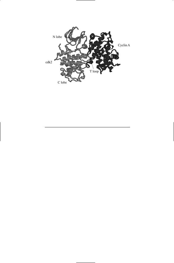
FIGURE 14.7. Crystal structure of the Cdk2—Cyclin A complex: Binding of Cyclin A to the kinase results in partial activation. Binding of the cyclin results in the extension of the catalytically important T loop. A further extension and full activation of the cdk is achieved through phosphorylation by a separate kinase. The figure was generated using Protein Explorer. The Brookhaven PDB accession number is 1FIN.
TABLE 14.4. Cyclins and cyclin-dependent kinases (cdks).
Cyclin |
Protein kinase |
Cell-cycle phase |
Cyclins D1 to D3 |
Cdk4, Cdk6 |
Late G1 phase |
Cyclin E |
Cdk2 |
G1/S phase transition |
Cyclin A |
Cdk2 |
S-phase |
Cyclin A |
Cdk1 (Cdc2) |
S-phase, G2-phase |
Cyclin B |
Cdk1 (Cdc2) |
G2-phase, M-phase |
|
|
|
together in a tissue or organ. The retinoblastoma protein, pRb, functions in a checkpoint role by inhibiting activation of an array of transcription factors, collectively designated as E2Fs, required for cell cycle progression. It keeps the gate shut at the G1 checkpoint where most of the decisions are made whether to proceed to mitosis. The pRb gatekeeper integrates a variety of positive and negative mitogenic signals conveyed as phosphorylation events and if conditions are propitious it opens the gate and enables progression through to mitosis.
The retinoblastoma protein binds to members of the E2F family of transcription factors. During G0 and G1 stages of the cell cycle the E2Fs are maintained in an inactive state by their pRb binding. During this time, pRb is underphosphorylated (hypophosphorylated), and is able to form stable complexes with these transcription factors. Its phosphorylation state changes near the G1/S boundary when the protein becomes hyperphosphorylated. Cyclins and their associated cyclin-dependent kinases play