

Molecular and Cellular Signaling - Martin Beckerman
.pdfReferences and Further Reading |
215 |
References and Further Reading
Lymphocytes
Abbas AK, Murphy KM, and Sher A [1996]. Functional diversity of helper T lymphocytes. Nature, 383: 787–793.
Fearon DT, and Locksley RM [1996]. The instructive role of innate immunity in the acquired immune response. Science, 272: 50–54.
Glimcher LH, and Murphy KM [2000]. Lineage commitment in the immune system: The T helper lymphocyte grows up. Genes Dev., 14: 1693–1711.
Goldrath AW, and Bevan MJ [1999]. Selecting and maintaining a diverse T-cell repertoire. Nature, 402: 255–262.
Pulendran B, Palucka K, and Banchereau J [2001]. Sensing pathogens and tuning immune responses. Science, 293: 253–256.
Rissoan MC, et al. [1999]. Reciprocal control of T helper cell and dendritic cell differentiation. Science, 283: 1183–1186.
NF-kB Signaling Node
Hoffmann A, et al. [2002]. The IkB-NF-kB signaling module: Temporal control and selective gene activation. Science, 298: 1241–1245.
Li QT, and Verma IM [2002]. NF-kB regulation in the immune system. Nature Rev. Immunol., 2: 725–734.
Senftleben U, et al. [2001]. Activation by IKKa of a second, evolutionary conserved, NF-kB signaling pathway. Science, 293: 1495–1499.
Silverman N, and Maniatis T [2001]. NF-kB signaling pathways in mammalian and insect innate immunity. Genes Dev., 15: 2321–2342.
MAP Kinase Modules
Cowan KJ, and Storey KB [2003]. Mitogen-activated protein kinases: New signaling pathways functioning in cellular response to environmental stress. J. Exp. Biol., 206: 1107–1115.
English J, et al. [1999]. New insights into the control of MAP kinase pathways. Exp. Cell Res., 253: 255–270.
TRAFs
Arch RH, Gedrich RW, and Thompson CB [1998]. Tumor necrosis factor receptorassociated factors (TRAFs)—A family of adapter proteins that regulate life and death. Genes Dev., 12: 2821–2830.
Chung JY, et al. [2002]. All TRAFs are not created equal: Common and distinct molecular mechanisms of TRAF-mediated signal transduction. J. Cell Sci., 115: 679–688.
Toll and Toll-Like Receptors
Aderem A, and Ulevitch RJ [2000]. Toll-like receptors in the induction of the innate immune response. Nature, 406: 782–787.
Hemmi H, et al. [2000]. A Toll-like receptor recognizes bacterial DNA. Nature, 408: 740–745.

216 9. Signaling by Cells of the Immune System
Medzhitov R, and Janeway C, Jr. [1997]. Innate immunity: The virtues of a nonclonal system of recognition Cell, 91: 295–298.
Medzhitov R, Preston-Hulburt P, and Janeway CA, Jr. [1998]. A human homologue of the Drosophila Toll protein signals activation of adaptive immunity. Nature, 388: 394–397.
TNFs
Bodmer JL, Schneider P, and Tschopp J [2002]. The molecular architecture of the TNF superfamily. Trends Biochem. Sci., 27: 19–26.
Locksley RM, Killeen N, and Lenardo MJ [2001]. The TNF and TNF receptor superfamilies: Integrating mammalian biology. Cell, 104: 487–501.
Jaks, STATS, and Hematopoietins
Guthridge MA, et al. [1998]. Mechanism of activation of GM-CSF, IL-3, and IL-5 family of receptors. Stem Cells, 16: 301–313.
Heinrich PC, et al. [2003]. Principles of interleukin (IL)-6-type cytokine signaling and its regulation. Biochem. J., 374: 1–20.
Levy DE, and Darnell JE, Jr. [2002]. STATs: Transcripional control and biological impact. Nature Rev. Mol. Cell Biol., 3: 651–662.
Ortmann RA, et al. [2000]. Janus kinases and signal transducers and activators of transcription: Their roles in cytokine signaling, development and immunoregulation. Arthritis Res., 2: 16–32.
The Interferon System
Goodbourn S, Didcock L, and Randall RE [2000]. Interferons: Cell signalling, immune modulation, antiviral responses and viral countermeasures. J. Gen. Virol., 81: 2341–2364.
Chemokines
Baggiolini M [1998]. Chemokines and leukocyte traffic. Nature, 392: 565–568. Cyster JG [1999]. Chemokines and cell migration in secondary lymphoid organs.
Science, 286: 2098–2102.
Rollins BJ [1997]. Chemokines. Blood, 90: 909–928.
T Cell Costimulation
Lanzavecchia A [1998]. License to kill. Nature, 393: 413– 414.
Schwartz RH [2001]. It takes more than two to tango. Nature, 409: 31–32.
Sharpe AH, and Freeman GJ [2002]. The B7-CD28 superfamily. Nature Rev. Immunol., 2: 116–126.
Viola A, et al. [1999]. T lymphocyte costimulation mediated by reorganization of membrane microdomains. Science, 283: 680–682.
Immunological Synapses
Coombs D, et al. [2002]. Activated TCRs remain marked for internalization after dissociation from pMHC. Nature Immunol., 3: 926–931.
Problems 217
Dustin ML, and Cooper JA [2000]. The immunological synapse and the actin cytoskeleton: Molecular hardware for T cell signaling. Nature Immunol., 1: 23–29.
Grakoui A, et al. [1999]. The immunological synapse: A molecular machine controlling T cell activation. Science, 285: 221–227.
Krummel MF, et al. [2000]. Differential clustering of CD4 and CD3z during T cell recognition. Science, 289: 1349–1352.
Lee KH, et al. [2003]. The immunological synapse balances T cell receptor signaling and degradation. Science, 302: 1218–1222.
Monks CRF, et al. [1998]. Three-dimensional segregation of supramolecular activation clusters in T cells. Nature, 395: 82–86.
Problems
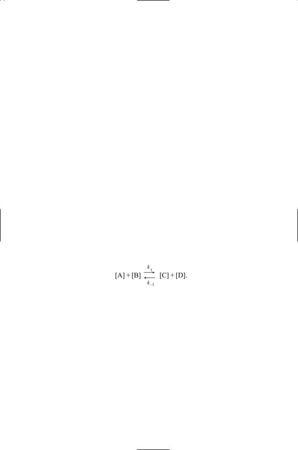
9.1There are two basic approaches used in theoretical studies of how signaling pathways operate: macroscopic and microscopic. The starting point for the macroscopic approaches is the law of mass action, and for the microscopic approach it is the principle of detailed balance. In both classes of approaches one constructs a set of equations that captures the salient features of the signaling process in terms of rates and other quantities that can be related to experiment. One then follows the evolution of the system on a computer to answer questions posed by the experimental data. In many situations, modeling a system and then simulating its behavior on a computer is the only way to see just what the biomolecules are doing over time.
The law of mass action is a statement that the rate of a chemical reaction is proportional to the concentrations of the reactants. To illustrate how this rule works, consider a reaction of the form
The rate of change of the product [C] is
d[C] |
= k1 [A][B] - k-1 [C][D]. |
(9.1) |
|
||
dt |
|
|
In the above, the rate of change of a reactant is expressed as the difference between its rate accumulation and its rate of loss through degradation and additional/reverse reactions. Expressions of this form can be simplified if some of the concentrations do not change appreciably over time, if some intermediates are formed rather transiently compared to the other steps, and if the reaction only proceeds in one direction or the other. For example, if the concentration of D does not change over time the second term in Eq. (9.1) becomes a linear one in which [D] is absorbed into the constant. An intermediate step, where a
218 9. Signaling by Cells of the Immune System
complex between A and B is formed, has been omitted in writing the above under the transient formation assumption.
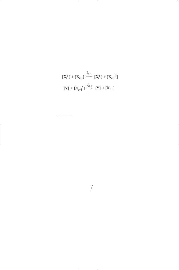
One can use the law of mass action to construct models of signaling pathways. Consider a set of kinases arranged in a pathway so that one kinase activates a second kinase and so. Let XiP denote the ith activated kinase in the pathway. Let Xi+1 be its substrate kinase, and let Y denote a protein phosphatase that acts on Xi+1P to return it to an unphosphorylated state. The following pair of expressions can describe the joint actions of XiP and Y on Xi+1:
The rate equation governing the buildup of the phosphorylated (i + 1)th kinase is
d Xi+1 |
p |
] = ki +1 [Xi p ][Xi+1 ] - ji¢+1 [Xi+1p ], |
|
|
|
||
[ dt |
|
(9.2) |
where [Y] has been assumed to be constant and was absorbed into the phosphatase rate constant j¢. Other equations can be constructed that describe interactions between ligands and receptors, between receptors and kinases/adapters, and between adapters and kinases. The result is a model of each of the steps in the signaling pathway.
(a)Derive the above expression, Eq. (9.2), for the rate of change in
[Xi+1P]. This equation can be solved to determine the steady-state (ss) concentration of Xi+1P.
(b)Show that the result, taking the concentration of the unphosporylated plus phosphorylated forms of Xi+1 to be a constant T, is
p |
|
|
|
[Xi p ]T |
|
|
[Xi+1 |
]ss |
= |
|
|
. |
(9.3) |
(ji¢+1 |
ki +1 )+ [Xi p ] |
|||||
This steady-state formula, Eq. (9.3), relates the stimulus [XiP] to the response [Xi+1P]. What is the behavior of the resulting stimulusresponse curve at low [XiP], and at high [XiP]? How does this compare to Michaelis–Menten kinetics discussed in the problems for Chapter 7?
9.2The reaction kinetics models based on the law of mass action make up a macroscopic description of the biochemical processes taking place. Alternatively one could employ a microscopic description that deals with the movement and interactions at the molecular and atomic levels rather than dealing with macroscopic quantities such as the concentra-

Problems 219
tions. The starting place for the microscopic approaches is the principle of detailed balance, which states that the rate at which a system makes a transition into a particular microstate from a preceding state is equal to the rate that the system will transition back out of that particular microstate to its predecessor. In mathematical terms the principle is
pi Pij = pj Pji , |
(9.4) |
where pi is the probability that the ith microstate is occupied (i.e., the occupation probability), and Pij is the transition probability from microstate i to microstate j. The above expression is a statement that the laws of physics are the same going forward or backward. In particular, the expression states that the probability that a state is occupied times the rate at which there is a transition out of that state to another is equal to the probability that the entered state is occupied times the rate at which that state is exited back to its predecessor. The occupation probabilities can be defined in terms of the Boltzmann factor b:
pi = 1 e -bEi , |
(9.5) |
Z |
|
where b = 1/kBT, Ei is an energy that characterizes the microstate i, and Z is a normalization factor. With this definition one can introduce a sampling algorithm that allows a researcher using a computer to simulate the evolution of a biochemical system over time in order to study its properties and see how it works. The sampling algorithm for the transitions is
Ïe -bDEij, |
Ej > Ei , |
(9.6) |
|
pij = Ì |
Ej £ Ei . |
||
Ó1, |
|
||
In the simulations, one starts out with the system in some initial state, then randomly picks a new microstate and calculates the energy difference DEij = Ej - Ei. If the energy difference is negative then the transition leads to a state of lower energy and the move is allowed. If the energy difference is positive, then the transition increases the energy of the system. One then selects a random number between 0 and 1. If that random number is less than the calculated Pij, the transition is allowed. Otherwise the move is rejected and another proposed transition is selected. Note that in this approach moves that increase the energy become possible allowing the system to transition over barriers and out of pockets in the energy landscape. This would not be possible in situations where only energy decreasing steps were allowed.
This algorithm, known as the Metropolis algorithm, was introduced in a landmark paper in 1953. The general method of randomly sampling a set of moves of a microsystem according to a probability distribution is known as the Monte Carlo method and is extensively used in several

220 9. Signaling by Cells of the Immune System
different forms to study how complex physical and chemical systems evolve over time.
Show that the Metropolis algorithm, Eq. (9.6), satisfies the detailed balance condition, Eq. (9.4), using Eq. (9.5). What are the results of this sampling procedure when the energy of state j just barely exceeds that of state i? What happens when the energy differences between the two states are large? The quantity Z appearing in the definition of the occupation probabilities is known as the partition function in statistical physics. Write an expression for it in terms of the Boltzmann factor b.
Theoretical (Modeling) Studies
Asthagiri AR, and Lauffenburger DA [2001]. A computational study of feedback effects on signal dynamics in a mitogen-activated protein kinase (MAPK) pathway model. Biotechnol. Prog., 17: 227–239.
Heinrich R, Neel BG, and Rapoport TA [2002]. Mathematical models of protein kinase signal transduction. Mol. Cell, 9: 957–970.
Tyson JJ, Chen KC, and Novak B [2003]. Sniffers, buzzers, toggles and blinkers: Dynamics of regulatory and signaling pathways in the cell. Curr. Opin. Cell Biol., 15: 221–231.

10
Cell Adhesion and Motility
Movement and adhesive contacts between cells and opposing surfaces are necessary for embryonic development and normal adult functioning of cells in the body. During development, cells do more than grow, multiply, and differentiate. They also segregate, migrate, and aggregate in forming tissues and organs. In adult life, vascular endothelial cells and fibroblasts migrate during wound healing, leukocytes migrate in response to infections, and cancerous cells migrate during metastasis.
Cells establish and maintain contacts with other cells and with the extracellular matrix. These contacts stabilize the aggregates of cells and enable them to work together to carry out their functions. Adhesive contacts underlie axon outgrowth during development of the nervous system. Paralleling the steps taken by migrating cells, growth cones, motile sensory structures enriched in adhesion molecules, guide the formation of connections in the nervous system.
Several different families of cell adhesion molecules—integrins, cadherins, immunoglobulin superfamily cell adhesion molecules (IgCAMs), and selectins—mediate adhesive contacts between surfaces in the body.
These molecules, acting as receptors and counterreceptors/ligands on opposing surfaces, are responsible for establishing and maintaining physical contact and communication between the extracellular matrix, the cell surface, and the cytoskeleton. These families of adhesion molecules, along with several different kinds of diffusible molecules and their receptors, also mediate axon outgrowth in the nervous system. Leukocyte adhesion and mobility will be examined in the first part of this chapter and axon outgrowth in the second.
10.1Cell Adhesion Receptors: Long Highly Modular Glycoproteins
The extracellular regions of cell adhesion receptors are mosaics of domains, strung together in a linear fashion. The domains may be composed of a number of short identical subdomains, referred to as repeats, or they may
221
222 10. Cell Adhesion and Motility
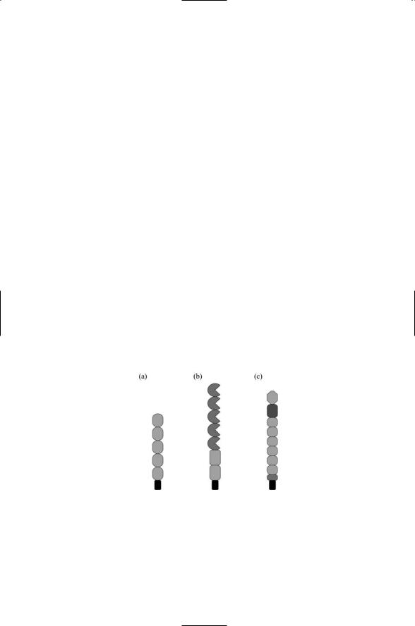
be erected from a mixture of two or more different domains, or they may be built from both repeats and single domains. Some of the domains in the extracellular regions are specific to a particular family and define membership in that family. Examples are cadherin domains in cadherins, and lectin domains in selectins. Other domains, most notably the immunoglobulin- (Ig) like domains and fibronectin type III repeats, occur in many different proteins. A representative sampling of extracellular regions of adhesionpromoting proteins is presented in Figure 10.1.
The extracellular regions of these proteins are attached to several different kinds of membane-associated segments. The most commonly encountered form of attachment is to a single-pass transmembrane segment that is followed by a short cytoplasmic tail. All of the proteins appearing is Figure 10.1 attach in this manner. There are three families of selectins— vascular endothelial (E), leukocyte (L), and platelet (P). All of these attach by means of transmembrane segments. So do 5/2 IgCAMs such as NCAM, 6/5 IgCAMs such as L1, and 4/6 IgCAMS such as deleted in colorectal cancer (DCC), but IgCAMS such as Contactin attach by means of a GPI anchor. Most cadherins attach through a single-pass TM segment, but cadherins such as Flamingo attach by means of a seven-pass transmebrane chain and may signal through G proteins.
During an inflammatory/immune response, leukocytes emigrate out of the bloodstream and converge upon the injured tissue. The emigration process occurs in several stages. Leukocytes first establish a loose contact with the endothelial cells lining the vessel walls and begin rolling. Next, a hard adhesive contact is established, and finally the leukocytes crawl out of the blood vessel and migrate into the injured tissue. Adhesion receptors
FIGURE 10.1. Representative extracellular regions of proteins involved in cell adhesion: (a) Classical cadherins containing 5 cadherin domains N-terminal to the transmembrane segment shown in black. (b) Neural cell adhesion molecules (NCAMs) containing 5 Ig-like domains and 2 fibronectin type III (FNIII) repeats. (c) E- selectins containing an amino terminal lectin domain, an EGF domain, 6 short consensus repeats (SCRs), and a membrane proximal cleavage region.

10.2 Integrins as Bidirectional Signaling Receptors |
223 |
work synergistically with receptors for cytokines and soluble growth factor. They signal through many of the same pathways as growth factors and together promote cell survival, proliferation, and differentiation. The cytoplasmic domains of cell adhesion receptors make contact with the cytoskeleton, and the receptors help regulate cell shape and polarization, and cytoskeleton organization and motility. During metastasis cancer cells utilize a similar ensemble of mechanical and adhesion receptor signaling processes. In metastasis, cells detach from the surrounding tissue, migrate to remote sites elsewhere in the body using the lymphatic and circulatory systems, reattach, and then establish a colony. Signaling between the cell surface and the nucleus coordinates the expression of specific cell adhesion molecules at different stages in metastasis.
Cell adhesion receptors are both large and flexible and because of these properties present challenges in their study using high resolution NMR and X-ray crystallography methods. The solution to this technical challenge is to take advantage of their inherent modularity and characterize portions— fragments and domains—of these molecules. The resulting NMR and X-ray crystallography data together with electron microscopy results provide a core body of structural information on these essential proteins.
10.2 Integrins as Bidirectional Signaling Receptors
Integrins are membrane-spanning glycoproteins composed of noncovalently attached a and b subunits. In vertebrates, 18 distinct alpha subunits and 8 different beta subunits have been identified so far. Not all of the 18 ¥ 8 = 144 combinations of alpha and beta subunits can be formed. Instead, a far smaller number, namely, 24, ab heterodimers can occur. Each integrin subunit contains a large extracellular domain, a single transmembrane segment, and a short cytoplasmic domain. There is one exception to this rule: The b4 subunit has a large cytoplasmic domain. The a subunits are larger than the b subunits. The a subunits vary in size from 120 to 170 kDa (up to 1114 amino acid residues) while b subunits range in size from 90 to 100 kDa (up to 678 residues).
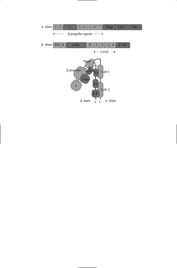
Integrins are multidomain proteins. The extracellular parts of the alpha and beta chains each contain five or more domains and some of these domains may consist of multiple subdomains. The domain organization of a representative integrin alpha chain and of a typical beta chain are depicted in Figure 10.2. In part (c) the two chains of the integrin molecule are bent in a V-shape that supports low affinity ligand binding. Not all integrin molecules contain inserted (I) or I-like domains. These ligand-binding domains are included in the model integrin depicted in the figure. The most important feature of this figure is the bent shape. The integrin molecule undergoes massive rearrangements in response to allosteric signals, and the far more open shapes that result mediate intermediate and high affinity
224 10. Cell Adhesion and Motility
(a)
(b)
(c)
FIGURE 10.2. Domain organization and assembly of the extracellular portions of an integrin containing inserted (I) and I-like ligand-binding domains: (a) Domain organization of the alpha subunit. (b) Domain organization of the beta subunit.
(c) Assembly of the alpha and beta chains in a V-shaped conformation showing the exposed ligand binding I and I-like domains at the end of the molecule. Abbreviations: b-tail (bT); hybrid (H); integrin-epidermal growth factor (I-EGF, or E); plexin/semaphorin/integrin (PSI).
ligand binding. X-ray crystallography data revealing this bent shape are presented in Figure 10.2.
Integrins and cadherins differ from most transmembrane signal transducers that transmit signals in one direction, from outside the cell inward. Integrin and cadherin receptors transmit signals in both directions—from outside the cell inward (outside-in) and from inside the cell outward (inside-out). In outside-in signaling, binding of the integrins to the ECM triggers changes in the pattern of gene expression. Inside-outside signals produce changes in the integrin conformation resulting in changes in adhesiveness. Integrins tie the ECM to the cellular cytoskeleton and anchor cells in a fixed position, while cadherins are a primary element of cell-to-cell junctions.
10.3 Role of Leukocyte-Specific Integrin
The leukocyte-specific integrin LFA-1 mediates the migration of T cells. The migration of T cells occurs in several stages. In the first stage, lamellipodia, broad flat structures, form and extend out from the leading edge of the cell. This stage is followed by a step in which new adhesive contacts are established and stabilized. The cell body then contracts, and the following edge, or tail, detaches. T cells use the aLb2 integrin, also called the leukocyte