

Molecular and Cellular Signaling - Martin Beckerman
.pdf144 7. Two-Component Signaling Systems
presence of a nucleophile to break bonds. A nucleophile is an atom or molecule that can donate an electron pair. A nucleophilic (nucleus-loving) process is one in which an electron pair is donated to another atom or molecule. There are a number of common nucleophiles. Deprotonated water molecules can serve as a nucleophiles. Alternatively, solvent-exposed oxygen atoms in hydroxyl groups in amino acid residue side chains can function as nucleophiles. The nucleophile in histidine kinase action is the imidazole ring structure on the histidine side chain. In this instance, the imidazole ring is said to attack the terminal phosphoryl group of the ATP molecule.
A key feature in the catalytic actions of the kinases is the presence of Mg2+ ions that facilitates the breaking of the bond holding terminal phosphoryl groups in place in ATPs. The Mg2+ion binds the ATP molecule prior to catalysis. During catalysis, the Mg2+-bound ATP and the substrate come together in a small region of space, usually the groove, pocket, or cleft formed by the kinase that was mentioned in the last paragraph. The combined actions of physical positioning and electrostatic shaping greatly accelerate the phosphotransfer reaction. These actions are said to stabilize the transition state, lowering the barrier for transfer of the phosphoryl group from donor to acceptor. (See Problem 7.1 for a further discussion of catalysis.)
7.4 The GHKL Superfamily
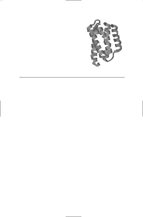
Histidine kinases along with a large number of ATPases form the GHKL superfamily that is quite distinct from the eukatyotic kinase STTE superfamily. The phosphoaccepting histidine, His48, is surrounded by glutamate, and lysine residues. The histidine, glutamate, and lysine residues form a hydrogen-bonding network. This arrangement and that of the four-helix bundle (Figure 7.4) also characterize the histidine-containing phosphotransfer (HPt) domains that are key components of phosphotransfer systems. The amino acid residues that make up the HPt domains fold in an up-down, up-down manner into four-helix bundles. The second helix in the bundle contains a highly conserved histidine residue that is exposed to the solvent and serves as the acceptor site for transfer of the phosphoryl group. The phosphoryl group covalently attached to the histidine residue is subsequently transferred to an aspartate residue located in a response regulator protein.
The ensemble of alpha helices and beta sheets that forms the threedimensional structure of the kinase core differs markedly from that of the serine/threonine and tyrosine kinases (STTKs). The histidine kinases closely resemble a number of ATPases. ATPases are proteins that couple metabolic energy stored in the high-energy bonds of phosphoryl groups to operate as pumps and motor proteins, all of which require that work be done. These proteins, including the histidine kinases, form the GHKL super-
7.5 Activation of Response Regulators by Phosphorylation |
145 |
FIGURE 7.4. The CheA P1 domain: The structure revealed by X-ray crystallography is that of a fourhelix bundle (A through D), plus a flanking helix
(E). A box shows the location of the conserved His48 residue plus conserved flanking residues on helix B. The figure was prepared using Protein Explorer with atomic coordinates from PDB file under accession code 1i5n.
TABLE 7.3. Domain structures of the response regulators.
Family |
Domain structures |
CheY |
Receiver domain |
OmpR |
Receiver domain, winged helix-turn-helix output domain |
FixJ |
Receiver domain, four-helix bundle output domain |
NtrC |
Receiver domain, ATPase domain, DNA-binding domain |
|
|
family. They each have a characteristic fold consisting of five helices and three sheets, and possess the conserved residues that define the N, G1, F, and G2 boxes. Crystal structures for ATPases such as DNA gyrase B, Hsp90, and MutL closely resemble those for CheA and EnvZ.
7.5Activation of Response Regulators by Phosphorylation
Response regulators can be placed into families according to their domain structure. Most transcription factors contain at least two domains. There is an N-terminal domain that functions as a receiver, and there is a C- terminal domain that operates as an output unit (Table 7.3). Receivers function as protein kinases that catalyze the transfer of the phosphoryl groups from the conserved histidines on histidine kinases to one of their own conserved aspartate residues. Output domains typically contain DNA-binding and regulatory sequences that enable the response regulators to function as transcription factors. Some response regulators, such as the chemotactic
CheY and CheB proteins (to be discussed later in this chapter) contain only the receiver domain and do not bind DNA. However, most proteins diffuse to DNA control points once the phosphoryl group is transferred to the receiver.
Proteins are continually in motion and undergoing shape (conformational) changes. There are atomic, group, and residue movements, backbone

146 7. Two-Component Signaling Systems
motions, side-chain motions, shifts in secondary structure elements, and domains movements. These motions enable a protein to continually sample a population of states, and this ability is important for binding and release, catalysis, and signaling. Rapid motions occur on picosecod-nanosecond time scale, involving only a few atoms and small energy changes. Slower motions occurring on longer microsecond to millimicrosecond time scales, involving appreciable numbers of amino acid residues and far larger energy changes.
NMR spectroscopy has been used to explore protein motions and conformational changes. Using these techniques, it has been observed that the active site of an enzyme undergoes conformation changes taking place on a time scale of microseconds to milliseconds. The regions that undergo these motions correlate with the region involved in protein-protein interactions. In examining what happens to the NtrC protein, it was found that NtrC dimers form in response to phosphorylation and then oligomers form that activate gene transcription. The picture that emerges from studies of NtrC and several other response regulators is one in which response regulators and activated in an allosteric manner by phosphorylation.
There are two populations of states. One population of states corresponds to the inactive protein, the other to the active form. Phosphorylation shifts the equilibrium between the two populations of states. Both populations preexist and are continually sampled, but phosphorylation shifts the balance in favor of the active form. Shifts in equilibria between two preexisting populations of conformational states, produced either by ligand binding or by covalent attachment of phosphoryl groups, are known as allosteric modifications. In an allosteric modification, binding at one location in the molecule alters how other portions of the molecule respond to their binding partners. These alterations may be thought of as a consequence of the conformational changes accompanying the shifts in equilibrium.
7.6Response Regulators Are Switches Thrown at Transcriptional Control Points
The commonality with pumps and other ATPases and GTPases seen with histidine kinases extends to response regulators. One of the key observations of how the pumps work is that they utilize a highly conserved aspartate residue in the active site along with several other residues. Like the pump ATPases, an aspartate functioning as the nucleophile, working along with two other aspartate residues, a lysine, a threonine or serine, and a pair of water molecules, form a dense network of bonds in this region of the molecule. Phosphorylation of the conserved aspartate is a key intermediate step in the energy-generating hydrolysis process carried out by the pumps. As they did for the histidine kinases, the common properties of the residues provide useful insights into how the response regulators function.
7.7 Structure and Domain Organization of Bacterial Receptors |
147 |
Response regulators are switches. The energy stored in the high-energy acyl phosphate bond is released when the switch is thrown. The response regulators involved in transcription activation form complexes that jointly drive a series of conformational changes leading to transcription activation. The throwing of the switch is achieved through hydrolysis of the bond, that is, the response regulators catalyze their own dephosphorylation. This occurs when response regulators and their target proteins come together so that the energy released through hydrolysis is used to drive conformational changes in the complexes that activate transcription.
7.7Structure and Domain Organization of Bacterial Receptors
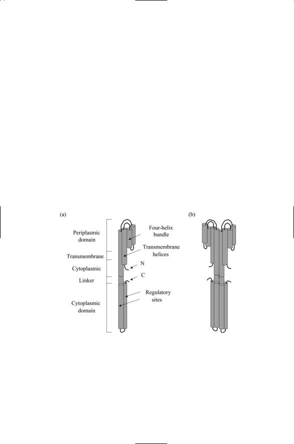
Bacterial receptors have a stereotypic structure and domain organization. Most bacterial sensor units are constructed from a single polypeptide chain that passes through the plasma membrane twice. The N-terminal lies in the cytoplasm at the end of a short segment. The chain threads out and then back through the plasma membrane as shown in Figure 7.5. The extra-
FIGURE 7.5. Tar receptor: (a) Tar monomer with labels denoting the various regions—The presence of several regulatory (methylation) sites in the cytoplasmic domain is denoted. (b) Tar dimer—The basic structure is that of a homodimer that appears as a 35-nm long helical bundle oriented perpendicular to, and passing through, the plasma membrane. The outer, ligand-binding, portion has the form of two four-helix bundles, one per dimer subunit. Two helices from each bundle span the plasma membrane. The cytoplasmic portion is arranged into a four-helix bundle, formed by two helical hairpins, one from each subunit.

148 7. Two-Component Signaling Systems
cytoplasmic looping portion serves as the ligand-binding region. The portion that is C-terminal to the transmembrane segments contains a linker followed by a long signaling domain. While most receptors conform to this plan, there are some receptors, most notably those involved in cell-to-cell signaling, such as the quorum-sensing (AgrC) receptor and the competence (ComD) receptor, that pass through the plasma membrane six times. The twoand six-pass receptors are bacterial counterparts to the oneand seven-pass receptors found in eukaryotes.
The EnvZ sensor unit is a representative example of the single chain, two-pass membrane topology. This form differs from the bacterial chemotaxis receptor Tar, which signals to a separate histidine kinase CheA protein. Tar is a two-pass receptor of the form described above, but some of the signaling pieces such as the histidine kinase domain are sequestered on a separate protein. NtrB represents a third kind of sensor unit; it is a soluble protein and does not attach to the plasma membrane at all.
7.8 Bacterial Receptors Form Signaling Clusters
Bacterial receptors form dimers, trimers, and higher order oligomers. The two subunits of the Tar dimer are linked through multiple bonds, and form a tightly packed and relatively inflexible set of parallel helices. The multiple bonds limit the possible relative movements of the signal helix in response to ligand binding, especially since the energy effects of ligand binding are small. The movements used to transmit a signal over a distance of 35 nm from the outside to the histidine kinase on the inside are modest. In response to ligand binding, the signal helix undergoes a 0.1 to 0.2 nm sliding, or piston-like, displacement towards the cytoplasm. The mechanism is an allosteric one in which ligand binding shifts the equilibrium towards a population of displaced conformations.
The cytoplasmic signaling module of the Tar receptor forms associations with two kinds of proteins. One of these is CheW that functions as a molecular scaffold. The other is CheA, the histidine kinase. CheA proteins are linked to the Tar signal helix through the CheW scaffold. Both CheW and CheA contain modules termed SH3 domains. These modules serve as interfaces for the protein-protein interactions leading to the assembly of not just a pair of receptors, scaffolds, and histidine kinases, but rather for the formation of an extended network of such units. The Tar receptors are not the only ones in these signaling clusters. Tar receptors are intermingled with another major receptor, the Tsr serine receptor, and with three less prominent chemotactic sensors. The signals sent through the receptors converge upon and are integrated by the CheA histidine kinase.
The net result of the interactions between receptors, scaffolds, and protein kinases is the formation at one pole of the bacterium of a signaling mesh. In this mesh, the extracellular portions of the receptors bind to

7.9 Bacteria with High Sensitivity and Mobility |
149 |
ligands, while the cytoplasmic portions of the receptors composed of sets of helical coils form a set of signaling “whiskers.” These whiskers bind to the scaffolds and through them to the protein kinases. The overall system functions much like a primitive nose, exhibiting considerable sensitivity to external stimuli over a broad range of concentrations.
The signaling complex operates through ligand-induced expansions and contractions. In the absence of ligand binding, the receptors bind CheA and thus stimulate the kinase (autophosphorylation) activity leading to structural changes within the array. Ligand binding inhibits these interactions. When ligands bind the receptors, the pistonlike movements of the signal helices cause the elements of the array to move apart, or disperse. This expansion is sufficient to shut down the autophosphorylation of histidine residues in CheA proteins and the attendant downstream signaling. The formation of large signaling complexes consisting of multiple transmembrane receptors is not restricted to bacteria. Lymphocytes belonging to the vertebrate immune system and neurons utilize extensive signaling complexes and meshes on their surfaces, too.
7.9 Bacteria with High Sensitivity and Mobility
Bacteria such as Escherichia coli and Salmonella typhimurium are highly mobile, free-swimming organisms. Bacteria such as Escherichia coli and Salmonella typhimurium are able to sense nutrients and noxious substances in their local environments, and, in response, swim towards the nutrients and away from dangerous chemicals. The essential components of the system that makes possible these chemotactic responses lie at the poles of the cell. A flagellar motor complex resides at one pole, and a sensor complex lies at the other. About 50 genes are involved in chemotaxis. Roughly 10 genes are needed to encode the sensor and signaling complex, and about 40 genes are needed to encode assembly and structural proteins of the flagellar motor. These two systems are coupled to one another. Signals are sent from the sensor system to control point at the flagellar motor switch.
The flagellar motor converts an electrochemical gradient into a mechanical torque that drives the bacterium forward at speeds up to 25 m/s. The propulsive force is provided by 6 to 10 flagella organized into a bundle. Each flagellum can either rotate clockwise (CW) or counterclockwise (CCW). A flagellum has an intrinsic handedness. When all flagella undergo CCW rotation, the bundle movement is concerted and the cell moves uniformly. If one or more flagella rotate in a CW fashion, the bundle flies apart, the movement is disorganized, and the cell tumbles. Because of its small size,
Brownian effects limit the effective straight-line distance that can be traversed without readjustments to the trajectory. The movement of a swimming bacterium consists of a series of smooth runs punctuated by periods of tumbling in which a new direction for running is chosen randomly. The
150 7. Two-Component Signaling Systems
seemingly random movements of the bacterium are biased towards attractants or away from repellents through modifications in the tumbling frequency arising from the sensory input signals.
7.10 Feedback Loop in the Chemotactic Pathway
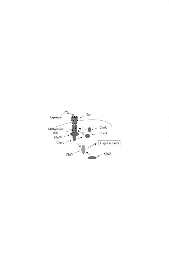
The chemotactic pathway contains a feedback loop that promotes robust behavior. The Tar, CheW, and CheA proteins constitute the sensor/transmitter unit of the two-component bacterial chemotaxis system. The CheY protein functions as the response regulator. The overall arrangement of the various units in the chemotactic system is depicted in Figure 7.6, and their functions are summarized in Table 7.4. CheY is a diffusible messenger molecule. Upon activation through phosphorylation it diffuses to the motor complex where it binds to motor switch protein FliM to promote CW motion and tumbling. CheY is phosphorylated by activated CheA, and dephosphorylated by the protein phosphatase CheZ. In the absence of
FIGURE 7.6. Tar chemotactic signaling pathway: The receptor (Tar) and histidine kinase (Che A) are encoded on separate chains. CheW is a scaffold that mediates the interactions between sensor and histidine kinase. CheY is the aspartate-bearing response regulator that mediates the cellular response (contact with flagellar motor).
TABLE 7.4. Tar chemotactic pathway.
Component |
Function |
Tar |
Receptor |
CheW |
Scaffold |
CheA |
Histidine kinase transmitter |
CheY |
Aspartyl response regulator |
CheZ |
Regulatory (phosphatase) |
CheR |
Regulatory (methyltransferase) |
CheB |
Regulatory (methylesterase) |
|
|

7.10 Feedback Loop in the Chemotactic Pathway |
151 |
CheZ, CheY dephosphorylation would take about 10 s, a period of time that is too long for rapid adaptation to changes in nutrient concentration. When CheZ is present it binds to CheY and reduces the time for dephosphorylation from 10 s to 1 s.
The remaining two proteins, CheB and CheR regulate the activity of the Tar receptor. Tar, like many receptors, is a conduit for two-way communication between outside and inside. Ligand binding on the outside alters the binding properties and catalytic actions of the cytoplasmic part of the protein. Binding events taking place in the cytoplasmic portion of the molecule influence the receptor’s outside ligand binding properties. The cytoplasmic part of the Tar signal helix contains binding sites for attachment of methyl groups. As a consequence, Tar and receptors like it are known as methyl-accepting chemotactic proteins (MCPs). CheR catalyzes the transfer of methyl groups to glutamate residues on the Tar signal helix using S- adenosyl methionine as the methyl donor. This activity is continual, and the addition of these methyl groups on the helix progressively reduces the binding affinity of the Tar receptors for their ligand. CheB does the opposite. It removes methyl groups from Tar and restores a high binding affinity.
CheB is activated by phosphorylated CheA, thereby forming a feedback loop—Tar signals to CheA, which signals to CheB, which signals back to Tar. As the ligand concentration builds up, and more and more Tar receptors are bound, CheA is less and less phosphorylated, CheB remains inactive, and only a few methyl groups are removed from Tar. Thus, at high ligand concentrations, the sensitivity of Tar to its ligand is reduced. In the absence of ligand binding, that is, at low ligand concentrations, CheA is phosphorylated and can activate CheB. CheB removes methyl groups from Tar, and returns Tar to a condition of high binding affinity and sensitivity to its ligand. By this means, the methylation level tracks the concentration. At low concentrations, Tar has few attached methyl groups, and it has a high affinity for its ligand. At high concentrations, many methyl groups are added, and Tar has a low binding affinity.
Tar can maintain its sensitivity to concentration gradients over some five orders of magnitude through this feedback mechanism. This process is called exact adaptation; the steady state tumbling frequency in a homogeneous ligand environment (no signal) is insensitive to the ligand concentration over a broad range of attractant or repellent concentrations. The chemotactic process is independent of specific values of the concentrations over a broad range. As a result, the bacterium can sense small spatial changes in concentration, i.e., it can detect small concentration gradients over many orders of magnitude in mean concentration. This ability is referred to as robustness. It is a consequence of the network connectivity, particularly that of the crucial feedback loop. This feedback loop compensates for the effect of ligand binding, thereby maintaining a robust performance regardless of the ligand concentration.
A bacterium is too small to be able to detect concentration gradients by comparing concentrations at different points along its body. Instead of using

152 7. Two-Component Signaling Systems
a spatial strategy, bacteria adopt a temporal one: They determine concentration gradients by comparing concentrations at slightly different times. This is equivalent to determining a spatial concentration gradient, since there is translational motion of the bacterium during the measurement time interval. The key to the temporal or time derivative method is to compare two different quantities, each measuring in some way concentration. One quantity is the concentration at an instant in time, represented as a percent of receptor occupancy. The second quantity is the receptor concentration a few seconds earlier, represented by the level of receptor methylation. There is a lag between receptor binding and the adjustment in methylation— CheA must be phosphorylated, and then CheB must be phosphorylated before there is an adjustment in receptor methylation. The lag between receptor binding and methylation of a few seconds serves as a memory that enables determination of temporal gradients.
7.11 How Plants Sense and Respond to Hormones
Plants use two-component systems and phosphorelays for sensing and responding to hormones. At certain times in its life a plant will undergo senescence, the state in which somatic tissues no longer required are broken down and their components reused in younger tissues. At other times a plant will undergo organ abscission, in which organs such as seeds and fruit are detached from the main body of the plant in a developmentally regulated manner. Plants, like animals, undergo wound-healing and mount defenses against pathogens. These developmental and defense processes are regulated by plant hormones such as ethylene (C2H4) and cytokinins.
Arabidopsis is a small weed, and it was the first plant species to have its genome sequenced. The Arabidopsis genome encodes 12 histidine kinases,
22 response regulators, and 5 HPt proteins. Histidine kinases used for sensing and responding to ethylene, cytokinins, and for osmosensing are listed in Table 7.5. All of these with the exception of ERS1 are hybrids. They contain a receiver domain and signal through HPt proteins.
The Arabidopsis response regulators (RRs) can be partitioned into two groups. A-type ARRs contain the required Asp phosphorylation site, as well
TABLE 7.5. Hormone and osmosensing in plants.
Histidine kinase |
Sensing function |
ETR1 |
ethylene |
ERS1 |
ethylene |
CRE1/AHK4/WOL |
cytokinins |
AHK2 |
cytokinins |
AHK3 |
cytokinins |
AtHK1 |
osmosensor |
7.11 How Plants Sense and Respond to Hormones |
153 |
as several other required residues, but lack the DNA-binding domain needed for operation as transcription factors. A-type ARRs are found in increased numbers shortly after a cell is exposed by cytokinins. B-type ARRs contain a DNA-binding domain in their C-terminal regions; nuclear localization signals (NLSs) needed for entry into the nucleus; and modules associated with Asp response regulation and signaling. Thus, the type-B ARRs contain all the components needed for operation as transcription factors.
Cytokinin ligands are recognized by the sensor histidine kinases CRE1,
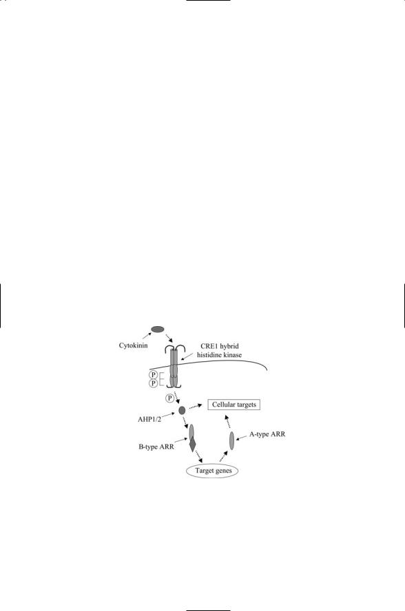
AKH2 and AKH3. Receptor-binding activates a phosphorelay from CRE1 to an HPt protein called AHP, which translocates to the nucleus and transfers its phosphoryl group to a type B response regulator. The type B RRs activate transcription of genes encoding Type A response regulators. The type A RRs operating in conjunction with AHPs and histidine kinases modulate the activities in other signaling pathways (Figure 7.7).
The ethylene-signaling pathway is erected in a way that combines histidine kinase action with MAP signaling cascades. Ethylene receptors are histidine kinases, but these proteins do not appear to transfer phosphoryl groups to aspartyl response regulators, but rather signal through one or more MAP-like serine/threonine kinases. The signaling mechanisms appearing in the Arabidopsis ethylene pathway bear a striking resemblance to those of the yeast high osmolarity (HOG) pathway discussed in the last chapter. In the ETR1 pathway, ethylene is a negative regulator of its recep-
FIGURE 7.7. Cytokinin signal transduction in Arabidopsis thaliana: Binding of the cytokinin ligand to the Cre1 receptor/histidine kinase activates a His-Asp phosphorelay. Signals are relayed through AHP1/2 types of HPt intermediates, and a B-type Arabidopsis response regulator (ARR), resulting in the expression of cytokinin-responsive genes including A-type of ARRs. These together with B-type ARRs and AHPs regulate downstream cellular targets.