

Molecular and Cellular Signaling - Martin Beckerman
.pdf30 2. The Control Layer
tures pack into one of a small number of geometries and the chains assume one of an equally small number of topologies. For example, beta sheets form layered structures with either alpha helices or other beta sheets on their faces. Alpha helices either distribute themselves about a core or form layered structures. The packing of helices about a core can be described by regular polyhedra. Three helices pack into an octahedron, four helices describe a dodecahedron, five helices form a hexadecahedron, and six helices pack into a icosahedron. The packing of sheets is more variable.
Most beta sheets pack into two-layered structures, with the sheets either aligned or orthogonal. Some beta sheets, notably in a/b proteins, form barrels. In barrel patterns the b sheets coil around to form a cylinder and the a helices are arranged on the cylinder surface.
The rules for chain topology state that knots in the polypeptide chain do not form nor do secondary structure elements cross through one another. Instead, the chain topology is minimally convoluted. Secondary structure elements that are adjacent in the polypeptide sequence prefer to pack in an antiparallel manner, and groupings of the form b-X-b, where X is either an a-helix or a b-strand in an adjacent sheet, are right handed. The helices pass through the folding domain and so the connections between helices form on the outside along the ribs of the polyhedra. Beta sheet topology forms hairpin, Greek key, or jellyrolls, depending on the number of strands (respectively, two, four, six).
2.7 Tertiary Structure of a Protein: Motifs and Domains
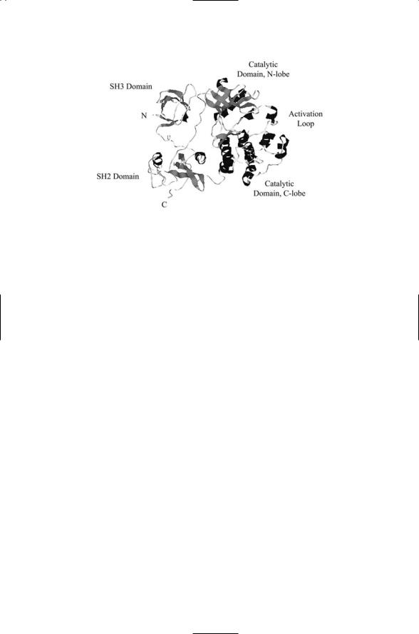
Proteins involved in signaling tend to be fairly large. They are constructed from a number of structural motifs and domains connected by loops that serve as flexible linkers. The Src protein shown in Figure 2.7 serves as an example of domain organization. Domains operate as functional units, and when proteins are formed with a specific set of domains they inherit those functions. Some signaling proteins contain just a few domains, while others contain large numbers of domains and because of that are termed mosaic proteins. Domain folds typically contain from 100 to 250 amino acids, but can be as small as 25 to 30 amino acids. In mosaic proteins, small domains along with larger ones often appear as tandem repeated sequences. Prokaryotic genes average 850 to 1000 bp in length while eukaryotic genes average 1400 to 1450 base pairs (bp) in size, and thus contain a greater number of these semiautonomous folding units.
The tertiary structure of a protein refers to the way the constituent domains and supersecondary structure elements come together to form the folded and physiologically functional protein. For most proteins this is the final layer of protein organization, and proteins are classified into structural families based on their domain composition. Listed in Table 2.2 are some of the more common families of proteins encoded in the human genome as
2.7 Tertiary Structure of a Protein: Motifs and Domains |
31 |
FIGURE 2.7. Domain organization of the Src protein, a nonreceptor tyrosine kinase: It consists of four domains—an N-terminal SH3 domain, a C-terminal SH2 domain, and two catalytic domains, connected by linker segments. Amino acid residues that form well-defined secondary structure elements are drawn as (dark) coiled ribbons and as (grey) planar ribbons or arrows. The coiled ribbons, like the cylinders used in the previous figure, denote alpha helices. The planar ribbons or arrows form layers that denote beta sheet secondary structures, either parallel or antiparallel. The SH2 domain is of the form a + b; its secondary structure resembles a twolayered sandwich with two short alpha helices plus a central beta sheet. The SH3 domain contains a pair of antiparallel beta sheets. Tyrosine kinases catalyze the transfer of phosphoryl groups to selected tyrosine residues. As can be seen in the figure the catalytic domains are mostly alpha helical, except for the N-lobe, where a prominent beta sheet component is present. Tyrosine kinases will be discussed in Chapter 11. The figure was generated using Protein Explorer with the Brookhaven Protein Data Bank (PDB) entry (accession number) 2 Src containing the atomic coordinates of Src determined by x-ray crystallography (to be discussed in Chapter 3).
defined by the presence of one or more of the domains listed in the first column. The sizes vary from as few as 25 to 33 amino acid residues for the minidomains to 120 amino acid residues for the PH domain. The domains are not mutually exclusive, and a single protein will usually contain one or more of several different kinds of domains. Minidomains are often arranged in the proteins as tandemly repeated elements. Including the term “repeat” as part of the name reflects this propensity.
Several kinds of changes have taken place in the human genome. New families of vertebrate genes appear that encode proteins belonging to the immune and nervous systems, and there is a new family (KRAB) of zinc finger transcription factors. A more widespread kind of change is the creation of new architectures, that is, of new arrangements of domains that can subsume new functions. A third type of change is the large expansion of existing families. Listed in Table 2.2 are representative examples of each of

32 2. The Control Layer
TABLE 2.2. Commonly encountered domains in the human: Sizes of the domains are given in terms of the numbers of amino acid residues. The fourth column lists the number of genes encoding the domains. All of these domains have signaling roles. The last column indicates the most prominent activities associated with proteins containing these domains. Im: immune system function; Extra: Extracellular adhesion; Trans: transcription regulation.
Domain |
Symbol |
Size |
Genes* |
Function |
Immunoglobulin |
Ig |
100 |
765 |
Im, Extra |
EGF-like |
EGF |
35 |
222 |
Im, Extra |
Fibronectin Type III |
Fn3 |
90 |
165 |
Im, Extra |
EF hand |
EF |
40 |
242 |
Im, Extra |
Ankyrin repeat |
Ankyrin |
33 |
276 |
Im, Extra, Signal |
Leucine-rich repeat |
LRR |
25 |
188 |
Im, Extra, Signal |
Cadherin |
Cadherin |
110 |
114 |
Im, Extra, Signal |
WD-40 repeat |
WD40 |
50 |
277 |
Signal |
Pleckstrin homology |
PH |
120 |
193 |
Signal |
Src homology 3 |
SH3 |
60 |
143 |
Signal |
Src homology 2 |
SH2 |
100 |
119 |
Signal |
PDZ |
PDZ |
80 |
162 |
Signal |
C2H2 zinc finger |
C2H2 |
30 |
706 |
Trans |
Homeobox |
Homeobox |
60 |
267 |
Trans |
RING finger |
RING |
50 |
210 |
Trans |
Krueppel-associated box |
KRAB |
75 |
204 |
Trans |
|
|
|
|
|
* Data from International Human Genome Sequencing Consortium [2001]. Nature 409: 860–921; Venter JC et al. [2001]. Science, 291: 1304–1351.
these categories. The table includes domain families prominently associated with immune and extracellular functions such as adhesion to the extracurricular matrix, the ECM, and also large numbers of domains that mediate interactions between control layer proteins and between proteins and
DNA. These domains and the signaling proteins that contain them will be examined in detail in later chapters.
2.8 Quaternary Structure: The Arrangement of Subunits
Signaling proteins are frequently assembled from distinct polypeptide chains. In these situations, the protein is said to have a quaternary structure. “Quaternary structure” describes how the individual chains, or subunits, are arranged in the protein. Several kinds of multichain proteins play important roles in the control layer. One type of multichain protein is the ion channel, where several subunits form a membrane-spanning pore that permits passage of ions and small molecules across the membrane. In these proteins, the subunits are tightly bound to one another. Other classes of proteins embedded in the plasma membrane, and serving as receivers of cell-to-cell signals, are assembled from multiple subunits that are much more loosely bound to one another. This kind of protein organization is

2.9 Many Signaling Proteins Undergo Covalent Modifications |
33 |
widely encountered in the immune system, and in these cases, unlike the ion channels, the different chains may perform distinct functions.
Some of the large signaling proteins that reside in the cytosol and serve as central organizers of the signal pathways are constructed from multiple subunits. Specific functions are sequestered in the different subunits of the proteins, and the ability of the protein’s subunits to associate and dissociate from one another in a key part of how the signaling system works. Alternative splicing is often used to generate different forms of specific subunits.
This makes possible the creation of many different combinations of subunits; each one specialized for a specific cell or tissue type. By this means many proteins of a similar kind, but each performing a slightly different task, can be constructed from a small instruction set.
2.9Many Signaling Proteins Undergo Covalent Modifications
Proteins can associate with the plasma membrane in several ways. If they pass completely through the plasma membrane they are able to convey a signal from one side to the other. Most receptors work this way relaying signals from outside the cell to the inside. Alternatively, the proteins may attach to either the extracellular side or the intracellular side by means of the tether. The tether, a type of post-translational modification, passes into one of the leaflets but does not pass all the way through to the other side of the plasma membrane. The proteins so anchored tend to form clusters of signaling elements, which may relay signals to nearby proteins that pass through the plasma membrane or not, depending on the composition of the cluster.
One of the most striking features of the control layer is its widespread use of post-translational modifications. Some of the modifications are made during protein processing and finishing as discussed in the last chapter. This type of modification is common in proteins that function in the plasma membrane. But many others are made later and are part of the signaling process itself. There are several different kinds of modifications all involving either the covalent addition of a group or structure, or the cleavage of the protein or a part thereof.
The main modifications are:
•Covalent attachment of anchors that tether signal proteins to one side or the other of the plasma membrane.
•Addition of sugar groups to the extracellular region of transmembrane signaling proteins.
•Proteolytic cleavage of anchors and proteins to free up the proteins for movement and conveyance of a message, or, alternatively, to keep them inactive and degrade them.
•Covalent addition and removal of phosphoryl, methyl, and acyl groups to cytosolic proteins.

34 2. The Control Layer
2.10 Anchors Enable Proteins to Attach to Membranes
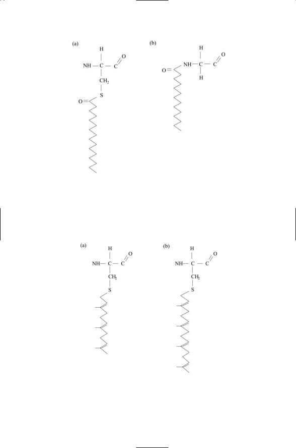
The region at and just below the plasma membrane is an important locus of signaling molecules. In response to the onset of signaling, many cytosolic proteins translocate to the plasma membrane where they are anchored to the cytosolic face and become activated. There are four types of modifications to cytosolic proteins that enable them to anchor to the plasma membrane and to the membranes of organelles: myristoylation, palmitoylation, farnesylation, and geranylgeranylation (Table 2.3, and Figures 2.8 and 2.9).
A crucial feature of acyl and prenyl anchors is that they are weak. Electrostatic interactions between N-terminal basic (+) residues and acidic (–) lipids contribute along with the anchor to the attachment. Because of the weak attachment the proteins can be detached easily by altering the electrostatic environment. This is usually done by phosphorylation, the covalent attachment of a phosphoryl group (with two negative charges). The modification to the basic residues weakens the electrostatic forces sufficiently to enable the protein to detach from the membrane and translocate to the cytoplasm to carry out its signaling function. This allows for reversible attachment and operation as an electrostatic switch.
The addition of the aforementioned fatty groups to proteins makes possible the proteins’ anchoring to the inner, or cytoplasmic, leaflet of the plasma membrane. A different kind lipid modification is made to proteins to allow them to be anchored to the outer, or exoplasmic, leaflet. These
TABLE 2.3. Membrane anchors: Abbreviations—cysteine (C); alipathic residue (residues with long hydrocarbon side chains such as leucine and isoleucine) (a); X = serine (S), methionine (M), alanine (A), or glutamine (Q); leucine (L).
Location and type of anchor |
Attachment |
Inner Leaflet |
|
Acyl type |
|
Myristoyl |
Preferentially attaches to glycine residues at the |
|
N-terminus |
Palmitoyl |
Preferentially attaches to cysteine residues variably |
|
located through a thioester link |
Prenyl type |
Covalently attaches through a thioester link to a cysteine |
|
residue located four residues from the C-terminus. The |
|
last three residues are removed and the new C-terminus |
|
group is methylated. |
Farnesyl |
C-terminal sequence CaaX |
Geranylgeranyl |
C-terminal sequence CaaL |
Outer Leaflet |
|
Glycosylphosphatidylinositol |
Attaches to the C-terminal amino acid; directed by a |
(GPI) |
signal peptide that is removed and replaced by the |
|
anchor |
|
|
2.10 Anchors Enable Proteins to Attach to Membranes |
35 |
FIGURE 2.8. Acyl anchors covalently attached to membrane proteins: (a) A 16-C palmitoyl anchor is attached to a cysteine residue. (b) A 14-C myristoyl anchor is covalently attached to a glycine residue.
FIGURE 2.9. Prenyl anchors covalently attached to membrane proteins: (a) A 15-C farnesyl anchor is attached to a cysteine residue. (b) A 20-C geranylgeranyl anchor is covalently attached to a cysteine residue.

36 2. The Control Layer
anchors are made from a complex sugar plus a phosphatidylinositol grouping, and are called GPI (glycosyl phosphatidyl inositol) anchors. The GPI anchors are preformed in the endoplasmic reticulum (ER) and attached to the newly synthesized proteins. Proteins bearing these anchors are localized on the cell exterior, where they can be easily freed up by proteolytic proteins called sheddases. These proteolytic proteins are given that particular name because they cleave, or shed, the exoplasmic domains, or ectodomains of their targets. The proteins, once freed of their anchors, function as soluble proteins. Dual form proteins, membrane-associated and soluble, are frequently encountered in immune and endocrine (hormonal) signaling.
2.11 Glycosylation Produces Mature Glycoproteins
Most proteins destined for insertion in the plasma membrane contain covalently linked oligosaccharides that extend out from their extracellular side. These proteins are referred to as glycoproteins. Their post-translational modifications are started in the ER and finished in the Golgi apparatus. There are two forms of modification, N-linked and O-linked. In N-linked glycoproteins, a carbohydrate is added to a side chain NH2 group of an asparagine amino acid residue. In O-linked glycoproteins, the oligosaccharide chain is appended to a side chain hydroxyl group of a serine or threonine amino acid residue.
These modifications alter the binding properties of the signaling proteins. By adding or removing the carbohydrate groups, the affinity for a particular ligand can be either increased or decreased, and can even be shifted to favor one ligand over another. These properties have been explored for a prominent signaling protein called Notch that is involved in embryonic development. Notch signaling and other developmentally important processes will be explored in Chapter 13. What is of interest here is that rather than genetically encoding a variety of Notchlike proteins, each with slightly different ligand-binding properties, a small number of Notch proteins are expressed and their properties are then altered post-translationally as the need arises.
2.12 Proteolytic Processing Is Widely Used in Signaling
Notch undergoes additional modifications subsequent to ligand binding.
It is proteolytically processed to form a diffusible messenger protein that translocates to the nucleus where it functions as a transcription factor. Proteolytic processing of membrane proteins to create mobile messengers is not limited to Notch, but instead is used in several signaling pathways. In

2.13 Reversible Addition and Removal of Phosphoryl Groups |
37 |
this way, a single protein performs multiple tasks within a single signaling pathway. Because several signaling intermediates are eliminated, the signaling process is a rapid one requiring few steps to go from the initiating sensing stage to a terminating control point.
Proteolytic processing is not restricted to membrane proteins. It is utilized heavily to change the properties of cytosolic proteins from immobilized forms to mobile ones. Proteolytic processing is a key component of signaling pathways involved in regulating the cell cycle, embryonic development, and immune function. The mechanism is similar in all of the pathways. A crucial signaling element is parked in a specific location in the cell through its binding to an inhibitory protein. In response to activating signals, proteolytic enzymes (the 26S proteosome) chop up the inhibitory proteins, thereby freeing the signaling proteins for movement into the nucleus where they promote gene transcription. These mechanisms will be discussed in more detail when the specific pathways in which they appear are examined.
2.13Reversible Addition and Removal of Phosphoryl Groups
Reversible protein phosphorylation is the preeminent mechanism used by all cells to convey a signal. It is used to direct cellular responses to the binding of hormones, growth factors, and neurotransmitters to receptors and ion channels at the cell surface. It is used to regulate metabolism, growth and differentiation, and learning and memory. In this process, a phosphoryl group is covalently attached to a specific residue on a target protein thereby modifying that protein’s activity. ATP serves as the donor of the phosphoryl group. Protein kinases are enzymes that catalyze the transfer of a phosphoryl groups from the ATP molecules to protein targets. Another class of signaling molecules, protein phosphatases, does the opposite. Protein phosphatases catalyze the removal of phosphoryl groups, that is, they catalyze their hydrolysis.
Different amino acid residues serve as the primary recipients of phosphoryl groups in bacteria and eukaryotes. In bacteria, phosphoryl groups are transferred to aspartate and histidine residues forming His-Asp phosphorelays. In eukaryotes, there are two classes of protein kinases. One group catalyzes the covalent attachment of phosphoryl groups to serine and threonine residues and the other promotes the attachment to tyrosine residues (Figure 2.10). Protein kinases are central elements in most signal pathways and are present in large numbers of metazoan and plant genomes. The human genome encodes close to 900 eukaryotic protein kinases. These signaling elements will be introduced again in Chapters 6 (bacteria) and 7 (yeasts and other eukaryotes).
38 2. The Control Layer
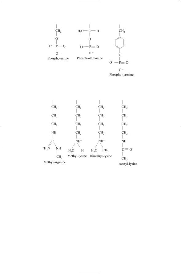
FIGURE 2.10. Phosphorylation of serine, threonine, and tyrosine side chains: Hydroxyl (OH) groups located at the ends of the side chains provide sites for covalent attachment of phosphoryl groups.
FIGURE 2.11. Covalent attachment of methyl and acetyl groups to arginine and lysine side chains: Amino groups located at the ends of arginine and lysine side chains provide sites for attachment of one or more methyl or acetyl groups.
2.14Reversible Addition and Removal of Methyl and Acetyl Groups
Phosphoryl groups are not the only groups that are added and removed from signaling proteins as part of their cellular function. Methyl and acetyl groups are added and removed, as well. Whereas side chain hydroxyls provide binding sites for the phosphoryl groups, side chain nitrogens do the same for the methyl and acetyl groups. The transfer targets are the amino groups lying at the ends of the side chains of lysine and arginine residues. The amino groups provide multiple attachment sites for methyl groups. Several examples of methylation are presented in Figure 2.11. Two

2.15 Reversible Addition and Removal of SUMO Groups |
39 |
nitrogens are present at the ends of arginine side chains, allowing for a symmetric distribution of methyl groups between the two nitrogens, or, alternatively, for one or the other of the nitrogens to have most if not all of the methyl groups.
The enzymes that catalyze the transfer of methyl groups to arginine and lysine residues are referred to as arginine methyltransferases and as lysine methyltransferases, respectively. These enzymes use an endogenous cellular molecule called S-adenosyl-L-methionine, or SAM, as the methyl group donor. SAM is synthesized in cells of the body from methionine and ATP, and readily donates its methyl group to the guanidine groups on the arginine side chains and to the amino groups on the lysine side chains.
The amino groups at the end of lysine side chains are the main attachment sites for acetyl groups. The enzymes responsible for catalyzing the transfer of acetyl groups to lysines are known as acetyltransferases. These enzymes use acetyl coenzyme A (acetyl CoA) as the acetyl group donor. The acetyl group is attached by a sulfur atom to CoA forming a high-energy thioster bond, making it easy to transfer to acceptors such as lysine.
2.15Reversible Addition and Removal of SUMO Groups
Small ubiquitin-related modifier (SUMO) is a member of the ubiquitin family of regulatory proteins. Like ubiquitin, it is covalently attached to a variety of proteins through the sequential actions of three sets of enzymes. Recall that ubiquitin is first activated in an ATP-dependent way by E1 enzymes. A thioester bond is formed between the C-terminal of the ubiquitin protein and the E1 ubiquitin-activating enzyme. The ubiquitin protein is then transferred to an E2 ubiquitin-conjugating enzyme, and then to the E3 ubiquitin protein ligase, which then transfers it to either the substrate or to multiubiquitin chains formed there. The substrate so tagged by one or more ubiquitin molecules is then degraded by the 26S proteosome.
The SUMO system of enzymes operates in a somewhat similar fashion. There is an E1 SUMO-activating enzyme, which consists of a heterodimer in place of the single polypeptide chain found for the ubiquitin E1. There is an E2 SUMO-conjugating enzyme, and there is an E3 SUMO protein ligase, which accelerates the direct transfer of SUMO from the E2 to the substrate. These attachments are illustrated in Figure 2.12.
Both ubiquitination and sumoylation have regulatory roles. Whereas ubiquitination of a protein generally tags that protein for destruction by the 26S proteosome, attachment of a SUMO group has a different role. It is a reversible process. It helps stabilize proteins and their interactions with