

Molecular and Cellular Signaling - Martin Beckerman
.pdf9.5 Role of NF-kB/Rel in Adaptive Immune Responses |
195 |
FIGURE 9.3. The NF-kB node: Extracellular stimuli and intracellular stresses activate upstream protein kinases, which phosphorylate the IKKs. In response, the IKKs phosphorylate the IkB proteins, triggering either ubiquitination/degradation and/or nucleocytoplasmic shuttling, depending on the upstream signals and subunits present. The NF-kB proteins form dimers that translocate to the nucleus where they stimulate transcription of NF-kB responsive genes.
removed from p105 and p100 contains a series of ankyrin repeats that are also present in the IkB proteins and mediates their sequestration. Ankyrin repeats are 33 amino acid residues, protein-protein interaction modules. They are found in many signaling proteins, usually as four or more tandem-arranged repeated copies. The chain of steps leading to mobilization of the NF-kB proteins is depicted schematically in Figure 9.3.
In the absence of activating signals, the IkB proteins bind to sites in the Rel homology domain (RHD) of the NF-kBs, thereby masking their nuclear localization sequences (NLSs) and forcing their retention in the cytosol. The IkBa proteins contain nuclear export sequences (NESs) and, when they are bound to nuclear NF-kB dimers, they promote their export to the cytosol. The inhibition on NF-kB by the IkB proteins is relived by phosphorylation, which induces the ubiquitin-mediated degradation of the IkBs by the 26S proteasome (Figure 9.3). IkB proteins contain N-terminal serines that are the substrates of IkB kinases (IKKs).
The NF-kB module turns on and then turns off. One of the genes upregulated by the NF-kB proteins encodes IkBa. Signaling through the TNF receptor leads to increased migration of NF-kB proteins to the nucleus where they stimulate transcription of the gene for IkBa, which once synthesized binds to NF-kB, thereby shutting down its response to TNF. Because of the time delays inherent in the signaling pathway the NF-kB protein activity levels move up and down over time. The other IkB subunits do not depend on the NF-kB proteins for their transcription. The subunits’ activity remains fairly constant over time, enabling them to smooth out the oscillations in NF-kB activity brought on by the negative feedback.

196 9. Signaling by Cells of the Immune System
9.6Role of MAP Kinase Modules in Immune Responses
MAP kinase modules convey cytokine and stress signals in the inflammatory, innate, and adaptive immune responses. Mitogen-activated protein (MAP) kinase modules convey cytokine, stress, and growth signals that are sent to them from the plasma membrane to the nucleus where they influence transcription of target genes. There are three mammalian MAP kinase pathways. One of these, the extracellular signal-regulated kinase (ERK) pathway, primarily carries growth signals. The other two, the c-Jun NH2- terminal kinase (JNK) and p38 MAP kinase pathways, relay inflammatory cytokine and stress signals.
As was the case for the yeast MAP kinases discussed in Chapter 6, there are three kinases in a MAP kinase module. The first of these is a serine/ threonine kinase. It phosphorylates the second kinase in the module, which is a dual specificity kinase. The middle kinase phosphorylates the third kinase in the module at threonine and tyrosine residues, hence the name dual specificity. The target residues are arranged as Thr-X-Tyr, where X is either Glu, Pro, or Lys for the ERK, JNK, and p38 pathways, respectively. Once they are phosphorylated, the third and last kinases in the module typically stimulate the transcriptional activity of members of several families of transcription factors. The three families, their upstream activating signals, and their downstream targets are depicted schematically in Figure 9.4.
The Ras and Rho GTPases function as molecular switches that relay growth, cytokine, and stress signals from receptors and their adapters and other intermediaries to the MAP kinase modules. Ras is a crucial regulator of growth signals. When its gene is mutated at certain residues it can get stuck in the “on” position and will continually send inappropriate growth messages to the ERK MAP kinase module. Ras will be discussed in more detail along with the other GTPases and their upstream growth factor receptors in Chapter 11 and again in Chapter 14, on cancer.
9.7 Role of TRAF and DD Adapters
TRAF and DD adapters transduce signals from TNF receptors into the cell. Scaffolds, anchors and adapters were introduced in Chapter 6. These proteins are not enzymes, but rather they organize the signaling nodes and control points where the enzymes can interact with one another and with the fixed infrastructure. Scaffolds that help organize MAP kinase cascades were examined in Chapter 6; anchors that tether serine/threonine kinases to membranes were discussed in the last chapter, and now an

9.7 Role of TRAF and DD Adapters 197
FIGURE 9.4. Mammalian MAP kinase modules:The upstream activators fall into two groups. (a) The ERK MAP kinase module is activated by growth and mitogenic signals relayed to it by the Ras GTPase. (b) The JNK and p38 MAP kinase modules are activated by proinflammatory cytokines and stress signals relayed to them by the Rho family of GTPases.
important class of adapters—the TRAF proteins—will be introduced. The tumor necrosis factor (TNF) receptor-associated factors, or TRAFs, help organize signaling nodes in pathways that mediate innate and adaptive immune responses and stress responses. These proteins contain a
TRAF domain in their C-terminus. The TRAF domain consists of two portions: a TRAF-C domain that interfaces to the receptors and a coiledcoil domain that mediates interactions with other TRAFs. These are shown in Figure 9.5.
The TRAF adapters act as key intermediaries between the receptors and downstream signaling elements. The N-terminal domains of the TRAFs contain motifs called zinc and RING fingers. These motifs consist of arrangements of four cysteine and/or histidine residues that bind zinc ions, facilitating the formation of a compact domain that, along with the C- terminus coiled-coil domain, mediates downstream signaling. There are six mammalian TRAFs, named TRAF1 through TRAF6. The TRAF2 and
TRAF6 proteins are crucially involved in TNF and Toll signaling, where they function as adapters that link the receptors to downstream NF-kBs, AP-1 signaling, and MAP kinase cascades.
198 9. Signaling by Cells of the Immune System
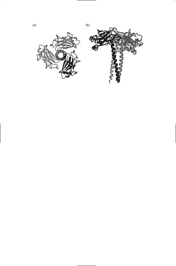
FIGURE 9.5. Structure of the TRAF domain of the TRAF2 adapter determined by X-ray crystallography: (a) Top view—Shown is a trimeric arrangement of TRAF domains that transduce signals from a 3 : 3 arrangement of TNF receptors and ligands. (b) Side view—The overall shape of the signaling unit resembles a mushroom with the C-TRAF domains forming the cap and the coiled-coil domains making up the stalk. The figure was prepared using Protein Explorer with atomic coordinates deposited in the PDB under accession number 1ca9.
9.8Toll/IL-1R Pathway Mediates Innate Immune Responses
A group of plasma membrane-bound receptors called the Toll/IL-1R family plays a key role in leukocyte responses to bacterial lipopolysaccharide (LPS). The Toll/IL-1R signaling pathway activated in response to ligand binding by receptors in this family is an ancient one. It has been identified in plants, in insects (Drosophila), where it is known as the Toll/Dorsal pathway, and in vertebrates where it is referred to as the IL-1R/NF-kB pathway.
LPS is a prominent component of the outer membrane of gram-negative bacteria such as E. coli. The sensing of the presence of LPS by leukocytes, most notably, macrophages, activates the Toll/IL-1R signaling pathway. The focus of this pathway is the nuclear factor-kB (NF-kB) and a MAP kinase cascade leading to activation of members of the AP-1 family of transcription factors such as c-Jun (Figure 9.6). Ligand binding results in the rapid activation and subsequent translocation of NF-kB to the nucleus, where it and c-Jun upregulate the expression of genes for IL-1, IL-6, interferons, TNF, the cell adhesion molecules ICAM-1 and E-selectin, and the chemokine IL-8 (not shown). Signaling through this pathway not only starts an infection-containing innate immune response, but also launches the adaptive immune response.
9.9 TNF Family Mediates Homeostasis, Death, and Survival |
199 |
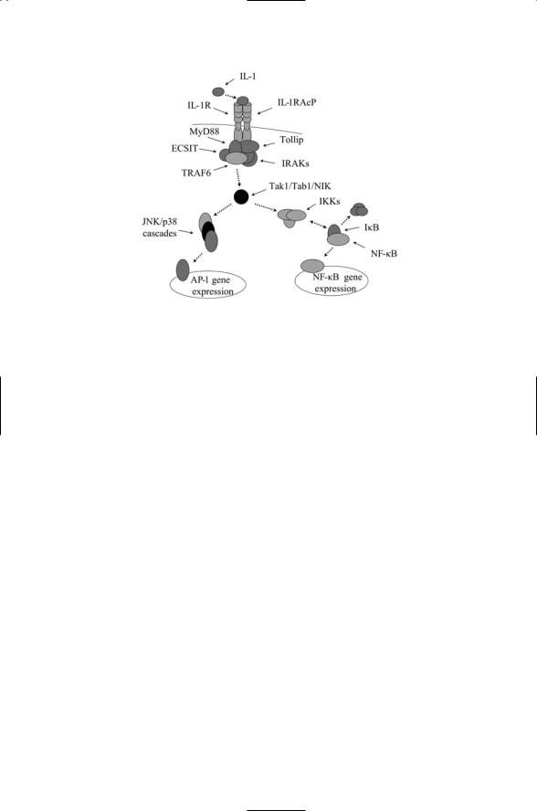
FIGURE 9.6. Signaling through the IL-1/Toll pathway: Shown are the IL-1R and its associated IL-1RAcP receptor along with several adapter proteins, namely, MyD88, Tollip, and ECSIT, which assist in recruiting kinases and other signaling elements to the plasma membrane. The first step in their activation is the recruitment of the adapter proteins to the plasma membrane. The serine/threonine kinase IRAK (IL- 1R-associated kinase) and the TRAF6 adapter are then assembled at the site. These proteins activate a number of upstream kinases collectively designated in the figure as Tak1/Tab1/NIK, which then activate the NF-kB pathway and the JNK/p38 MAP kinase pathways.
9.9TNF Family Mediates Homeostasis, Death, and Survival
Cell suicide, or apoptosis, instructions are an important part of the immune response. Immature T lymphocytes that do not respond properly to selfantigens are eliminated in the thymus through apoptosis. Other thymocytes that do not have the correct arrangement of their T cell receptors (TCRs) are disposed of in this way, too. At the end of an immune response, superfluous, mature, activated T cells are removed, and the immune system is returned to its basal level ready to respond to the next infection. The maintenance of basal levels of the different kinds of leukocytes when there are no infections are present is referred to as cellular homeostasis. It relies on apoptosis to remove the superfluous leukocytes.
Apoptosis signaling mechanisms are not only used for elimination of incorrectly functioning and superfluous cells. It provides a mechanism for ensuring that cells do not survive outside of the tissue to which they belong. It is used by cytotoxic T cells to kill virus-infected cells and cancerous cells.
200 9. Signaling by Cells of the Immune System
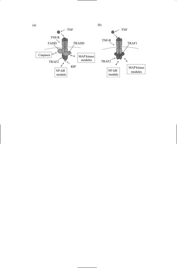
FIGURE 9.7. Upstream signaling in the TNF pathways: Trimeric arrangements of ligands, receptors, and adapters convey messages to signal enzymes. (a) Apoptosis signals are conveyed by death domain-bearing receptors and adapters among which are Fas-associated death domain (FADD) proteins, TNF-R-associated death domain (TRADD) proteins and receptor-interacting proteins (RIPs). (b) Inflammatory cytokine- (e.g., IL-6 and IFNg) promoting signals.
Some tissues such as the eye, testis, and parts of the central nervous system cannot tolerate inflammation. These are privileged sites that insulate themselves from immune responses. If a lymphocyte comes into contact with a cell in any of these sites it will rapidly undergo apoptosis. Tumor cells trying to evade the immune response use this mechanism, as well.
The TNF receptors can be divided into two groups according to the kind of adapter proteins they use. Members of one group of TNF receptors have TRAF-binding motifs in their cytoplasmic domain, which mediate binding to TRAFs, while members of the second group have death domain (DD) motifs and bind to DD-bearing adapters. Receptors belonging to the second group are called death receptors because they convey cell suicide (apoptosis) instructions to the recipient. Upstream signaling events in these two TNF pathways are depicted in Figure 9.7. Most TNF ligands are single-pass transmembrane proteins with their C-terminus outside the cell and their N- terminus in the cytoplasm. They contact TNF receptors expressed on the surface of an opposing cell. The arrangement of receptors and ligands is 3 : 3. Three receptors arranged symmetrically contact three ligands. This signaling arrangement is facilitated by a trimeric arrangement of adapters of the form illustrated in Figure 9.5.
9.10 Role of Hematopoietin and Related Receptors
Hematopoietin receptors bind most of the interleukins, while a closely related set of receptors binds the interferons. Members of the hematopoietin family of cytokines are leukocyte hormones and growth factors.
Although all members of this family of ligands and receptors have similar

9.10 Role of Hematopoietin and Related Receptors |
201 |
TABLE 9.4. Hematopoietic and interferon receptors, Jaks and STATs: Abbreviations—erythropoietin (EPO); prolactin (PRL); thrombopoietin (TPO); growth hormone (GH); granulocyte macrophage colony stimulating factor (GM-CSF).
Receptor |
Jak |
STAT |
Class I-Hematopoietin cytokines: |
|
|
Homodimeric receptors: |
|
|
EPO, PRL, TPO |
Jak2 |
STAT5a, STAT5b |
GH |
Jak2 |
STAT5a, STAT5b, STAT3 |
Receptors that share gc: |
|
|
IL-2, IL-7, IL-9, IL-15 |
Jak1, Jak3 |
STAT5a, STAT5b, STAT3 |
IL-4 |
Jak1, Jak3 |
STAT6 |
Receptors that share bc: |
|
|
GM-CSF, IL-3, IL-5 |
Jak2 |
STAT5a, STAT5b |
Receptors that share gp130: |
|
|
IL-6, IL-11 |
Jak1, Jak2, Tyk2 |
STAT3 |
IL-12: |
|
|
IL-12Rb1, IL-12Rb2 |
Jak2, Tyk2 |
STAT4 |
Class II-Interferon/IL-10: |
|
|
IFNa, IFNb |
Jak1, Tyk2 |
STAT1, STAT2 |
IFNg |
Jak1, Jak2 |
STAT1 |
IL-10 |
Jak1, Tyk2 |
STAT1, STAT3, STAT5 |
|
|
|
structures, there are some differences in structure and subunit composition that allow grouping the family members into subfamilies. This breakdown is presented in Table 9.4. Hematopoietic receptors are assembled from two or three subunits, each a single-pass glycoprotein. Each receptor contains a ligand-binding subunit that is unique for that receptor type, plus one or more subunits that are common, or shared, by several other receptor types.
A number of cytokines have a common beta chain (bc); others share a gamma chain (gc); still others have a common gp130 subunit. The IL-12 group of cytokines listed in the table departs somewhat from the overall pattern. This group consists of cytokines that bind to homodimeric chains. Interferon and IL-10 receptors (Class II) are constructed from multiple unique subunits and in this way differ from the other grouping in the table, which are collectively called Class I receptors.
Cells regulate the binding activity of their plasma membrane receptors by varying the subunit composition. Cells can switch between low, medium, and high affinity-binding by differentially expressing different receptor subunits. Several examples of this form of control are supplied by the cytokine receptors. IL-2 is composed of IL-2Ra, IL-2b, and IL-2g subunits. The IL-2g chain is expressed widely while the other two subunits are expressed in a restricted fashion. By varying the subunit composition the receptor affinity for its cognate ligand can change from high affinity to low affinity with one or more intermediate affinity complexes. A similar depend-
202 9. Signaling by Cells of the Immune System
ence of binding affinity upon receptor subunit composition occurs in the IL-12 and IL-6 receptor systems.
Cytokine ligands are typified by their structure, a four-helix bundle fold consisting of two pairs of antiparallel a-helices linked together by loops. Some are short chain cytokines; IL-2, 3, 4 are constructed from short a-helices, 8 to 10 residues in length. Long chain cytokines such as GH, EPO, G-CSF, and the gp130 cytokines are built from chains that are longer, 10 to 20 residues in length. A third group of cytokine ligands, notably IL-5 and
IFN-g, are formed as a doubled four-a-helix fold, and contain eight a-helices.
9.11 Role of Human Growth Hormone Cytokine
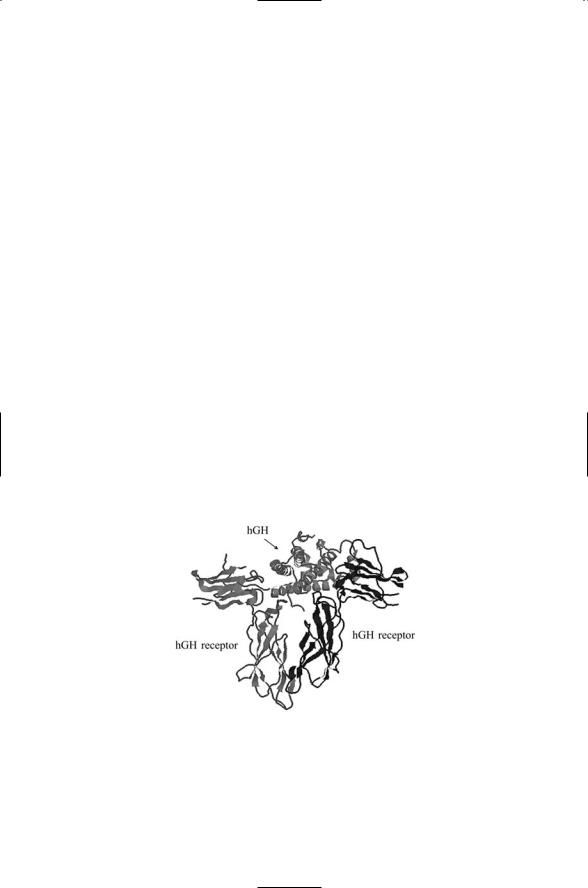
The human growth hormone (hGH) cytokine and its receptor serve as a model system for cytokine receptor-ligand recognition. The hematopoietin chains bind their ligands in characteristic ways. In Figure 9.8, one molecule of the growth hormone ligand simultaneously binds two growth hormone receptor chains. This 1 : 2 binding is fairly typical of the entire group. An examination of the human growth hormone provides some insights into how this happens. Each hGH molecule contains two receptor-binding sites. One interface is located in helix 4 and the other is formed from helices 1 and 3. Thus, a single molecule of hGH forms a homodimeric complex with a pair of hGH receptor molecules.
Not all residues in the hGH ligand-hGH receptor interface contribute equally to the binding energy. Instead, a few residues located in the vicinity
FIGURE 9.8. Structure of the human growth hormone (hGH) ligand bound to two hGH receptors: Shown are two extracellular domains of the hGH receptors bound to a single hGH ligand. The hGH ligand binds in the cleft between the two extracellular domains of the receptors. The figure was prepared using Protein Explorer with atomic coordinates deposited in the PDB under accession number 1hwg.

9.12 Signal-Transducing Jaks and STATs |
203 |
of the center of the interface contribute most of the binding energy. These residues form a “functional epitope,” or “hot spot,” that mediates ligand-receptor binding. Ligand engagement occurs when residues belonging to ligand and receptor portions of the hot spot come into close proximity. During the binding process an initial contact between the ligand and receptor surfaces is established. The contact is followed by a random diffusion stage where one surface moves (rolls) relative to the other until the motion is stabilized. This happens when the two portions of the functional epitope make contact. The maintenance of close surface contact through rolling diffusion greatly accelerates the association rate over that which would result from a single collision followed by a separation and then another elastic collision, and so on, until the correctly oriented surfaces are engaged.
The 1 : 2 association of the hGH ligand with its receptors is more efficient than a 2 : 2 association. The 1 : 2 complex is formed in a stepwise fashion. In the first step, a ligand molecule finds and establishes contact with one receptor molecule. The 1 : 1 complex then makes contact with the second (unbound) receptor molecule to create a stable 1 : 2 signaling unit. The advantage to this mechanism is that the second step involves diffusion in two dimensions—the second receptor molecule moves along membrane surface to contact the bound pair—rather than three. This may be compared to the case where the second step is another single ligand-single receptor binding event followed by association of the pairs. In this latter 2 : 2 scenario a second three-dimensional diffusion process would be required.
Many of the cytokines bind their receptors in a manner similar to that of the human growth hormone. Like hGH, the ligands for the bc family of receptors have two binding sites. Members of the bc family form high affinity complexes stepwise in the following manner. The ligand first binds the Ra subunit. The bc chain then binds to the bound pair to form a 1 : 1 : 1 complex. Two complexes then come together to form a dimerized complex that activates the intracellular components of the signaling machinery.
Hematopoietin receptors such as IL-2R that belong to the gc family are composed of three distinct subunits. A single ligand molecule possesses three binding sites and so is able to bind the three subunits when forming the high affinity 1 : 1 : 1 : 1 complex. A similar process is used by the gp130 cytokines to form stable complexes that are signaling-competent.
9.12 Signal-Transducing Jaks and STATs
Jaks and STATs transduce signals into the cell from the plasma membrane.
The reason for receptor dimerization can be understood by noting that there is considerable flexibility in the membrane-spanning polypeptide chains. Under these conditions the binding of a ligand to the extracellular portion of a chain will not produce a large and long-lasting (stable) shift in conformation of the cytoplasmic portion of the chain needed to serve as a
204 9. Signaling by Cells of the Immune System
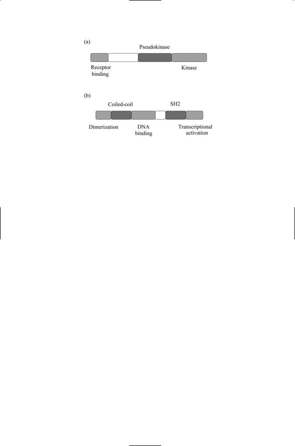
FIGURE 9.9. Domain organization of Jaks and STATs: (a) Janus tyrosine kinases, or Jak proteins. (b) Signal transducer and activator of transcription (STAT) proteins.
signal. The solution to this problem of how to transduce a signal into the cell is solved by dimerization. The combined effect of ligand binding and dimerization alters the intracellular electrostatic environment sufficiently to stabilize a different mix of conformational states that will activate the Jaks, triggering phosphorylation and the subsequent recruitment of the STATs and other signaling elements to the receptors. The Jaks (tyrosine kinases) and STATs (transcription factors) are then able to transduce the hematopoietin signals into the cell responses.
The domain organization of the Jak and STAT proteins is presented in Figure 9.9. As shown in part (a) of the figure, the Janus kinases possess kinase and pseudokinase domains that face one another, hence the name
Janus kinase (Jak), named after the Roman god of gates and doorways. They also possess a receptor-binding region that mediates their binding to recognition motifs in the cytoplasmic terminal of the cytokine receptors. Once recruited to the receptors, autophosphorylation occurs, resulting in their activation. The activated kinases then phosphorylate the receptors, thereby exposing docking sites for the STATs. As depicted in part (b) of the figure, the STATs possess a dimerization domain, an SH2 domain, a tyrosine phosphorylation site, and a DNA-binding domain. The signal transducer and activator of transcription proteins are transcription factors, and they are recruited to the activated signaling complex formed by the phosphorylated cytoplasmic receptor terminals and Jaks. They bind the phosphorylated tyrosine residues in the receptors through their SH2 domains, and they are phosphorylated on their tyrosine residues by the Jaks. Once these recruitment and activation steps take place, the STATs are able to form heterodimer and homodimers, and translocate to the nucleus where they stimulate transcription of their target genes.