

Biomolecular Sensing Processing and Analysis - Rashid Bashir and Steve Wereley
.pdf196 |
´ |
¨ |
HAIBO LI, RAFAEL GOMEZ-SJOBERG, AND RASHID BASHIR |
||
McClain et al. [48] reported a microfluidic device that integrated cell handling, rapid cell lysis, and electrophoretic separation and detection of fluorescent cytosolic dyes. Cell analysis rates of 7–12 cells/min were demonstrated and are >100 times faster than those reported using standard bench-scale capillary electrophoresis. Hong et al. [21] recently developed microfluidic chips with parallel architectures for automated nucleic acid purification from small numbers of bacterial or mammalian cells. All processes, such as cell isolation, cell lysis, DNA and mRNA purification, and recovery, can be carried out on each single microfluidic chip in nanoliter volumes without any preor post-sample treatment.
9.3.2. Mechanical Detection
Mechanical detection of biochemical entities and reactions has more recently been realized through the use of micro and nano-scale cantilever sensors on a chip. As shown in Fig. 9.7(a), these cantilever sensors (diving board type structures) can be used in two modes, namely stress sensing and mass sensing. In stress sensing mode one side of the cantilever is usually coated with a Self-Assembled Monolayer (SAM) of biomolecules that bind to the analyte being detected. The binding of the analyte to the SAM produces a change in surface free energy, resulting in a change in surface stress, which in turn leads to a measurable bending of the cantilever. The bending can then be measured using optical means (laser reflection from the cantilever surface into a quad position detector, like in an Atomic Force Microscope) or electrical means (piezo-resistors incorporated near the fixed edge of the cantilever). To increase the stress sensitivity of the cantilever, the spring constant should be reduced, while the overall surface of the cantilever determines the number of molecules that should attach to the surface to cause a given stress change. In the mass sensing mode, the resonant frequency of the cantilever is constantly monitored as it vibrates due to an external driving force (i.e. a piezo-electric transducer) or in response to the background thermal noise. When the species being detected binds to the cantilever, it changes the cantilever mass and hence its resonant frequency. The mass of the detected species can be calculated from the change in resonant frequency. The resonant frequency can be measured using electrical or optical means, in the same way that bending is detected in stress sensing. To increase the mass sensitivity, in general, the mass of the cantilever should be made smaller, the quality factor should be increased, the resonant frequency should be chosen such that it is easily measured, and the detection system should be designed to measure as small a frequency shift as possible. The quality factor is decreased with increased damping, for example in a fluid, and hence the minimum detectable mass is much higher in damping mediums (liquids) as compared to low-damping mediums (air). Thus, the stress detection mode is inherently preferred in a fluid.
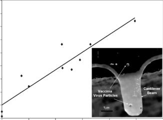
To have a significant change in surface stress, a large fraction of the cantilever area must be involved in the binding event that leads to detection, which precludes the use of the stress-based technique for detecting cells or large viruses. Even covering the whole cantilever surface (on one side) with cells bound to its surface via antibodies produces very small changes in surface stress because the effective binding area of each cell is just a small fraction of the total cantilever area. Detection of cells and microorganisms has been demonstrated using the mass detection method based on shifts in resonant frequency. Various examples of mass-based sensing are reported in the literature, for example, detection of the mass of E.coli O157:H7 using cantilevers [18, 28], detection of the mass of a single vaccinia
BIOMEMS FOR CELLULAR MANIPULATION AND ANALYSIS |
|
|
197 |
||||
|
225 |
|
|
|
|
|
|
(kHz) |
200 |
y = 33.6x + 25.4 |
|
|
|
|
|
|
|
|
|
|
|
||
|
|
|
|
|
|
|
|
|
175 |
R2 = 0.9 |
|
|
|
|
|
Shift |
|
|
|
|
|
|
|
150 |
|
|
|
|
|
|
|
Frequency |
|
|
|
|
|
|
|
125 |
|
|
|
|
|
|
|
|
|
|
|
|
|
|
|
Resonant |
100 |
|
|
|
|
|
|
75 |
|
|
|
|
|
|
|
|
|
|
|
|
|
|
|
|
50 |
|
|
|
|
|
|
|
25 |
|
|
|
|
|
|
|
0 |
|
|
|
|
|
|
|
0 |
1 |
2 |
3 |
4 |
5 |
6 |
Effective No. of Virus Particles
FIGURE 9.9. Shift (decrease) in resonant frequency with increasing number of virus particles. Inset shows an SEM of a nano-cantilever with a single Vaccinia virus particle (Reprinted with permission from Applied Physics Letters, vol. 84, no. 10, 2004 and with kind permission from R. Bashir).
virus particle, as shown in Fig. 9.9 [19], and mass change in a polymer upon absorption of a vapor [36].
9.3.3. Electrical Detection
Some of the earliest cell-related uses of micromachined devices were in the electrical probing of neurons by creating microscopic needles that could be inserted in vitro or in vivo in the neuron to stimulate it electrically and record the signals it produced [34]. Recent reports describe artificial structures where neurons are cultured and probed, while the configuration of interconnections between them is artificially patterned by microfabricated channels that guide axon growth [42, 47]. Devices for positioning and/or probing of other types of electrogenic cells, such as cardiac myocytes, were also reported in the literature [51, 65]. Most of these consist of arrays of electrodes, deposited either on a planar surface or at the bottom of cavities, on which the cells are located. In most cases the cells are placed on the electrodes manually, but Thielecke et al. [65] make use of an orifice at the center of each electrode, through which vacuum is created to move the cells towards the electrodes and hold them in place.
Monitoring the impedance of microfabricated electrodes over which adherent cells are cultured can reveal information about cellular motion, multiplication, metabolism, viability, etc. The signal produced in these devices arises from two main mechanisms: Cells growing
Parts of this section are reprinted from: R. Gomez´ -Sjoberg,¨ “Microfabricated device for impedance-based electronic detection of bacterial metabolism,” Ph.D thesis, School of Electrical and Computer Engineering, Purdue University, West Lafayette, IN, December 2003, with kind permission from the author.
198 |
´ |
¨ |
HAIBO LI, RAFAEL GOMEZ-SJOBERG, AND RASHID BASHIR |
||
attached to the electrodes act as insulators, blocking current flow between electrodes; and the difference in dielectric constant between the cells and the growth medium modifies the capacitance of the electrodes. For example, Keese and Giaever [32] built a cell biosensor for environmental monitoring. The impedance of two electrodes in a cell growth chamber was modified by changes in the cell population produced by phenomena such as cell motion, multiplication, death, and metabolic activity. Borkholder et al. [7] used an array of 10 µm diameter electrodes to study the response of cells to certain toxins that block membrane channels. Similarly, Ehret et al. [13] used microfabricated interdigitated electrodes (fingers are 50 µm wide) on a sapphire substrate to monitor the behavior of mammalian cells, by measuring the capacitance of the electrodes at a frequency of 10 kHz. Cells were grown adherently over the surface of the electrodes. Very clear signals were observed by the authors when the cells were destroyed by adding the detergent Triton X-100. The response of cells to different concentrations of the toxic ion Cd2+ could also be monitored over time. Building upon the work of Ehret et al. [13], Wolf et al. [78] and Lehman et al. [37] developed the so-called “PhysioControl-Microsystem” and “Cell-Monitoring-System” that incorporate microfabricated temperature, pH, oxygen, and ion sensors, along with interdigitated electrodes, to gather detailed information on cellular metabolism. Ion sensitive field-effect transistors (ISFET) were used as pH, oxygen, and ion sensors. In these transistors the gate is covered with a film selective to the ions that are detected, so that adsorption of the ions into the film causes a shift in the gate potential with a concomitant change in the current flowing through the channel of the transistor.
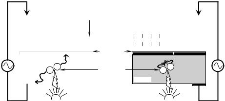
A very interesting commercial system for the electrical monitoring of cellular metabolism is the “Cytosensor Microphysiometer” developed by Molecular Devices GmbH in Germany [8, 20]. This device measures pH changes in the cell growth medium, produced by excreted metabolites, using light-addressable potentiometric sensors (LAPS). The principle on which LAPS works is depicted in Fig. 9.10. A silicon substrate is coated with
|
|
|
|
|
High |
|
|
|
Large negative |
||||||
|
|
|
- |
|
OH- |
|
|
|
|||||||
|
|
|
|
|
|
|
charge on surface |
||||||||
|
|
|
OH |
|
|
|
|
|
|
|
|||||
H2 |
H2 |
|
|
OH- |
|
|
|
|
|
|
|
||||
|
|
|
|
|
|
|
|
|
|
|
|||||
|
|
|
|
|
O- |
O- |
O- O- O- |
|
|
||||||
N O- |
N O- |
|
|
||||||||||||
|
|
|
|
|
|
|
|
|
|
|
|
|
|
|
Silicon |
Si Si |
Si |
Si |
Si |
Si |
Si Si Si |
|
|||||||||
|
|
|
|
|
|
|
|
|
|
|
|
|
|
|
Oxynitride |
|
|
|
|
|
|
|
|
h+ |
|
|
|
|
|
|
|
|
|
|
|
|
- |
|
|
|
|
|
|
|
Photo- |
||
|
|
|
|
|
|
e |
|
|
|
|
|
|
|
|
generated |
|
|
|
|
|
|
|
|
|
|
|
|
|
|
|
|
|
|
|
|
|
|
|
|
|
|
|
|
Silicon |
|
electrons |
|
|
|
|
|
|
|
|
|
|
|
|
|
|
|
|
and holes |
H+
H+
H+
H3 H H3+ H N O N O Si Si Si Si
Low
H+
|
H+ |
|
|
|
|
+ |
|
|
|
|
|
|
|
|
|
H |
|
|
|
H |
H |
H |
H |
H |
|||||
|
|
|
|
|
|
|
|
|
|
O |
O |
O |
O |
O |
|||||
|
|
|
|
|
|
|
|
|
|
Si |
Si |
Si |
Si |
Si |
|||||
e- h+
Silicon
Large current |
|
LED |
LED |
|
Negligible current |
|
|
|
|
|
|
FIGURE 9.10. Operating principle for the light-addressable potentiometric sensor used to measure changes in the pH of cell cultures produced by cell metabolism (Adapted with permission from Biosensors & Bioelectronics, vol. 15, no. 3–4, 2000 and with kind permission from F. Hafner).
BIOMEMS FOR CELLULAR MANIPULATION AND ANALYSIS |
199 |
a silicon oxynitride film that will be in contact with the liquid medium being monitored. When hydrated, silanol (Si-OH) and silamine (Si-NH2) groups will appear at the surface of the film. An ohmic contact is established with the silicon substrate and a reference electrode is immersed in the liquid medium, so that a voltage can be applied between the liquid and the silicon. Free electrons and holes are generated in the silicon by illuminating it with a pulsating LED. The silamine and silanol groups are ionized to different levels depending on the pH of the medium, affecting the surface charge on the film. When the pH of the medium is high, most of the silanol groups are ionized (they have donated a H+ ion to the medium) so that a large negative charge exists on the surface. The electric field generated by this charge will separate the photo-generated holes and electrons, increasing their recombination time, and thus producing a large current across the electrodes. When the pH is low, most of the surface groups are neutral and little separation of electrons and holes occur, leading their rapid recombination and hence to a low current. The voltage between the silicon and the liquid is adjusted to have a constant photo-current, so that the required changes in voltage are proportional to changes in pH. This technique can detect changes as small as 5 × 10⁻⁴ pH units. Cells are kept in a chamber over the microfabricated LAP sensor, through which growth medium can flow. The flow of medium is stopped when the metabolic rate is being measured, so that metabolites accumulate in the chamber and change the pH. A very important feature of the LAPS technique is that local measurements of pH can be done by limiting the illumination to the area of interest. In this way, a pH map of the cell culture can be constructed by having an array of LEDs, each one illuminating a small section of the sensor. It is also worth mentioning the use of microcalorimetry for measuring metabolism, as exemplified by the device built by Verhaegen et al. [66]. This microcalorimeter uses aluminium/p+-polysilicon junction thermopiles to measure the heat generated by metabolizing cells cultured in chambers microfabricated on a silicon substrate.
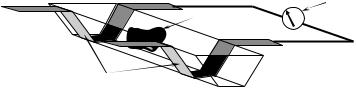
The electrical impedance of individual cells located between two microelectrodes formed on opposite sides of a microchannel can provide information such as the capacitance of the cell membrane and the dielectric constant and conductivity of the cytoplasm [1, 2, 57, 66]. Differences in impedance could be used to discriminate between different types of cells for sorting and counting. The same principle and geometry were used by Sohn et al. [60] to create a microfabricated cytometer for mammalian cells flowing one by one between electrodes in a microchannel. Since the capacitance is greatly affected by the DNA content of the cells, due to the large number of charges in DNA molecules, it can be used to track the multiplication phases of the cells. Suehiro et al. [63, 64] used DEP to trap E. coli cells and detect their presence over the DEP electrodes by monitoring the electrode impedance. The presence of cell bodies changes the impedance of the electrodes because their dielectric properties are different from those of the suspension medium, and the impedance can be correlated to the number of trapped cells. Koch et al. [33] fabricated a device to detect the passage of cells through fully enclosed microfluidic channels, based on the Coulter Counter principle. Two electrodes are placed across the channels, perpendicular to them, spaced 40 µm apart. When a cell passes between the electrodes the resistance measured across them changes (shown in Fig. 9.11) because cells are significantly less conductive than the liquid in which they are suspended (they can in fact be modeled as non-conductive particles).
Detecting viable bacteria is a very important goal in the development of novel biosensors, and microscale impedance-based monitoring of metabolic activity has the potential
200 |
´ |
¨ |
HAIBO LI, RAFAEL GOMEZ-SJOBERG, AND RASHID BASHIR |
||
|
|
Resistance |
|
Cell |
monitor |
|
R |
|
 Microchannel
Electrodes
FIGURE 9.11. Microfabricated Coulter Counter used to detect the passage of cells through microchannels (Adapted with permission from Proceedings of Ninth Micromechanics Europe Workshop—MME‘98, 1998 and with kind permission from M. Koch).
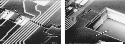
of realizing that goal in a simple and cost-effective way. Macroscale impedance-based detection is relatively slow when low numbers of cells are present. The lower the initial population of microorganisms, the longer it takes for the impedance to change by a measurable amount. This should be obvious, since it will take longer for a small number of organisms to produce enough ions to modify the impedance by a certain amount, than for a larger number of them. Eden & Eden [12] and Dupont et al. [11] showed that the detection time (incubation time needed to have the impedance change by a certain predefined value) decreases exponentially with increasing bacterial concentration. Consequently, the impedance method has the potential to detect bacterial contamination in a very short time if the bacterial concentration is somehow increased by several orders of magnitude. The number of bacterial cells in a given amount of food sample is an uncontrollable parameter, so the only other controllable parameter is the volume in which cells are placed for performing the measurement. The typical volumes in which impedance measurements are done range from 1ml to 100ml in the macro-scale impedance-based detection systems currently available commercially. With such large volumes, typical detection times are on the order of 8 hours or more for initial bacterial loads on the order of 10CFU/ml. By confining the same number of cells in a microfabricated volume, their effective concentration becomes very high and the detection time drops dramatically. Implementing the impedance monitoring method on a biochip, where detection chambers with volumes of even 0.1 nl can be readily fabricated, is an ideal way of exploring its potential for fast detection. Gom´ez et al. [15, 16, 17] have developed microfabricated devices (Fig. 9.12) with nanoliter-scale chambers used to study this bacterial detection method. A metabolic signal could be detected in off-chip incubated samples at cell concentrations equivalent to about 50 cells in a 5.27nl measuring volume in an initial biochip prototype [16, 17].
9.4. CONCLUSIONS AND FUTURE DIRECTIONS
Considerable progress has been made in the field of BioMEMS, especially in systems for cellular analysis as described above, and with the current drive towards nano-scale devices, microand nano-technologies are being combined with the emerging field of bionanotechnology [76]. BioMEMS are enabling us to probe, measure, and explore the microand nano-machinery in the biological world, including the inner workings of single cells. Such micro and nano-scale systems and sensors could allow us to precisely measure
BIOMEMS FOR CELLULAR MANIPULATION AND ANALYSIS |
201 |
|||||||
Cavities with |
|
|
|
|
||||
Pt electrodes |
|
|
|
|
||||
|
|
20 m wide |
|
|
|
|
||
|
|
channel |
|
|
|
|
||
Input port |
Groove for insertion |
|||||||
of tube (~360 m deep) |
||||||||
|
|
|
|
|||||
|
|
|
|
|
|
|
300 m |
|
|
|
|
80 m |
|
||||
|
|
|
|
|
|
|||
|
|
|
|
|
(b) |
|||
(a) |
|
|||||||
FIGURE 9.12. Scanning Electron Micrograph of a biochip used to explore the impedance-based detection of bacterial metabolism. (a) Chambers where bacterial cells are incubated for detection; (b) input/output port created by DRIE. (Reprinted with permission from Ph.D. thesis of R. Gomez´ -Sjoberg,¨ 2004, School of Electrical and Computer Engineering, Purdue University, and with kind permission from R. Gomez´ -Sjoberg)¨ .
the protein, mRNA, and chemical profiles of cells in real time, as a function of controlled stimuli and increase understanding of signaling pathways inside the cell. These issues will also be the focus of the post-genomic era and also in the applications of systems theories to biology, also referred to as systems biology [22].
ACKNOWLEDGMENTS
The sponsorship of NIH (NIBIB), USDA Center for Food Safety Engineering at Purdue, NASA Institute of Nanoelectronics and Computing (INAC), and NSF Career Award is greatly appreciated. The authors would also like to thank all members of the Laboratory of Integrated Biomedical Micro/Nanotechnology and Applications (LIBNA) in the School of Electrical and Computer Engineering, Department of Biomedical Engineering, Purdue University, for providing the motivation for this review.
REFERENCES
[1]H. Ayliffe, A. Frazier, and R. Rabbitt. Am. Soc. Mech. Eng., Bioeng. Div. (Publication) BED, 35:485–486, 1997.
[2]H. Ayliffe, A. Frazier, and R. Rabbitt. IEEE J. Microelectromech., 8:50–57, 1999.
[3]R. Bashir. Advanc. Drug Del. Rev., 56:1565–1586, 2004.
[4]R. Bashir and S. Wereley (eds.). Biomolecular sensing, processing, and analysis. In BioMEMS and Biomedical Nanotechnology. Kluwer Academic Publishers, 2004.
[5]F.F. Becker, X.-B.Wang, Y. Huang, R. Pethig, J. Vykoukal, and P.R.C. Gascoyne. J. Phys. D: Appl. Phys., 27:2659–2662, 1994.
[6]F.F. Becker, X.-B. Wang, Y. Huang, R. Pethig, J. Vykoukal, and P.R.C. Gascoyne. Separation of Human Breast Cancer Cells from Blood by Differential Dielectric Affinity. Proceedings of National Academy of Science, 1995.
[7]D. Borkholder, I. Opris, N. Maluf, and G.Kovacs. Planar Electrode Array Systems for Neural Recording and Impedance Measurements. Proceedings of Annual International Conference of the IEEE Engineering in Medicine and Biology, 1996.
202 |
´ |
¨ |
HAIBO LI, RAFAEL GOMEZ-SJOBERG, AND RASHID BASHIR |
||
[8]L. Bousse, R.J. Mcreynolds, G. Kirk, T. Dawes, P. Lam, W.R. Bemiss, and J.W. Farce. Sens. Actuat. B-Chem., 20(2–3):145–150, 1994.
[9]H. Chang, A. Ikram, F. Kosari, G. Vasmatzis, A. Bhunia, and R. Bashir. J. Vac. Sci. Technol. B., 20(5):2058– 2064, 2002.
[10]J.A. Chediak, Z.S. Luo, J.G. Seo, N. Cheung, L.P. Lee, and T.D. Sands. Sens. Actu. A-Phys., 111(1):1–7, 2004.
[11]J. Dupont, D. Menard, C. Herve, F. Chevalier, B. Beliaeff, and B. Minier. J. Appl. Bacteriol., 80(1):81–90, 1996.
[12]R. Eden and G. Eden. Impedance microbiology. Research Studies Press Ltd., John Wiley & Sons Inc., 1984.
[13]R. Ehret, W. Baumann, M. Brischwein, A. Schwinde, K. Stegbauer, and B. Wolf. Biosens. Bioelectron., 12(1):29–41, 1997.
[14]J. Gimsa, P. Marszalek, U. Loewe, and T.Y. Tsong. Biophys. J. 60:749–60, 1991.
[15]R. Gomez. Microfabricated Device for Impedance-based Electronic Detection of Bacterial Metabolism. Ph.D thesis, School of Electrical and Computer Engineering, West Lafayette, IN, Purdue University, 2003.
[16]R. Gomez, R. Bashir, and A.K. Bhunia. Sens. Actu B: Chem., 86:198–208, 2002.
[17]R. Gomez, R. Bashir, A. Sarikaya, M. Ladisch, J. Sturgis, J. Robinson, T. Geng, A. Bhunia, H. Apple, and S. Wereley. Biomed. Microdev., 3(3):201–9, 2001.
[18]A. Gupta, D. Akin, and R. Bashir. Resonant Mass Biosensor For Ultrasensitive Detection Of Bacterial Cells.
Microfluidics, Biomems, and Medical Microsystems Conference at Spie’s Photonics West Micromachining and Microfabrication 2003 Symposium. Proceedings of SPIE—The International Society for Optical Engineering, San Jose, CA, 2003.
[19]A. Gupta, D. Akin, and R. Bashir. Appl. Phys. Lett., 84(11):1976–1978, 2004.
[20]F. Hafner. Biosen. Bioelectron., 15(3–4):149–158, 2000.
[21]J.W. Hong, V. Studer, G. Hang, W.F. Anderson, and S.R. Quake. Nat. Biotechnol., 22(4):435–439, 2004.
[22]L. Hood and D. Galas. Nature, 421(6921):444–448, 2003.
[23]Y. Huang, K.L. Ewalt, M. Tirado, R. Haigis, A. Forster, D. Ackley, M.J. Heller, J.P. O’Connell, and M. Krihak. Anal. Chem., 73(7):1549–1559, 2001.
[24]Y. Huang, R. Holzel, R. Pethig, and X.-B. Wang. Phys. Med. Biol., 37(7):1499–1517, 1992.
[25]Y. Huang and R. Pethig. Measurement Sci. Technol., 2:1142–46, 1991.
[26]M. P. Hughes. Nanotechnology, 11:124–132, 2000.
[27]M.P. Hughes. Electrophoresis, 23:2569–82, 2002.
[28]B. Ilic, D. Czaplewski, H.G. Craighead, P. Neuzil, C. Campagnolo, and C. Batt. Appl. Phys. Lett., 77(3):450– 452, 2000.
[29]T.B. Jones and J.P. Kraybill. J. Appl. Phys., 60:1247–52, 1986.
[30]A.M. Jorgensen, K.B. Mogensen, J.P. Kutter, and O. Geschke. Sens. Actuat. B-Chem., 90(1–3):15–21, 2003.
[31]K.V.I.S. Kaler and T.B. Jones. Biophys. J., 57:173–82, 1990.
[32]C. Keese and I. Giaever. A biosensor that monitors cell morphology with electrical fields. IEEE Eng. Med. Biol. Mag., 3:402–408, 1994.
[33]M. Koch, A. Evans, and A. Brunnschweiler. Design and Fabrication of a Micromachined Coulter Counter.
Proceedings of Ninth Micromechanics Europe Workshop—MME’98, 155–158, 1998.
[34]G.T.A. Kovacs. Micromachined Transducers Sourcebook, Boston, MA, WCB/Mcgraw-Hill, 1998.
[35]L.J. Kricka. Clinica. Chimica. Acta, 307(1):219–223, 2001.
[36]D. Lange, C. Hagleitner, A. Hierlemann, O. Brand, and H. Baltes. Anal. Chem., 74(13):3084–3095, 2002.
[37]M. Lehmann, W. Baumann, M. Brischwein, H.J. Gahle, I. Freund, R. Ehret, S. Drechsler, H. Palzer, M. Kleintges, U. Sieben, and B. Wolf. Biosens. Bioelectron., 16(3):195–203, 2001.
[38]H. Li and R. Bashir. Sens. Actuat. B: Chem., 86:215–221, 2002.
[39]H. Li and R. Bashir. Biomed. Microdev., 6(4):289–295, 2004.
[40]H. Li, Y. Zheng, D. Akin, and R. Bashir. IEEE J. Microelectromech. Syst., 14(1):105–111 2005.
[41]M.J. Madou. Fundamentals of Microfabrication: the Science of Miniaturization. Boca Raton, FL, CRC Press, 2002.
[42]M.P. Maher, J. Pine, J. Wright, and Y.C. Tai. J. Neurosci. Meth., 87(1):45–56, 1999.
[43]G.H. Markx, Y. Huang, X.-F. Zhou, and R. Pethig. Microbiology, 140:585–91, 1994a.
[44]G.H. Markx and R. Pethig. Biotechnol. Bioeng., 45:337–343, 1995.
[45]G.H. Markx, M.S. Talary, and R. Pethig. J. Biotechnol., 32:29–37, 1994b.
[46]P. Marszalek, J.J. Zielinski, and M. Fikus. Biotechnol. Bioeng., 22:289–98, 1989.
BIOMEMS FOR CELLULAR MANIPULATION AND ANALYSIS |
203 |
[47]S. Martinoia, M. Bove, M. Tedesco, B. Margesin, and M. Grattarola. J. Neurosci. Meth., 87(1):35–44, 1999.
[48]M.A. McClain, C.T. Culbertson, S.C. Jacobson, N.L. Allbritton, C.E. Sims, and J.M. Ramsey. Anal. Chem., 75(21):5646–5655, 2003.
[49]J.N. Mehrishi and J. Bauer. Electrophoresis, 23(13):1984–1994, 2002.
[50]W.E. Moerner and M. Orrit. Science, 283(5408):1670–1676, 1999.
[51]A. Mohr, W. Finger, K.J. Fohr, W. Gopel, H. Hammerle, and W. Nisch. Sens. Actuat. B-Chem., 34(1–3):265– 269, 1996.
[52]T. Muller, G. Gradl, S. Howitz, S. Shirley, T. Schnelle, and G. Fuhr. Biosens. Bioelectron., 14:247–256, 1999.
[53]S.M. Nie and R.N. Zare. Ann. Rev. Biophys. Biomol. Struct., 26:567–596, 1997.
[54]K.E. Petersen. Silicon as a Mechanical Material. Proceedings of the IEEE, 1982.
[55]H.A. Pohl. J. Appl. Phys., 22:869–871, 1951.
[56]D. Polla, P. Krulevitch, A. Wang, G. Smith, J. Diaz, S. Mantell, J. Zhou, S. Zurn, Y. Nam, L. Cao, J. Hamilton, C. Fuller, and P. Gascoyne. MEMS based Diagnostic Microsystems. Proceedings of 1st Annual International IEEE-EMBS Special Topic Conference on Microtechnology in Medicine and Biology, 2000.
[57]D.L. Polla. Microdev. Med. Annual Rev. Biomed. Engin., 2:551–576, 2000.
[58]A.T. Poortinga, R. Bos, and H.J. Busscher. Biotechnol. Bioengin., 67(1):117–120, 2000.
[59]T. Schnelle, T. Muller, and G. Fuhr. J. Electrostat., 50:17–29, 2000.
[60]L. Sohn, O. Saleh, G. Facer, A. Beavis, and D. Notterman. Capacitance Cyotmetry: Measuring Biological Cells One by One. Proceedings of the National Academy of Science, 2000.
[61]M. Stephens, M.S. Talary, R. Pethig, A.K. Burnett, and K.I. Mills. Bone Marrow Transplant, 18:777–782, 1996.
[62]D.L. Stokes, G.D. Griffin, and V.D. Tuan. Fresenius J. Analyt. Chem., 369(3-4):295–301, 2001.
[63]J. Suehiro, R. Hamada, D. Noutomi, M. Shutou, and M. Hara. J. Electrostat., 57(2):157–168, 2003.
[64]J. Suehiro, R. Yatsunami, R. Hamada, and M. Hara. J. Phys. D-Appl. Phys., 32(21):2814–2820, 1999.
[65]H. Thielecke, T. Stieglitz, H. Beutel, T. Matthies, and J. Meyer. A Novel Cellpositioning Technique for Extracellular Recording and Impedance Measurements on Single Cells using Planar Electrode Substrates.
Proceedings of the 20th Annual Conference of the IEEE Engineering in Medicine and Biology Society, 1998.
[66]K. Verhaegen, J. Simaels, W.V. Driessche, K. Baert, W. Sansen, B. Puers, L. Hermans, and R. Mertens. Biomed. Microdev., 2(2):93–98, 1999.
[67]T. Vo-Dinh and B. Cullum. Fresenius J. Analyt. Chem., 366(6–7):540–551, 2000.
[68]J. Voldman, R.A. Braff, M. Toner, M.L. Gray, and M.A. Schimidt. Biophys. J., 80(1):531–41, 2001a.
[69]J.Voldman, R.A. Braff, M.Toner, M.L. Gray, and M.A. Schmidt. Quantitative design and analysis of singleparticle dielectrophoretic traps. In V.D.B. et al. Micro Total Analysis Systems 2000, Kluwer Academic Publishers: 431–434, 2000.
[70]J. Voldman, M. Toner, M. L. Gray, and M.A. Schmidt. Transducers, 01:322–325, 2001b.
[71]J. Voldman, M. Toner, M.L. Gray, and M.A. Schmidt. J. Electrostat., 57:69–90 2003.
[72]X.-B. Wang, Y. Huang, J.P.H. Burt, G.H. Markx, and R. Pethig. J. Phys. D: Appl. Phys., 26:1278–1285, 1993.
[73]X.-B. Wang, Y. Huang, P.R.C. Gascoyne, and F.F. Becker. IEEE Transact. Indust. Appl., 33(3):660–9, 1997.
[74]M. Washizu, T. Nanba, and S. Masuda. IEEE Transact. Indust. Appl., 26:352–358, 1990.
[75]B.H. Weigl, R.L. Bardell, and C.R. Cabrera. Advanc. Drug Del. Rev. 55(3):349–377, 2003.
[76]G.M. Whitesides. Nat. Biotechnol. 21(10):1161–1165, 2003.
[77]K.D. Wise and K. Najafi. Science, 254:1335–1342, 1991.
[78]B. Wolf, M. Brischwein, W. Baumann, R. Ehret, and M. Kraus. Biosens. Bioelectron., 13(5):501–509, 1998.
[79]J. Yang, Y. Huang, X.-B.Wang, F.F. Becker, and P.R.C. Gascoyne. Analyt. Chem., 71(5):911–918, 1999.
10
Implantable Wireless
Microsystems
Babak Ziaie
School of Electrical and Computer Engineering, Purdue University,
W. Lafayette, IN 47907
10.1. INTRODUCTION
The ability to use wireless techniques for measurement and control of various physiological parameters inside human body has been a long-term goal of physicians and biologists going back to the early days of wireless communication. From early on, it was recognized that this capability could provide effective diagnostic, therapeutic, and prosthetic tools in physiological research and pathological intervention. However, this goal eluded scientists prior to the discovery of transistor in 1947. Vacuum tubes were too bulky and power hungry to be of any use in implantable systems. During the late 50’s, MacKay performed his early pioneering work on what he called “Endoradiosonde” [1]. This was a single-transistor blocking oscillator designed to be swallowed by a subject and was able to measure pressure and temperature in the digestive track. Following this early work came a number of other simple discrete systems each designed to measure a specific parameter (temperature, pressure, force, flow, etc.) [2]. By the late 60’s, progress in the design and fabrication of integrated circuits provided an opportunity to expand the functionality of these early systems. Various hybrid single and multichannel telemetry systems were developed during the 70’s and the 80’s [3]. In addition, implantable therapeutic and prosthetic devices started to appear in the market. Cardiac pacemakers and cochlear prosthetics proved effective and reliable enough to be implanted in thousands of patients. Recent advances in microelectromechanical (MEMS) based transducer and packaging technology, new and compact power sources (high efficiency inductive powering and miniature batteries), and CMOS low-power wireless integrated circuits have provided another major impetus to the development of wireless implantable microsystems [4–9]. These advances have created new
206 |
BABAK ZIAIE |
opportunities for increased reliability and functionality, which had been hard to achieve with pervious technologies. Furthermore, the burgeoning area of nanotechnology is poised to further enhance these capabilities beyond what have been achievable using MEMS techniques. This is particularly true in the biochemical sensing and chemical delivery areas and will undoubtedly have a major impact on the future generations of implantable wireless microsystems. In this article, we present some of these recent advances in the context of several devices currently being pursued in academia and industry. The fact that these systems are designed to be implanted creates regulatory concerns, which has contributed to their late arrival in the clinical market. In the following sections, after discussing several major components of such microsystems such as transducers, interface electronics, wireless communication, power sources, and packaging; we will present some selected examples to demonstrate the state of the art. Although we have separated these microsystems under diagnostic, therapeutic, and prosthetic categories; this division is not always representative and systems that can be considered diagnostic in one situation may represent a therapeutic device under another circumstance.
10.2. MICROSYSTEM COMPONENTS
For the purpose of current discussion implantable wireless microsystems can be defined as a group of medical microdevices that: 1) incorporate one or several MEMS-based transducers (i.e., sensors and actuators), 2) have an on-board power supply (i.e., battery) or are powered from outside using inductive coupling, 3) can communicate with outside (bi-directional or uni-directional) through an RF interface, 4) have on-board signal processing capability, 5) are constructed using biocompatible materials, and 6) use advanced MEMS-based packaging techniques. Although one microsystem might incorporate all of the above components, the demarcation line is rather fluid and can be more broadly interpreted. For example, passive MEMS-based microtransponders do not contain on-board signal processing capability but use advanced MEMS packaging and transducer technology and are usually considered to be wireless microsystems. We should also emphasize that the above components are inter-related and a good system designer must pay considerable attention from the onset to this fact. For example one might have to choose a certain power source or packaging scheme to accommodate the desired transducer, interface electronics, and wireless communication.
10.2.1. Transducers
Transducers are interfaces between biological tissue and readout electronics/signal processing and their performance is critical to the success of the overall microsystem [10–14]. Current trend in miniaturization of transducers and their integration with signal processing circuitry has considerably enhanced their performance. This is particularly true with respect to MEMS-based sensors and actuators where the advantages of miniaturization have been prominent. Development in the area of microactuators has been lagging behind the microsensors due to the inherent difficulty in designing microdevices that efficiently and reliably generate motion. Although some transducing schemes such as electrostatic force generation has advantageous scaling properties in the microdomain, problems associated