

Biomolecular Sensing Processing and Analysis - Rashid Bashir and Steve Wereley
.pdf84 |
CENGIZ S. OZKAN ET AL. |
(A) |
|
|
|
Osteblast-Edta Frequency Spectrum |
|
|
|
||||
|
|
|
|
|
(514, 1.404) |
|
|
|
|
||
|
1818 |
|
|
|
|
|
|
|
|
||
|
t=0s |
|
|
|
|
|
|
|
|
|
|
|
|
|
|
|
|
|
|
|
|
|
|
|
1616 |
|
|
|
|
|
(514, 1.465) |
|
|
|
|
|
t=30s |
|
|
|
|
|
(667, 0.343) |
|
|
|
|
|
|
|
|
|
|
|
|
|
|
||
|
1414 |
|
|
(258, 1.484) |
|
|
|
|
(872, 0.651) |
|
|
|
t=60s |
|
|
|
|
|
(667, 0.606) |
|
|
||
|
|
|
|
|
|
|
|
|
|
|
|
|
1212 |
|
|
((258, 1.834) |
|
|
(667, 0.975) |
|
(872, 0.744) |
|
|
|
t=90s |
|
|
(401, 0.207) |
|
|
|
||||
Amplitude Amplitude |
|
|
|
|
|
|
|
|
|||
1010 |
|
(129, 1.579) |
|
(437, 0.680) |
(565, 0.358) |
|
|
(872, 0.769) |
|
||
t=120s |
|
|
(667, 0.334) |
|
|
||||||
|
|
|
|
|
|||||||
88 |
|
|
|
|
|
|
|
|
(872, 2.242) |
|
|
|
|
(334, 0.851) |
(565, 0.814) |
|
|
|
|
||||
|
|
t=150s |
|
(667, 0.437) |
|
|
|
||||
|
66 |
|
|
|
|
|
|
|
|
|
|
|
|
(129, 1.449) |
|
|
|
|
|
|
(872, 1.124) |
|
|
|
|
|
|
(437. 0.671) (565, 0.356) |
(667, 0.546) |
|
|
||||
|
44 |
t=180s |
|
|
|
|
|
||||
|
|
(129, 1.409) |
|
|
|
|
|
|
(872, 1.08) |
|
|
|
|
|
(334, 0.282) (437, 0.690) (565, 0.395) |
|
|
|
|||||
|
22 |
t=210s |
|
(667, 0.301) |
|
|
|
||||
|
|
|
|
|
|
(514, 1.654) |
|
|
|
|
|
|
|
|
|
|
|
|
(667, 0.262) |
|
|
|
|
|
|
t=240s |
|
|
|
|
|
|
|
|
|
|
00 |
100 |
200 |
300 |
400 |
500 |
600 |
700 |
800 |
900 |
1000 |
|
0 |
||||||||||
|
0 |
100 |
200 |
300 |
400 |
500 |
600 |
700 |
800 |
900 |
1000 |
|
|
|
|
|
|
Frequency(Hz) |
|
|
|
|
|
|
|
|
|
|
|
Frequency(Hz) |
|
|
|
|
|
(B) |
0.5 x 10⁻³ |
|
|
|
|
|
|
|
|
|
|
|
0 |
|
|
|
|
|
|
|
|
|
|
|
-0.5 |
|
|
|
|
|
|
|
|
|
|
Amplitude |
-1 |
|
|
|
|
|
|
|
|
|
|
-1.5 |
|
|
|
|
|
|
|
|
|
|
|
|
|
|
|
|
|
|
|
|
|
|
|
|
-2 |
|
|
|
|
|
|
|
|
|
|
|
-2.5 |
|
|
|
|
|
|
|
|
|
|
|
-3 |
|
|
|
|
|
|
|
|
|
|
|
|
|
TR = 0.14s |
|
|
|
|
|
|
|
|
-3.5
0 |
0.1 |
0.2 |
0.3 |
0.4 |
0.5 |
0.6 |
0.7 |
0.8 |
0.9 |
1 |
t(s)
FIGURE 4.17. A. Signature Pattern Vector of single osteoblast due to the action of EDTA at 180 ppm. B. Response time of a single osteoblast due to the action of EDTA [105].
CELL BASED SENSING TECHNOLOGIES |
85 |
TABLE 4.2. Comparison of chemical concentrations and response times.
|
Ethanol |
Peroxide |
EDTA |
Pyrethroids |
|
|
|
|
|
Response |
0.41 |
0.71 |
0.14 |
0.23 |
time(s) |
|
|
|
|
Concentration |
19 ppm |
25 ppm |
180 ppm |
890 ppm |
|
|
|
|
|
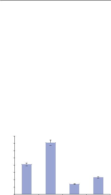
sensitive to pyrethroid (890 ppm). Also, single osteoblast cells respond the fastest to EDTA (0.14 sec) whereas they take the maximum time to respond to hydrogen peroxide (0.71 sec). This data is summarized in Table 4.2. Figure 4.18 is a graphical representation of the response times obtained for each specific chemical agent. The graph shows the repeatability of the response. The response time for each chemical was determined by testing a specific agent in three cycles and each cycle comprised of three runs.
4.4.7.6. Effect of Varying Concentration of Chemical Agents It was observed for all the chemical agents that the amplitude of the response decreased as the concentration of the chemical agent in the local microenvironment increased. WT analysis was performed where local time domain characterization of the amplitude was performed as a function of concentration. This analysis identified the amplitude shifts corresponding to the varying concentration. WT analysis indicated that at a higher concentration (1000 ppm), there was a large decrement in the amplitude of the time domain signal of the extracellular potential. For low levels of concentration near the detection limit, the decrement of the amplitude was much smaller, by a factor of about 80%. Figures 4.19(A) and (B) represent the variation in amplitude due to low (180 ppm) and high (1000 ppm) concentrations of EDTA. It was also observed that there is no noticeable difference in the response times due to varying concentrations for a specific chemical agent.
Response Time(s)
0.8 |
|
0.71s |
|
|
0.7 |
|
|
|
|
0.6 |
|
|
|
|
0.5 |
0.41s |
|
|
|
0.4 |
|
|
|
|
0.3 |
|
|
|
0.23s |
0.2 |
|
|
0.14s |
|
0.1 |
|
|
|
|
0 |
Ethanol |
Peroxide |
|
Pyrethroids |
|
EDTA |
FIGURE 4.18. Representation of response times for specific chemical agents [105].

86 |
CENGIZ S. OZKAN ET AL. |
FIGURE 4.19. A. Variation in amplitude of single osteoblast due to the action of EDTA at 180 ppm. B. Variation in amplitude of single osteoblast due to the action of EDTA at 1000 ppm [105].
CELL BASED SENSING TECHNOLOGIES |
87 |
4.4.7.7. Cascaded Sensing of Chemical Agents Using Single Osteoblast |
To simulate |
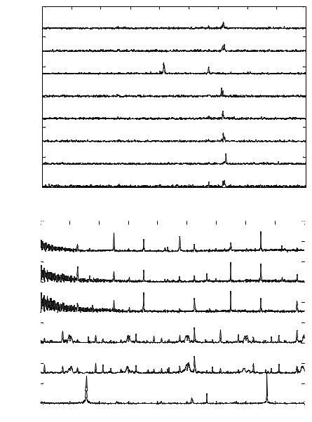
real time field conditions, the selectivity of single osteoblast sensors was tested. The ability of the sensor was examined to identify specific chemical agents when introduced in cascade by exhibiting the SPV corresponding to each chemical agent. Here, cascaded sensing of ethanol and hydrogen peroxide was described by single osteoblast cells. After determining the detection limits for both of the chemical agents, first, ethanol at 19 ppm was introduced into the chip sensor and the modified extracellular potential was recorded. As observed previously the osteoblast cell then regains its initial spectrum after undergoing modulation. Hydrogen peroxide at 25 ppm was then introduced into the chip sensor and the modulated response was recorded. FFT analysis of the acquired data indicates that the SPV obtained in the cascaded sensing exactly correlated with the SPVs obtained from individual sensing of ethanol and hydrogen peroxide. The Eigen vectors corresponding to ethanol (514 Hz and 722 Hz) and those corresponding to hydrogen peroxide (257 Hz, 576 Hz and 852 Hz) can be correlated to those obtained during individual chemical sensing. There is a slight shift in two of the Eigen vectors of hydrogen peroxide from 565 Hz to 576 Hz and from 873 Hz to 852 Hz which can be accounted for by the interaction between ethanol and hydrogen peroxide. Figures 4.20(A) and (B) represent the SPVs of a single osteoblast cell due to the cascaded action of ethanol and hydrogen peroxide respectively (for n = 15).
4.5. DISCUSSION AND CONCLUSION
The surge of interest in bioanalysis over the last decade has resulted in an ingenious of new proposals and a series of solutions to earlier problems. We have seen the reduction to commercial reality of some of the more speculative proposals from earlier years. Among the most rapidly advancing of these fronts is the area of biosensing, whether it is single analyte detection methods or multiarray-based biochip technology. The 1990s have seen the development of biosensors for many different analyses, and even seen them begin to advance to clinical and in some cases commercially available technologies [55, 60].
Cell-based biosensors constitute a promising field that has numerous applications ranging from pharmaceutical screening to environmental monitoring. Cells provide an array of naturally evolved receptors and pathways that can respond to an analyte in a physiologically relevant manner. Enzymes, receptors, channels, and other signaling proteins that may be targets of an analyte are maintained and, as necessary, regenerated by the molecular machinery present in cells. The array of signaling systems characteristic of cell-based sensors yields generic sensitivity that is a distinguishing feature in comparison to other molecular biosensor approaches. In addition, cell-based sensors offer an advantage of constituting a function-based assay that can yield insight into the physiologic action of an analyte of interest. Three important issues that constitute barriers for the use of cell-based sensors have been presented and discussed. There are certainly other areas that will require attention, as cell-based sensors move from the laboratory environment; namely, cell delivery and/or preservation technologies. As further progress is made to address fundamental challenges, cell-based biosensors and related cellular function based assays will undoubtedly become increasingly important and useful. It is of popular belief that such function-based assays will become an indispensable tool for monitoring in environmental, medical, and defense applications. Strategies relying on a single population of excitable cells appear most well
88 |
CENGIZ S. OZKAN ET AL. |
(a) |
|
1212 |
|
|
|
|
|
|
|
|
|
|
(A) |
|
|
|
|
|
|
|
|
|
|
|
|
|
|
|
t=0s |
|
|
|
|
|
(722, 0.468) |
|
|
|
|
|
|
|
|
|
|
(667, 0.207) |
|
|
|
||
|
|
1010 |
|
|
|
|
|
|
(722, 0.487) |
|
|
|
|
|
|
t=30s |
|
|
|
|
|
|
|
||
|
|
|
|
|
|
|
(667, 0.156) |
|
|
|
||
|
|
88 |
|
|
|
(514, 0.760) |
(667, 0.491) |
|
|
|
||
|
|
t=60s |
|
|
|
|
|
|
|
|||
|
|
|
|
|
|
|
|
|
|
|
||
|
|
|
|
|
|
|
|
|
|
|
|
|
Amplitude |
Amplitude |
|
t=90s |
|
|
|
|
|
(712, 0.577) |
|
|
|
|
|
|
|
|
|
|
|
|
|
|||
66 |
|
|
|
|
|
|
|
|
|
|
||
|
t=120s |
|
|
|
|
|
(716, 0.565) |
|
|
|||
|
|
|
|
|
|
|
|
|
|
|
|
|
|
|
44 |
|
|
|
|
|
|
(718, 0.661) |
|
|
|
|
|
|
t=150s |
|
|
|
|
|
|
|
||
|
|
|
|
|
|
|
|
|
|
|
|
|
|
|
|
t=180s |
|
|
|
|
|
(722, 0.728) |
|
|
|
|
|
22 |
|
|
|
|
|
|
|
|
|
|
|
|
|
t=210s |
|
|
|
|
|
(722, 0.536) |
|
|
|
|
|
|
|
|
|
|
|
|
|
|
|
|
|
|
00 |
200 |
300 |
400 |
500 |
|
600 |
700 |
800 |
900 |
1000 |
|
|
100 |
|
|||||||||
|
|
|
|
|
|
Frequency(Hz) |
|
|
|
|
||
(b) |
|
99 |
|
|
|
|
|
|
|
|
|
|
(B) |
|
|
|
(351, 0.929) |
|
|
|
|
|
(852, 1.001) |
|
|
|
|
|
|
|
(576, 0.757) |
|
|
|
||||
|
|
|
|
|
(452, 0.595) |
(752, 0.433) |
|
|
||||
|
|
88 |
t=240s |
|
|
|
|
|
||||
|
|
|
|
|
|
|
|
|
|
|
||
|
|
|
|
|
|
|
|
|
|
|
|
|
|
|
77 |
|
|
|
|
|
|
(752, 0.962) |
(852, 0.908) |
|
|
|
|
t=270s |
( 351, 0.445) (452, 0.514) |
|
|
|
|
|
|
|||
|
|
|
(576, 0.253) |
|
|
|
|
|||||
|
|
|
|
|
|
|
|
|
|
|
||
|
66 |
|
|
|
|
|
|
|
|
|
Amplitude |
44 |
|
|
|
(452, 0.897) |
(626, 0.624) |
(752, 1.023) |
(852, 0.596) |
|
|
|
|
t=300s |
(351, 0.453) |
|
(626, 0.603) |
|
|
|
||
|
55 |
|
|
|
|
|
|
|||
|
|
|
|
|
|
|
|
|
|
|
|
|
t=330s |
|
|
|
|
|
|
|
|
|
33 |
|
|
|
|
|
|
|
|
|
|
|
|
|
|
|
(626, 0.635) |
|
|
|
|
|
22 |
t=360s |
|
|
|
|
|
|
|
|
|
|
|
|
|
|
|
|
|
|
|
|
11 |
|
( 257, 1.226) |
|
|
|
|
|
( 852, 1.346) |
|
|
|
|
|
|
(668, 0.505) |
|
|
|
||
|
|
t=390s |
|
|
|
|
|
|
||
|
00 |
|
|
|
|
600 |
700 |
800 |
900 |
1000 |
|
100 |
200 |
300 |
400 |
500 |
|||||
|
600 |
700 |
800 |
900 |
1000 |
|||||
Frequency(Hz)
FIGURE 4.20. A. Signature pattern vector of single osteoblast due to the cascaded action of ethanol-hydrogen peroxide at 19 ppm and 25 ppm respectively. B. Signature Pattern Vector of single osteoblast due to the cascaded action of ethanol-hydrogen peroxide at 19 ppm and 25 ppm, respectively [105].
CELL BASED SENSING TECHNOLOGIES |
89 |
suited for measurements of acute and direct effects of receptor agonist/antagonists. Compounds that fall within this category include ion channel modulators, metals, ligand–receptor blockers, and neurotransmitters. In fact, the detection of acute and direct effects of compounds may be sufficient and relevant for certain operational situations, such as a battlefield environment or the floor of an assembly plant, where cognitive function is absolutely critical. The prospect of detecting all physiologically active analytes using a single cell or tissue type is improbable. It is possible that particular analytes may undergo biotransformation, resulting in a secondary or tertiary compound of substantial physiologic effect. In spite of that drawback single cell based sensors are highly reliable. This has been shown by the newly developed single cell based sensing technique. This technique functions on the principle of integrating a fundamental biological tool like dielectrophoresis to biochip technology. Single cell arrays of the same biological state and differentiation can be developed using this method. Simultaneous sensing can be achieved, which reduces false alarms. Unique identification tags have been generated for identifying specific chemical analytes using this technique. These are known as Signature Patterns. The greatest advantage of this technique is its high sensitivity and speed of response. Chemical analytes of concentrations in the order of parts per billion have been detected. To determine the veracity and reliability of the sensor simultaneous fluorescence detection techniques have also been implemented at the detection limit obtained from the single cell based sensor. The physiological behavior corroborates the sensing. This establishes the viability of this technique for potential commercial implementation.
Finally, the threat of biological weapons has become a major concern to both the civilian and military populations. All of the present biological warfare and environmental agent rapid detection systems, in field use or under prototype development, rely on structural recognition approaches to identify anticipated agents. Cell based sensor technology utilizing biochip capability can be thought to be one potentially reliable solution. It is now only a matter of time before this technology will impinge on a wide range of commercial situations.
REFERENCES
[1]T. Akin, K. Najafi, R.H. Smoke, and R.M. Bradley. IEEE Trans. Biomed. Eng., 41:305, 1994.
[2]B.M. Applegate, S.R. Kermeyer, and G.S. Sayler. Appl. Environ. Microbiol., 64:2730, 1998.
[3]S. Belkin, D.R. Smulski, S. Dadon, A.C. Vollmer, T.K. Van Dyk, and R.A. Larossa. Wat. Res., 31:3009, 1997.
[4]R.A. Bissell, A.P. de Silva, H.Q.N. Gunaratne, P.L.M. Lynch, G.E.M. Maguire, and K.R.A.S. Sandanayake. Chem. Soc. Rev., 21:187–195, 1992.
[5]L. Bousse, R.J. McReynolds, G. Kirk, T. Dawes, P. Lam, W.R. Bemiss, and J.W. Parce. Sens. Actu. B, 20:145, 1994.
[6]L.J. Breckenridge, R.J.A.Wilson, P. Connolly, A.S.G. Curtis, J.A.T. Dow, S.E. Blackshaw, and C.D.W. Wilkinson. J. Neurosci. Res., 42:266, 1995.
[7]R.S. Burlage, A.V. Palumbo, A. Heitzer, and G. Sayler. Appl. Microbiol. Biotechnol., 45:731, 1994.
[8]J.C. Chang, G.J. Brewer, and B.C. Wheeler. J. Biomed. Microdev., 2(4):245, 2000.
[9]P. Clark, P. Connolly, A.S.G. Curtis, J.A.T. Dow, and C.D.W. Wilkinson. J. Cell Sci., 99:73, 1991.
[10]B.A. Cornell, V.L. Braach-Maksvytis, L.G. King, P.D. Osman, B. Raguse, L. Wieczorek, and R.J. Pace, Nature, 387:580, 1997.
[11]P. Connolly, G.R. Moores, W. Monaghan, J. Shen, S. Britland, and P. Clark. Sens. Actu., B6:113, 1992.
90 |
CENGIZ S. OZKAN ET AL. |
[12]K.S. Cole and H.J. Curtis. J. Gen. Physiol., 22:649, 1939.
[13]J. Csicsvari, D.A. Henze, B. Jamieson, K.D. Harris, A. Sirota, P. Bartho, K.D. Wise, and G. Buzsaki. J. Neurophysi., 90:1314, 2003.
[14]A.W. Czarnik and J.P. Desvergne. Chemosensors for Ion and Molecule Recognition. Kluwer, Dordrecht, The Netherlands, 1997.
[15]A.P. De Silva and R.A.D.D. Rupasinghe. J. Chem. Soc. Chem. Commun., 166:14, 1985.
[16]A.P. De Silva, H.Q.N. Gunaratne, T. Gunnlaugsson, A.J.M. Huxley, C.P. McCoy, J.T. Rademacher, and T.E. Rice. Chem. Rev., 97:1515, 1997.
[17]A.M. Dijkstra, B.H. Brown, A.D. Leathard, N.D. Harris, D.C. Barber, and L. Edbrooke. J. Med. Eng. Technol.,. 17:89, 1993.
[18]C.S. Dulcey, J.H. Georger, A. Krauthamer, Jr., D.A. Stenger, T.L. Fare, and J.M. Calvert. Science, 252:551, 1991.
[19]D.J. Edell, V.V. Toi, V.M. McNeil, and L.D. Clark. IEEE Trans. Biomed. Eng., 39:635, 1992.
[20]C.F. Edman, D.E. Raymond, D.J., Wu, E.G. Tu, R.G. Sosnowski,W.F. Butler, M. Nerenberg, and M.J. Heller. Nucleic Acids Res., 25:4907, 1997.
[21]G.A. Evtugyn, E.P. Rizaeva, E.E. Stoikova, V.Z. Latipova, and H.C. Budnikov. Electroanalysis, 9:1–5, 1997.
[22]H. Fricke and S. Morse. J. Gen. Physiol., 9:153, 1926.
[23]H. Fricke and H.J. Curtis. J. Gen. Physiol., 18:821, 1935.
[24]P. Fromherz, A. Offenhausser, T. Vetter, and J. Weis. Science, 252:290, 1991.
[25]G. Fuhr, H. Glasser, T. Muller, and T. Schnelle. Biochim. Biophys. Acta., 1201:353, 1994.
[26]L. Giaever and C.R. Keese. IEEE Trans. Biomed. Eng., 33:242, 1986.
[27]L. Griscom, P. Degenaar, B. LePioufle, E. Tamiya, and H. Fujita. Sens. Actu. B, 83(1–3):15, 2002.
[28]G.W. Gross, B.K. Rhoades, and R. Jordan. Sen. Actu. B, 6:1, 1992.
[29]G.W. Gross, B.K. Rhoades, H.M.E. Azzazy, and M.C. Wu. Biosens. Bioelectron., 10:553, 1995.
[30]G.W. Gross, W. Wen, and J. Lin. J. Neurosci. Meth., 15:243, 1985.
[31]G.W. Gross, B.K. Rhoades, D.L. Reust, and F.U. Schwalm. J. Neurosci. Meth., 50:131, 1993.
[32]G.W. Gross. Internal dynamics of randomized mammalian neuronal networks in culture. In Enabling Tedmologiesfor Cultured Neural Networks. Academic Press, Vol. 277, 1994.
[33]W. Gopel, J.L. Hesse, and J.N. Zemel. A Comprehensive Survey of Sensors, Trends in Sensor Technology/ Sensor Markets. (ed.), Elseiver, Netherlands, 1995.
[34]L.L. Hause, R.A. Komorowski, and F. Gayon. IEEE Trans. Biomed. Eng., BME-28:403, 1981.
[35]R.P. Haugland. Handbook of Fluorescent Probes and Research Chemicals, 6th Ed. Molecular Probes, Eugene, OR, 1996.
[36]A. Heitzer, K. Malachowsky, J.E. Thonnard, P.R. Bicnkowski, D.C. White, and G.S. Sayler. Appl. Environ. Microbiol., 60:1487, 1994.
[37]R. Heim and R.Y. Tsien. Curr Biol., 1:178–182, 1996.
[38]A.W. Hendricson, M.P. Thomas, M.J. Lippmann, and R.A. Morrisett. J. Pharmacol. Exp. Ther., 307(2):550, 2003.
[39]A.L. Hodgkin and A.F. Huxley. J. Physiol., 117:500, 1952.
[40]Y. Huang, R. Holzel, R. Pethig, and X.B. Wang. Phys. Med. Biol., 37(7):1499–1517, 1992.
[41]M.E. Huston, K.W. Haider, and A.W. Czarnik. J. Am. Chem. Soc., 110:4460, 1988.
[42]M. Jenker, B. Muller, and B. Fromherz. Biol. Cybernetics., 84:239, 2001.
[43]I.S. Kampa and P. Keffer. Clin. Chem., 44:884, 1998.
[44]Y. Kitagawa, M. Ameyama, K. Nakashima, E. Tamiya, and I. Karube. Analyst, 112:1747, 1987.
[45]G.T.A. Kovacs. IEEE Trans. Biomed. Eng., 1992.
[46]M. Kowolenko, C.R. Keese, D.A. Lawrence, and I. Giaever. J. Immunol. Methods, 127:71, 1990.
[47]Y.I. Korpan, M.V. Gonchar, N.F. Starodub, A.A. Shul’ga, A.A. Sibirny, and A.V. El’skaya. Anal. Biochem., 215:216, 1993.
[48]S. Lacorte, N. Ehresmann, and D. Barcelo. Environ. Sci. Technol., 30:917, 1996.
[49]H.B. Li and R. Bashir. Sens. Actu. B, 86:215, 2002.
[50]N. Li, H. Endo, T. Hayashi, T. Fujii, R. Takal, and E. Watanahe. Biosens. Bioelectron., 9:593, 1994.
[51]B. Luc. Sens. Actu. B, 34:270, 1996.
[52]M.P. Maher, J. Pine, J. Wright, and Y.C. Tai. J. Neurosci. Meth., 87:45, 1999.
[53]G.H. Markx, M.S. Talary, and R. Pethig. J. Biotechnol., 32:29, 1994.
CELL BASED SENSING TECHNOLOGIES |
91 |
[54]G.H. Markx and R. Pethig. Biotech. Bioeng., 45:337, 1995.
[55]M. Malmquist. M Biochem. Soc. T., 27:335, 1999.
[56]H.M. McConnell, J.C. Owicki, J.W. Parce, D.L. Miller, G.T. Baxter, H.G. Wada, and S. Pitchford. Science, 257:1906, 1992.
[57]T. Muller,¨ A. Gerardino, T. Schnelle, S.G. Shirley, Bordoni, Gasperis, G. De, R. Leoni, and G.J. Fuhr. Phys. D: Appl. Phys., 29:340, 1996.
[58]K. Najafi. Sens. Actu. B, 1:453, 1990.
[59]A. Offenhausser, C. Sprossler, M. Matsuzawa, and W. Knoll. Biosens. Bioelectron., 12(8):819, 1997.
[60]A. Ota and S. Hybridoma. Ueda, 17:471, 1998.
[61]J.C. Owicki and J.W. Parce. Biosen. Bioelectron., 7:255, 1992.
[62]J.C. Owicki, J.W. Parce, K.M. Kercso, G.B. Sisal, V.C. Muir, J.C. Vcnter, C.M. Fraser, and H.M. McConnell.
Proc. Natl. Acad. Sci., 87:4007, 1990.
[63]B.M. Paddle. Biosens. Bioelectron., 11:1079, 1996.
[64]J.W. Parce, J.C. Owicki, K.M. Kercso, G.B. Sigal, H.G. Wada, V.C. Muir, L.J. Bousse, K.L. Ross, B.I. Sikic, and H.M. McConnell. Science, 246:243, 1989.
[65]D. Pollard-Knight, E. Hawkins, D. Yeung, D.P. Pashby, M. Simpson, A. McDougall, P. Buckle, and S.A. Charles. Ann. Biol. Clin., 48:642, 1990.
[66]H.A. Pohl. Dielectrophoresis, the Behavior of Neutral Matter in Nonuniform Electric Fields. Cambridge University Press, 1978.
[67]H.A. Pohl and I. Hawk. Science, 152:647, 1966.
[68]S. Prasad, M. Yang, X. Zhang, C.S. Ozkan, and M. Ozkan. Biomed. Microdev., 5(2):125, 2003.
[69]W.G. Regehr, J. Pine, and D.B. Rutledge. IEEE Trans. Biomed. Eng., 35:1023, 1998.
[70]B.K. Rhoades and G.W. Gross. Brain Res., 643:310, 1994.
[71]J. Ruhe, R. Yano, J.S. Lee, P. Koberle, W. Knoll, and A. Offenhausser. J. Biomater. Sci. Polym. Ed., 10(8):859, 1999.
[72]T.G. Ruardij, M.H. Goedbloed, and W.L.C. Rutten. Med. Biol. Eng. Comput., 41(2):227, 2003.
[73]H.P. Schwan. C.A. Academic Press, New York, Vol. 5, pp. 147, 1957.
[74]G. Schmuck,A. Freyberger, H.J. Ahr, B. Stahl, and M. Kayser. Neurotoxicology, 24:55, 2003.
[75]M. Scholl, C. Sprossler, M. Denyer, M. Krause, K. Nakajima, A. Maelicke, W. Knoll, and A. Offenhausser.
J. Neurosci. Meth., 104:65, 2000.
[76]O. Selifonova, R.S. Burlage, and T. Barkay. Appl. Environ. Micobiol., 59:3083, 1993.
[77]D.E. Semler, E.H. Ohlstein, P. Nambi, C. Slater, and P.H. Stern. J. Pharmacol. Exp. Ther., 272:3:1052, 1995.
[78]J.B. Shear, H.A. Fishman, N.L. AIIbritton, D. Garigan, R.N. Zare, and R.H. Scheller. Science, 267:74, 1995.
[79]J. Singh, P. Khosala, and R.K. Srivastava. Ind. J. Pharmacol., 32:206, 2000.
[80]R.S. Skeen, W.S. Kisaalita, and B.J. Van Wie Biosens. Bioelectronic., 5:491, 1990.
[81]D.A. Stenger, G.W. Goss, E.W. Keefer, K.M. Shaffer, J.D. Andreadis,W. Ma, and J.J. Pancrazino. Trends in Biotechnol., 19:304, 2001.
[82]M. Stephens, M.S. Talary, R. Pethig, A.K. Burnett, and K.I. Mills. Bone Narrow Transpl., 18:777, 1996.
[83]P. Sticher, M.C.M. Jaspers, K. Stemmler, H. Harms, A.J.B. Zehnder, and vand der Meer. J.R. Appl. Environ. Microbiol., 63:4053–4060, 1997.
[84]A.A. Suleiman and G.G. Guilbault. Analyst., 119:2279, 1994.
[85]F.J. Swenson. Sens. Actu. B., 11:315, 1992.
[86]D.W. Tank, C.S. Cohan, and S.B. Kater. IEEE Conf. on Synthetic Microstructures, Airlie House, Arlington, Virginia, IEEE, New York, 1986.
[87]H.M. Tan, S.P. Chcong, and T.C. Tan. Biosens. Bioelectron., 9:1, 1994.
[88]T. Takayasu, T. Ohshima, and T. Kondo. Leg. Med., (Tokyo). 3:(3):157, 2001.
[89]P. Thiebaud, L. Lauer, W. Knoll, and A. Offenhausser. Biosens. Bioelectron., 17(1–2):87, 2002.
[90]C. Tiruppathi, A.B. Malik, P.J. Del Vecchio, C.R. Keese, and I. Giaever. Proc. Natl. Acad. Sci., 89:7919, 1992.
[91]R.Y. Tsien. Annu. Rev. Neurosci., 12:227–253, 1989.
[92]D.W. Tank, C.S. Cohan, and S.B. Kater. IEEE Conf. on Synthetic Microstructures. Airlie House, Arlington, Virginia, IEEE, New York, 1986.
[93]R.Y. Tsien. Annu. Rev. Neurosci., 12:227–253, 1989.
[94]S.J. Updike and G.P. Hicks. Nature, 214:986, 1967.
92 |
CENGIZ S. OZKAN ET AL. |
[95]D. Van der Lelie, P. Corbisier, W. Baeyens, S. Wnertz, L. Diels, and M. Mergeay. Res. Microbiol., 145:67, 1994.
[96]H.J. Watts, D. Yeung, and H. ParkesAnal. Chem., 67:4283, 1995.
[97]X. Wang, Y. Huang, P.R.C. Gascoyne, and F.F. Becker. IEEE Tran. Ind. Appl., 33:660, 1997.
[98]B.C. Wheeler, J.M. Corey, G.J. Brewer, and D.W. Branch. J. Biomech. Eng., 121:73, 1999.
[99]J.P. Whelan, L.W. Kusterbeck, G.A., Wemhoff, R. Bredehorst, and F.S. Ligler. Anal. Chem., 65:3561, 1993.
[100]P. Wilding, J. Pfahler, H.H. Ban, J.N. Zemcl, and L.J. Kricka. Clin. Chem., 40:43, 1994.
[101]C. Wyart, C. Ybert, L. Bourdieu, C. Herr, C. Prinz, and D.J. Chattenay. J. Neurosci. Meth., 117(2):23, 2002.
[102]M. Yang, X. Zhang, K. Vafai, and C.S. Ozkan. J. Micromech. Microeng., 13:864, 2003.
[103]M. Yang, X. Zhang, and C.S. Ozkan. Biomed. Microdev., 5:323, 2003.
[104]M. Yang, S. Prasad, X. Zhang, M. Ozkan, and C.S. Ozkan. Sensor Letters, 2:1, 2004.
[105]M. Yang, S. Prasad, X. Zhang, A. Morgan, M. Ozkan, and C.S. Ozkan. Sens. Mater., 15(6):313, 2003.
[106]M. Yang, X. Zhang, Y. Zhang, and C.S. Ozkan. Sens. Actu. B, (in print), 2004.
[107]G. Zeck and P. Fromherz.. Proc. Natl. Acad. Sci. U.S.A., 98(18):10457, 2001.
[108]I. Giaever and C.R. Keese. Proc. Natl. Acad. Sci. U.S.A., 88:7896, 1991.
[109]J. Pancrazio, S.A. Gray, Y.S. Shubin, N. Kulagina, D.S. Cuttino, K.M. Shaffer, K. Kisemann, A. Curran, B. Zim, G.W. Gross, and T.J. O’Shaughnessy. Biosens. Bioelectron., 18(11):1339, 2003.
5
Fabrication Issues of Biomedical
Micro Devices
Nam-Trung Nguyen
School of Mechanical and Production Engineering, Nanyang Technological University, 50. Nanyang Avenue, Singapore 639798
5.1. INTRODUCTION
Biomedical micro devices (BMMD) are microsystems, which can be used in surgery, biomedical diagnostics, and therapeutic management [28]. These devices allow precise surgical procedures with spatial control in the micrometer range. The minimal invasive approach enables faster recovery for patients through shorter access pathways and reduced operation trauma. BMMDs for biomedical diagnostics and therapeutic management utilizes microfluidic technology that allows faster screening of common diseases as well as painless and effective drug delivery. The successful development and introduction of these technologies in health care will have a great impact on the living quality of patients, and significantly lower the total cost of medical treatment.
Conventional fabrication method evolving from microelectronics were used for fabricating micro electromechanical systems (MEMS). MEMS-technology was successfully commercialized in products such as micro sensors and micro actuators. However, the extension of silicon-based devices to biomedical applications have certain limitations. The majority of developed devices have been realized on silicon and glass, because the fabrication technologies for them are matured and widely available [2, 28]. Almost all conventional micromachining techniques such as wet etching, dry etching, deep reactive ion etching, sputter, anodic bonding, and fusion bonding were used for fabricating BMMDs. Key components of a BMMD such as flow channels, flow sensors, chemical detectors, separation capillaries, mixers, filters, pumps, and valves have been developed based on silicon technology [26]. These devices have many advantages over their macro counterparts. They