

Biomolecular Sensing Processing and Analysis - Rashid Bashir and Steve Wereley
.pdfI
Micro and Nanoscale Biosensors
and Materials
1
Biosensors and Biochips
Tuan Vo-Dinh
Center for Advanced Biomedical Photonics, Oak Ridge National Laboratory, Bethel Valley Road; MS-6101, P.O. Box 2008, Oak Ridge, TN 37831-6101, U.S.A.
This chapter provides an overview of the various types of biosensors and biochips that have been developed for biological and medical applications, along with significant advances and over the last several years in these technologies. Various classification schemes that can be used for categorizing the different biosensor and biochip systems are also discussed.
1.1. INTRODUCTION
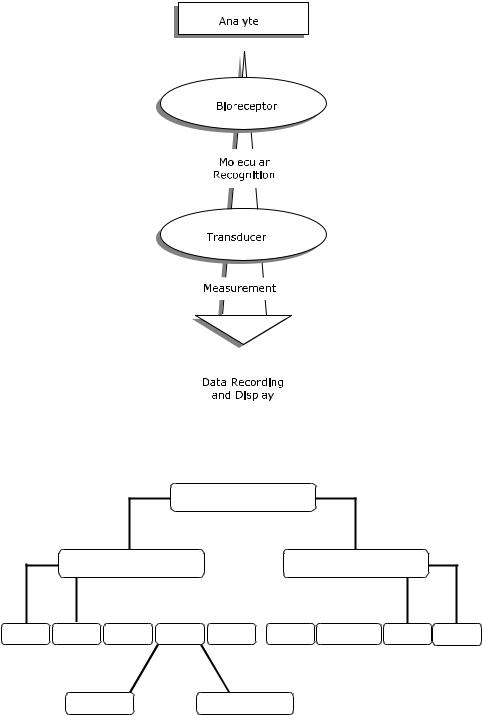
A biosensor can be generally defined as a device that consists of a biological recognition system, often called a bioreceptor, and a transducer. In general, a biochip consists of an array of individual biosensors that can be individually monitored and generally are used for the analysis of multiple analytes. The interaction of the analyte with the bioreceptor is designed to produce an effect measured by the transducer, which converts the information into a measurable effect, such as an electrical signal. Figure 1.1 illustrates the conceptual principle of the biosensing process. Biosensors that include transducers based on integrated circuit microchips are often referred to as biochips.
There are several classification schemes possible. Biosensors and biochips can be classified either by their bioreceptor or their transducer type (see Figure 1.2). A bioreceptor is a biological molecular species (e.g., an antibody, an enzyme, a protein, or a nucleic acid) or a living biological system (e.g., cells, tissue, or whole organisms) that utilizes a biochemical mechanism for recognition. The sampling component of a biosensor contains a bio-sensitive layer. The layer can either contain bioreceptors or be made of bioreceptors covalently attached to the transducer. The most common forms of bioreceptors used in biosensing are based on 1) antibody/antigen interactions, 2) nucleic acid interactions,
4 |
|
|
|
|
|
|
TUAN VO-DINH |
|
|
|
|
|
|
|
|
|
|
|
|
|
|
|
|
|
|
|
|
|
|
|
|
|
|
|
|
|
|
|
|
|
|
|
|
|
|
|
|
|
|
|
|
|
|
|
|
|
|
|
|
|
|
|
|
|
|
|
|
|
|
|
|
|
|
|
|
|
|
|
|
FIGURE 1.1. Conceptual diagram of the biosensing principle.
BIOCHIPS
Bioreceptor |
|
|
Transducer |
|||
|
||||||
|
|
|
|
|
|
|
|
|
|
|
|
|
|
Antibody |
Enzyme |
DNA |
Cell |
Biomimetic |
Optical |
Electrochemical Mass-Based |
Other |
Cellular Systems |
Non-Enzymatic Proteins |
FIGURE 1.2. Schematic of biosensor/biochip classification schemes.
BIOSENSORS AND BIOCHIPS |
5 |
3) enzymatic interactions, 4) cellular interactions (i.e. microorganisms, proteins) and 5) interactions using biomimetic materials (i.e., synthetic bioreceptors). For transducer classification, conventional techniques include: 1) optical measurements (i.e. luminescence, absorption, surface plasmon resonance, etc.) 2) electrochemical and 3) mass-sensitive measurements (i.e. surface acoustic wave, microbalance, etc.).
The development of biosensors was first reported in the early 1960s [6]. Biosensors have now seen an explosive growth and seen a wide variety of applications primarily in two major areas, biological monitoring and environmental sensing applications.
1.2. BIOSENSORS
1.2.1. Different Types of Bioreceptors
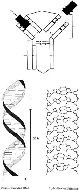
The key to specificity for biosensor technologies involves bioreceptors. They are responsible for binding the analyte of interest to the sensor for the measurement. These bioreceptors can take many forms and the different bioreceptors that have been used are as numerous as the different analytes that have been monitored using biosensors. However, bioreceptors can generally be classified into five different major categories. These categories include: 1) antibody/antigen, 2) enzymes, 3) nucleic acids/DNA, 4) cellular structures/cells and 5) biomimetic. Figure 1.3 shows a schematic diagram of two types of bioreceptors: the structure of an immunoglobulin G (IgG) antibody molecule (Fig. 1.3A), and DNA and the principle of base pairing in hybridization (Fig. 1.3B).
1.2.1.1. Antibody Bioreceptors An antibody is a complex biomolecule, made up of hundreds of individual amino acids arranged in a highly ordered sequence. Antibodies are biological molecules that exhibit very specific binding capabilities for specific structures. For an immune response to be produced against a particular molecule, a certain molecular size and complexity are necessary: proteins with molecular weights greater then 5000 Da are generally immunogenic. The way in which an antigen and its antigen-specific antibody interact may be understood as analogous to a lock and key fit, by which specific geometrical configurations of a unique key enables it to open a lock. In the same way, an antigen-specific antibody “fits” its unique antigen in a highly specific manner. This unique property of antibodies is the key to their usefulness in immunosensors where only the specific analyte of interest, the antigen, fits into the antibody binding site.
Radioimmunoassay (RIA) utilizing radioactive labels have been applied to a number of fields including pharmacology, clinical chemistry, forensic science, environmental monitoring, molecular epidemiology and agricultural science. The usefulness of RIA, however, is limited by several shortcomings, including the cost of instrumentation, the limited shelf life of radioisotopes, and the potential deleterious biological effects inherent to radioactive materials. For these reasons, there are extensive research efforts aimed at developing simpler, more practical immunochemical techniques and instrumentation, which offer comparable sensitivity and selectivity to RIA. In the 1980s, advances in spectrochemical instrumentation, laser miniaturization, biotechnology and fiberoptic research have provided opportunities for novel approaches to the development of sensors
6
A
ANTIGEN (LOCK AND KEY FIT)
NH2 TERMINUS
Fc
LIGHT |
|
|
|
CHAIN |
|
|
|
s |
|
|
|
s |
|
|
|
|
|
|
|
|
s |
|
s |
|
s |
|
|
|
|
s |
|
CHAIN HEAVY |
|
|
|
|
|
||
s s
DOMAIN VARIABLE
TUAN VO-DINH
Fab
COOH TERMINUS
B
C
G
3.4 Å
A T
G C
T A
C G
36 Å |
C |
G |
|
|
A T
T A
C G
20 Å
Double-Stranded DNA |
|
Hybridization Principle |
FIGURE 1.3. Schematic diagrams of two types of bioreceptors: A) IgG antibody. B) DNA and the hybridization principle.
BIOSENSORS AND BIOCHIPS |
7 |
for the detection of chemicals and biological materials of environmental and biomedical interest.
The first fiberoptic immunosensor was developed for in situ detection of the chemical carcinogen benzo[a]pyrene [52]. Nowadays, antibodies are often used in biosensors today. Biomolecular interactions can be classified in two categories, according to the test format performed (i.e., direct and indirect). In a direct format the immobilized target molecule interacts with a ligand molecule or the immobilized ligand interacts with a target molecule directly. For immunosensors, the simplest situation involves in situ incubation followed by direct measurement of a naturally fluorescent analyte [52]. For nonfluorescent analyte systems, in situ incubation is followed by development of a fluorophor-labeled second antibody. The resulting antibody sandwich produces a fluorescence signal that is directly proportional to the amount of bound antigen. The sensitivity obtained when using these techniques increases with increasing amounts of immobilized receptor. The indirect format involves competition between fluorophor-labeled and unlabeled antigens [42]. In this case, the unlabeled analyte competes with the labeled analyte for a limited number of receptor binding sites. Assay sensitivity therefore increases with decreasing amounts of immobilized reagent.
Antibody-based biosensors have been developed for use in an electrochemical immunoassay for whole blood [4]. The assay is performed on a conducting redox hydrogel on a carbon electrode on which avidin and choline oxidase have been co-immobilized. Biotinylated antibody was then bound to the gel. When the antigen binds to the sensor, another solution of complementary horseradish peroxidase labeled antibody is bound to the antigen, thus creating an electrical contact between the redox hydrogel and the peroxidase. The hydrogel then acts as an electrocatalyst for the reduction of hydrogen peroxide water.
Binding of the bioreceptor to the measurement support or the transducer is an important aspect of biosensor fabrication. A method for the immobilization of histidine-tagged antibodies onto a gold surface for surface plasmon resonance measurements was reported [15]. A synthetic thioalkane chelator is self-assembled on a gold surface. Reversible binding of an anti-lysozyme F-ab fragment with a hexahistidine modified extension on the C terminal end is then performed. Infrared spectroscopy was used to determine that the secondary structure of the protein was unaffected by the immobilization process. Retention of antibody functionality upon immobilization was also demonstrated. Due to the reversible binding of such a technique, this could prove a valuable method for regeneration of biosensors for various applications. Enzyme immunoassays can further increase the sensitivity of detection of antigen-antibody interactions by the chemical amplification process, whereby one measures the accumulated products after the enzyme has been allowed to react with excess substrate for a period of time [51].
With the use of nanotechnology, submicron fiberoptic antibody-based biosensors have been developed by Vo-Dinh and coworkers for the measurements of biochemicals inside a single cell [1, 8, 50]. Nanometer scale fiberoptic biosensors were used for monitoring biomarkers related to human health effects that are associated with exposure to polycyclic aromatic hydrocarbons (PAHs). These sensors use a monoclonal antibody for benzo[a]pyrene tetrol (BPT), a metabolite of the carcinogen benzo[a]pyrene, as the bioreceptor. Excitation light is launched into the fiber and the resulting evanescent field at the tip of the fiber is used to excite any of the BPT molecules that have bound to the antibody. The fluorescent light is then collected via a microscope. Using these antibody-based
8 |
TUAN VO-DINH |
nanosensors, absolute detection limits for BPT of ca. 300 zeptomol (10−21 moles) have been reported [1]. These nanosensors allow the probing of cellular and subcellular environments in single cells [8, 50] as well as monitoring signaling processes in single cells [18, 46].
1.2.1.2.Enzyme Bioreptors Another type of commonly used bioreceptors involves enzymes, which are often chosen as bioreceptors based on their specific binding capabilities as well as their catalytic activity. In biocatalytic recognition mechanisms, the detection is amplified by a reaction catalyzed by macromolecules called biocatalysts. With the exception of a small group of catalytic ribonucleic acid molecules, all enzymes are proteins. Some enzymes require no chemical groups other than their amino acid residues for activity. Others
require an additional chemical component called a cofactor, which may be either one or more inorganic ions, such as Fe2+, Mg2+, Mn2+, or Zn2+, or a more complex organic or metalloorganic molecule called a coenzyme. The catalytic activity provided by enzymes allows for much lower limits of detection than would be obtained with common binding techniques. The catalytic activity of enzymes depends upon the integrity of their native protein conformation. If an enzyme is denatured, dissociated into its subunits, or broken down into its component amino acids, its catalytic activity is destroyed. Enzyme-coupled receptors can also be used to modify the recognition mechanisms. For instance, the activity of an enzyme can be modulated when a ligand binds at the receptor. This enzymatic activity is often greatly enhanced by an enzyme cascade, which leads to complex reactions in the cell [9].
Multiple enzymes have been immobilized onto an array of optical fibers for use in the simultaneous detection of penicillin and ampicillin [29]. These biosensors provide an indirect technique for measuring penicillin and ampicillin based on pH changes during their hydrolysis by penicillinase. Immobilized onto the fibers with the penicillinase is a pH indicator, phenol red. As the enzyme hydrolyzes the two substrates, shifts in the reflectance spectrum of the pH indicator are measured. Various types of data analysis of the spectral information were evaluated using a multivariate calibration method for the sensor array containing biosensors of different compositions.
The development and use of a micrometer-sized fiber-optic biosensor were reported for the detection of glucose [30]. These biosensors are 100 times smaller than existing glucose optodes and represent the beginning of a new trend in nanosensor technology [2]. These sensors are based on the enzymatic reaction of glucose oxidase that catalyses the oxidation of glucose and oxygen into gluconic acid and hydrogen peroxide. To monitor the reaction, an oxygen indicator, tris(1,10-phenanthroline)ruthenium chloride, is immobilized into an acrylamide polymer with the glucose oxidase, and this polymer is attached to the fiberoptic via photopolymerization. A comparison of the response of glucose sensors created on different size fibers was made, and it was found that the micrometer size sensors have
response times at least 25 times faster (only 2 s) than the larger fibers. In addition, these sensors are reported to have absolute detection limits of ca. 10−15 mol and an absolute sensitivity 5–6 orders of magnitude greater than current glucose optodes [30].
1.2.1.3.Nucleic Acid Bioreceptors Nucleic acids have received increasing interest as bioreceptors for biosensor and biochip technologies. The complementarity of adenine:thymine (A:T) and cytosine:guanosine (C:G) pairing in DNA (Fig. 1.2b) forms the basis for the specificity of biorecognition in DNA biosensors, often referred to as genosensors.
BIOSENSORS AND BIOCHIPS |
9 |
If the sequence of bases composing a certain part of the DNA molecule is known, then the complementary sequence, often called a probe, can be synthesized and labeled with an optically detectable compound (e.g., a fluorescent label). By unwinding the double-stranded DNA into single strands, adding the probe, and then annealing the strands, the labeled probe will hybridize to its complementary sequence on the target molecule.
DNA biosensors have been developed for the monitoring of DNA-ligand interactions [26]. Surface plasmon resonance was used to monitor real-time binding of low molecular weight ligands to DNA fragments that were irreversibly bound to the sensor surface via Coulombic interactions. The DNA layer remained stable over a period of several days and was confirmed using ellipsometry. The sensor was capable of detecting binding effects between 10 and 400 pg/mm2. Binding rates and equilibrium coverages were determined for various ligands by changing the ligand concentration. In addition, affinity constants, association rates and dissociation rates were also determined for these various ligands.
Another type of biosensor uses a peptide nucleic acid as the biorecognition element [33]. The peptide nucleic acid is an artificial oligo amide that is capable of binding very strongly to complimentary oligonucleotide sequences. Using a surface plasmon resonance sensor, the direct detection of double stranded DNA that had been amplified by a polymerase chain reaction (PCR) has been demonstrated.
Vo-Dinh and coworkers have developed a new type of DNA gene probe based on surface-enhanced Raman scattering (SERS) detection [16, 49]. The SERS probes do not require the use of radioactive labels and have great potential to provide both sensitivity and selectivity via label multiplexing due to the intrinsically narrow bandwiths of Raman peaks. The effectiveness of the new detection scheme is demonstrated using the gag gene sequence of the human immunodefficiency (HIV) virus [16]. The development of a biosensor for DNA diagnostics using visible and near infrared (NIR) dyes has been reported [48]. The system employed a two-dimensional charge-coupled device and was used to detect the cancer suppressor p53 gene.
1.2.1.4. Cellular Bioreceptors Cellular structures and cells have been used in the development of biosensors and biochips [12]. These bioreceptors are either based on biorecognition by an entire cell/microorganism or a specific cellular component that is capable of specific binding to certain species. There are presently three major subclasses of this category: 1)cellular systems, 2) enzymes and 3) non-enzymatic proteins. Due to the importance and large number of biosensors based on enzymes, these have been given their own classification and were previously discussed. One of the major benefits associated with using this class of bioreceptors is that often the detection limits can be very low because of signal amplification. Many biosensors developed with these types of bioreceptors rely on their catalytic or pseudocatalytic properties.
Microorganisms offer a form of bioreceptor that often allows a whole class of compounds to be monitored. Generally these microorganism biosensors rely on the uptake of certain chemicals into the microorganism for digestion. Often, a class of chemicals is ingested by a microorganism, therefore allowing a class-specific biosensor to be created. Microorganisms such as bacteria and fungi have been used as indicators of toxicity or for the measurement of specific substances. For example, cell metabolism (e.g., growth inhibition, cell viability, substrate uptake), cell respiration or bacterial bioluminescence have been used to evaluate the effects of toxic heavy metals. Many cell organelles can be isolated and used as
10 |
TUAN VO-DINH |
bioreceptors. Since cell organelles are essentially closed systems, they can be used over long periods of time. Whole mammalian tissue slices or in vitro cultured mammalian cells are used as biosensing elements in bioreceptors. Plant tissues are also used in plant-based biosensors because they are effective catalysts as a result of the enzymatic pathways they possess [9].
A microbial biosensor has been developed for the monitoring of short-chain fatty acids in milk [34]. Arthrobacter nicotianae microorganisms were immobilized in a calciumalginate gel on an electrode surface. To this gel was added 0.5 mM CaCl2 to help stabilize it. By monitoring the oxygen consumption of the anthrobacter nicotianae electrochemically, its respiratory activity could be monitored, thereby providing an indirect means of monitoring fatty acid consumption. Detection of short-chain fatty acids, ranging from 4 to 12 carbons in length, in milk was accomplished with butyric acid being the major substrate. A linear dynamic range from 9.5–165.5 µM is reported with a response time of 3 min. Methods for shortening the response time and recovery time of microbial sensors are also discussed.
Many proteins often serve the purpose of bioreception for intracellular reactions that will take place later or in another part of the cell. These proteins could simply be used for transport of a chemical from one place to another, such as a carrier protein or channel protein on a cellular surface. In any case, these proteins provide a means of molecular recognition through one or another type of mechanism (i.e. active site or potential sensitive site). By attaching these proteins to various types of transducers, many researchers have constructed biosensors based on non-enzymatic protein biorecognition.
Detection of endotoxin using a protein bioreceptor based biosensor has been reported [17]. The liposaccharide endotoxin is a causative agent in the clinical syndrome known as sepis, which causes more than 100,000 deaths annually. This work describes an evanescent wave fiber optic biosensor that makes use of a covalently immobilized protein, polymyxin B, as the biorecognition element. The sensor is based on a competitive assay with fluorescently tagged lipopolysaccharide. When this sensor was applied to the detection of lipopolysaccharides in E. coli, detection of concentrations of 10 ng/mL in 30 s was reported.
Lipopeptides have been used as bioreceptors for biosensors [3]. A lipopeptide containing an antigenic peptide segment of VP1, a capsid protein of the picornavirus that causes foot-and-mouth diseases in cattle, was evaluated as a technique for monitoring antigen antibody interactions. The protein was characterized via circular dichroism and infrared spectroscopy to verify that upon self-assembly onto a solid surface it retained the same structure as in its free form. Based on surface plasmon resonance measurements, it was found that the protein was still fully accessible for antibody binding. This technique could provide an effective means of developing biomimetic ligands for binding to cell surfaces.
1.2.1.5. Biomimetic Receptors An artificial (man-made) receptor that is fabricated and designed to mimic a bioreceptor is often termed a biomimetic receptor. Several different methods have been developed over the years for the construction of biomimetic receptors. These methods include: genetically engineered molecules, artificial membrane fabrication and molecular imprinting. The molecular imprinting technique, which has recently received great interest, consists of mixing analyte molecules with monomers and a large amount of crosslinkers. Following polymerization, the hard polymer is ground into a powder and the analyte molecules are extracted with organic solvents to remove them from the polymer network. As a result the polymer has molecular holes or binding sites that are complementary to the selected analyte.
BIOSENSORS AND BIOCHIPS |
11 |
Recombinant techniques, which allow for the synthesis or modification of a wide variety of binding sites using chemical means, have provided powerful tools for designing synthetic bioreceptors with desired properties. Development of a genetically engineered single-chain antibody fragment for the monitoring of phosphorylcholine has been reported [27]. In this work, protein engineering techniques are used to fuse a peptide sequence that mimics the binding properties of biotin to the carboxyterminus of the phosphorylcholine-binding fragment of IgA. This genetically engineered molecule was capable of being attached to a streptavidin monolayer and total internal reflection fluorescence was used to monitor the binding of a fluorescently labeled phosphorylcholine analog.
Bioreceptor systems also used artificial membranes for many different applications. Stevens and coworkers have developed an artificial membrane by incorporating gangliosides into a matrix of diacetylenic lipids (5–10% of which were derivatized with sialic acid) [5]. The lipids were allowed to self-assemble into Langmuir-Blodgett layers and were then photopolymerized via ultraviolet irradiation into polydiacetylene membranes. When cholera toxins bind to the membrane, its natural blue color changes to red and absorption measurements were used to monitor the toxin concentration. Using these polydiacetylenic lipid membranes coupled with absorption measurements, concentrations of cholera toxin as low as 20 µg/mL were capable of being monitored.
Bioreceptors based on molecular imprinting have been used for the construction of a biosensor based on electrochemical detection of morphine [20]. A molecularly imprinted polymer for the detection of morphine was fabricated on a platinum wire using agarose and a crosslinking process. The resulting imprinted polymer was used to specifically bind morphine to the electrode. Following morphine binding, an electroinactive competitor, codeine, was used to wash the electrode and thus release some of the bound morphine. One of the major advantages of the molecular imprinting technique is the rugged nature of a polymer relative to a biological sample. The molecularly imprinted polymer can withstand harsh environments such as those experienced in an autoclave or chemicals that would denature a protein. On the other hand, due to their rigid structures, molecular imprint probes do not have the same flexibility and selectivity as compared to actual bioreceptors.
1.2.2. Types of Transducers
Transduction can be accomplished via a great variety of methods. Biosensors can also be classified based upon the transduction methods they employ. Most forms of transduction can be categorized in one of three main classes. These classes are: 1) optical detection methods, 2) electrochemical detection methods and 3) mass detection methods. However, new types of transducers are constantly being developed for use in biosensors. Each of these three main classes contains many different subclasses, creating a nearly infinite number of possible transduction methods or combination of methods.
1.2.2.1. Optical Techniques Optical biosensors can use many different types of spectroscopy (e.g., absorption, fluorescence, phosphorescence, Raman, SERS, refraction, dispersion spectrometry, etc.) with different spectrochemical properties recorded. For this reason, optical transduction, which offers the largest number of possible subcategories, have been developed in our laboratory over the last two decades [1, 2, 8, 9, 16, 29, 30, 42, 48–52]. These properties include: amplitude, energy, polarization, decay time and/or phase.