

Biomolecular Sensing Processing and Analysis - Rashid Bashir and Steve Wereley
.pdf12 |
TUAN VO-DINH |
Amplitude is the most commonly measured parameter of the electromagnetic spectrum, as it can generally be correlated with the concentration of the analyte of interest. The energy of the electromagnetic radiation measured can often provide information about changes in the local environment surrounding the analyte, its intramolecular atomic vibrations (i.e. Raman or infrared absorption spectroscopies) or the formation of new energy levels. Measurement of the interaction of a free molecule with a fixed surface can often be investigated based on polarization measurements. Polarization of emitted light is often random when emitted from a free molecule in solution, however, when a molecule becomes bound to a fixed surface, the emitted light often remains polarized. The decay time of a specific emission signal (i.e. fluorescence or phosphorescence) can also be used to gain information about molecular interactions since these decay times are very dependent upon the excited state of the molecules and their local molecular environment. Vo-Dinh and coworkers reported the development of a phase-resolved fiberoptic fluoroimmunosensor (PR-FIS), which can differentiate the carcinogen benzo[a]pyrene and its metabolite benzopyrene tetrol based on the difference of their fluorescence lifetimes [19]. Another property that can be measured is the phase of the emitted radiation. When electromagnetic radiation interacts with a surface, the speed or phase of that radiation is altered, based on the refractive index of the medium (i.e. analyte). When the medium changes, via binding of an analyte, the refractive index may change, thus changing the phase of the impinging radiation.
Absorption measurements of a pH sensitive dye are used to quantify the amount of urea present [23]. A lipophilic carboxylated polyvinyl chloride membrane containing a pH sensitive dye was used as the sensor transducer. Urease was covalently bound to this membrane, forming a very thin layer. As various concentrations of urea were tested using the sensor, the effective pH change caused a shift in the absorbance profile of the dye that was measured. This sensor allowed for the rapid determination of urea over the concentration range 0.3–100 mM.
A fiber-optic evanescent wave immunosensor for the detection of lactate dehydrogenase has been developed [28]. Two different assay methods, a one-step and a two-step assay process, using the sensor based on polyclonal antibody recognition were described. The response of this evanescent wave immunosensor was then compared to a commercially available surface plasmon resonance based biosensor for lactate dehydrogenase detection using similar assay techniques and similar results were obtained. It was also demonstrated that although the same polyclonal antibody can be used for both the oneand two-step assay techniques, the two-step technique is significantly better when the antigen is large.
1.2.2.2. Electrochemical Techniques Electrochemical detection is another possible means of transduction that has been used in biosensors [11, 31, 41]. This technique is very complementary to optical detection methods such as fluorescence, the most sensitive of the optical techniques. Since many analytes of interest are not strongly fluorescent and tagging a molecule with a fluorescent label is often labor intensive, electrochemical transduction can be very useful. By combining the sensitivity of electrochemical measurements with the selectivity provided by bioreception, detection limits comparable to fluorescence biosensors are often achievable. Electrochemical flow-through enzyme-based biosensors for the detection of glucose and lactate have been developed by Cammann and coworkers [32]. Glucose oxidase and lactate oxidase were immobilized in conducting polymers generated from pyrrole, N-methylpyrrole, aniline and o-phenylenediamine on platinum surfaces. These various
BIOSENSORS AND BIOCHIPS |
13 |
sensor matrices were compared based on amperometric measurements of glucose and lactate and it was found that the o-phenylenediamine polymer was the most sensitive. This polymer matrix was also deposited on a piece of graphite felt and used as an enzyme reactor as well as a working electrode in an electrochemical detection system. Using this system, a linear dynamic range of 500 µM − 10 mM glucose was determined with a limit of detection of <500 µM. For lactate, the linear dynamic range covered concentrations from 50 µM − 1 mM with a detection limit of <50 µM.
A biosensor for protein and amino acid estimation is reported [14]. A screen-printed biosensor based on a rhodinized carbon paste working electrode was used in the three electrode configuration for a two-step detection method. Electrolysis of an acidic potassium bromide electrolyte at the working electrode produced bromine which was consumed by the proteins and amino acids. The bromine production occurred at one potential while monitoring of the bromine consumption was performed using a lower potential. The method proved very sensitive to almost all of the amino acids, as well as some common proteins and was even capable of measuring L- and D- praline, which give no response to enzyme based biosensors. This sensor has been tested by measuring proteins and amino acids in fruit juice, milk and urine and consumes approximately 10 µL of sample for direct detection.
An electrochemical biosensor has been developed for the indirect detection of L-phenylalanine via NADH [25]. This sensor is based on a three-step multienzymatic/electrochemical reaction. Three enzymes, L-phenylalanine dehydrogenase, salicylate hydroxylase and tyrosinase, are immobilized in a carbon paste electrode. The principle behind this reaction/detection scheme is as follows. First, the L-phenylalanine dehydrogenase upon binding and reacting with L-phenylalanine produces NADH. The second enzyme, salicylate hydroxylase, then converts salicylate to catechol in the presence of oxygen and NADH. The tyrosinase then oxidizes the catechol to o-quinone which is electrochemically detected and reduced back to catechol with an electrode potential of −50 mV vs. a Ag/AgCl reference electrode. This reduction step results in an amplification of signal due to the recycling of catechol from o-quinone. Prior to the addition of the L-phenylalanine dehydrogenase to the electrode, it was tested for its sensitivity to NADH, its pH dependence and its response to possible interferents, urea and ascorbic acid. From these measurements, it was found that the sensor sensitivity for NADH increased 33 fold by introducing the recycling step over just the salicylate hydroxylase system alone.
1.2.2.3. Mass-sensitive Techniques Measurement of small changes in mass is another form of transduction that has been used for biosensors [24, 40]. The principle means of mass analysis relies on the use of piezoelectric crystals. These crystals can be made to vibrate at a specific frequency with the application of an electrical signal of a specific frequency. The frequency of oscillation is therefore dependent on the electrical frequency applied to the crystal as well as the crystal’s mass. Therefore, when the mass increases due to binding of chemicals, the oscillation frequency of the crystal changes and the resulting change can be measured electrically and be used to determine the additional mass of the crystal.
A quartz crystal microbalance biosensor has been developed for the detection of Listeria monocytogenes [43]. Several different approaches were tested for immobilization of Listeria onto the quartz crystal through a gold film on the surface. Once bound, the microbalance was then placed in a liquid flow cell where the antibody and antigen were
14 |
TUAN VO-DINH |
allowed to complex, and measurements were obtained. Calibration of the sensor was accomplished using a displacement assay and was found to have a response range from 2.5 × 10⁵ − 2.5 × 10⁷ cells/crystal. More recently, Guilbault and coworkers have developed a method for covalently binding antibodies to the surface of piezoelectric crystals via sulfur based self-assembled monolayers [53]. Prior to antibody binding, the monolayers are activated with 1-ethyl-3-[3-(dimethylamino)propyl] carbodiimide hydrochloride and N-hydroxysulfosuccinimide. Using this binding technique, a real time capture assay based on mouse IgG was performed and results were reported.
A horizontally polarized surface acoustic wave biosensor has been reported [10]. This sensor has a dual path configuration, with one path acting as an analyte sensitive path and the other path acting as a reference path [10]. Antibodies were immobilized onto the sensor via protein A, with a mass density of 0.4 ng/mm2. A theoretical detection limit of 33 pg was calculated based on these experiments, and a sensitivity of 100 kHz/(ng/mm2) is reported. In addition, a means of inductively coupling a surface acoustic wave biosensor to its RF generating circuitry has been reported recently [21]. This technique could greatly reduce wire bonding associated problems for measurements made in liquids, since the electrodes are coated with a layer of SiO2.
1.3. BIOCHIPS
1.3.1. Microarray Systems
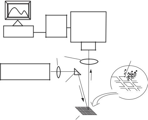
Within the last couple of decades, the development of integrated biosensors for the detection of multiple biologically relevant species has begun to take place. These integrated biosensor arrays that use the same excitation source for all of the elements and the same measurement process have been termed many things; gene chips, DNA-chips, etc. Most of the different array chips have been based on the use of nucleic acids (i.e. DNA) as the bioreceptors. Figure 1.4 illustrates an example of DNA microarray system with its associated detection system. Other types of bioreceptors such as antibodies, enzymes and cellular components can also be used. It is noteworthy that substrates having microarrays of bioreceptors are often referred to as biochips although most of these systems do not have integrated microsensor detection systems. A few of the more recent applications and advances in biochip technology will be discussed in this review.
A microarray of electrochemical biosensors has been developed for the detection of glucose and lactate on line [54]. This array of electrochemical biosensors was prepared using photolithographic techniques, using glucose oxidase and lactate oxidase as the bioreceptors. The glucose oxidase or lactate oxidase at each of the different sites in the array produces hydrogen peroxide when its appropriate substrate, glucose or lactate, is present. The hydrogen peroxide produced was measured at each element amperometrically.
An optical microarray system using a charge-coupled device (CCD) detector and DNA probes has been developed by Vo-Dinh and coworkers [48]. The evaluation of various system components developed for the DNA multi-array biosensor was discussed. The DNA probes labeled with visible and near infrared (NIR) dyes are evaluated. Examples of application of gene probes in DNA hybridization experiments and in biomedical diagnosis (detection of the p53 cancer suppressor gene) illustrated the usefulness and potential of the DNA
BIOSENSORS AND BIOCHIPS |
15 |
CCD |
MICROSCOPE |
DETECTOR |
SYSTEM |
COMPUTER
MICROSCOPE
OBJECTIVE
OPTICS |
DNA |
|
BIORECEPTORS |
||
|
LASER
SCANNING
MIRROR
HIGH-DENSITY MICROARRAY
CHIPS"
FIGURE 1.4. Schematic diagram of a DNA microarray with detection system.
multiarray device. An optical microarray for the detection of toxic agents using a planar array of antibody probes was described by Ligler and coworkers [13]. Their system was composed of a CCD for detection, an excitation source and a microscope slide with a photoactivated optical adhesive. Antibodies against three different toxins, staphylococcal enterotoxin B (SEB), ricin, and Yersinia pestis, were covalently attached to small wells in the slide formed by the optical adhesive. The microscope slide was then mounted over the CCD with a gradient refractive index (GRIN) lens array used to focus the wells onto the CCD. Toxins were then introduced to the slide followed by Cy5-labeled antibodies. The bound antibodies were then excited and the resulting fluorescence from all of the sensor locations were monitored simultaneously. Concentrations ranging from 5–25 ng/mL were capable of being measured for the different toxins.
High-density oligonucleotide arrays, consisting of greater than 96 000 oligonucleotides have been designed by Hacia et al. for the screening of the entire 5.53 kb coding region of the hereditary breast and ovarian cancer BRCA1 gene for all possible variations in the homozygous and heterozygous states [35]. Single stranded RNA targets were created by PCR amplification followed by in vitro transcription and partial fragmentation. These targets were then tested and fluorescence responses from targets containing the four natural bases to greater than 5 592 different fully complimentary 25 mer oligonucleotide probes were found.
16 |
TUAN VO-DINH |
To examine the effect of uridine and adenosine on the hybridization specificity, 33 200 probes containing centrally localized base pair mismatches were constructed and tested. Targets that contained modified 5-methyluridine showed a localized enhancement in fluorescence hybridization signals. In general, oligonucleotide microarrays, often referred to as “DNA chips”, are generally made by a light-directed chemical reaction that uses photographic masks for each chip [35]. A maskless fabrication method of light-directed oligonucleotide microarrays using a digital microarray has been reported [47]. In this method, a maskless array synthesizer replaces the chrome mask with virtual masks generated on a computer, which are relayed to a digital microarray.
1.3.2. Integrated Biochip Systems
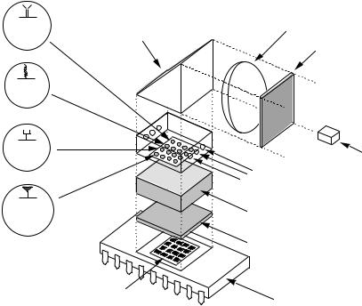
The development of a truly integrated biochip having a phototransistor integrated circuit (IC) microchip has been reported by Vo-Dinh and coworkers [47, 48]. This work involves the integration of a 4 × 4 and 10 × 10 optical biosensor array onto an integrated circuit (Figure 1.5). Most optical biochip technologies are very large when the excitation source and detector are considered, making them impractical for anything but laboratory usage. In this biochip the sensors, amplifiers, discriminators and logic circuitry are all built onto the chip. In one biochip system, each of the sensing elements is composed of 220 individual phototransistor cells connected in parallel to improve the sensitivity of the instrument. The
Antibody |
Reflective |
Focusing Lens |
Probe |
Optic |
|
|
|
Bandpass Filter |
DNA
Probe
Enzyme |
|
Probe |
Light Source |
|
(LED, Diode Laser) |
|
Sample Delivery |
|
Platform |
Cell- |
|
Based |
GRIN Lens Array |
Probe |
|
|
Detection Wavelength |
|
Selection Filter |
Photosensor |
|
Microarray |
Integrated |
|
Electrooptic Chip |
FIGURE 1.5. Schematic diagram of an integrated biochip system with microchip sensor.
BIOSENSORS AND BIOCHIPS |
17 |
ability to integrate light emitting diodes (LEDs) as the excitation sources into the system is also discussed. An important element in the development of the multifunctional biochip (MFB) involves the design and development of an IC electro-optic system for the microchip detection elements using the complementary metal oxide silicon (CMOS) technology. With this technology, highly integrated biochips are made possible partly through the capability of fabricating multiple optical sensing elements and microelectronics on a single system. Applications of the biochip are illustrated by measurements of the HIV1 sequence-specific probes using the DNA biochip device for the detection of a gene segment of the AIDS virus [47]. Recently, a MFB which allows simultaneous detection of several disease endpoints using different bioreceptors, such as DNA, antibodies, enzymes, cellular probes, on a single biochip system was developed [22]. The MFB device was a self-contained system based on an integrated circuit including photodiode sensor arrays, electronics, amplifiers, discriminators and logic circuitry. The multi-functional capability of the MFB biochip device is illustrated by measurements of different types of bioreceptors using DNA probes specific to gene fragments of the Mycobacterium Tuberculosis (TB) system, and antibody probes targeted to the cancer related tumor suppressor gene p53.
A biochip equipped with a microfluidics sample/reagent delivery system for on-chip monitoring of bioassayshas been developed for E. coli detection [39]. The microfluidics system includes a reaction chamber which houses a sampling platform that selectively captures detection probes from a sample through the use of immobilized bioreceptors. The independently operating photodiodes allow simultaneous monitoring of multiple samples. In this study the sampling platform is a cellulosic membrane that is exposed to E. coli organisms and subsequently analyzed using a sandwich immunoassay involving a Cy5labeled antibody probe. Studies show that the biochip has a linear dynamic range of three orders of magnitude observed for conventional assays, and can detect 20 E. coli organisms. Selective detection of E. coli in a complex medium, milk diluent, is also reported for both off-chip and on-chip assays.
A CMOS biochip coupled to multiplex capillary electrophoresis (CE) system has been developed [36, 37]. This combination of multiplex capillary gel electrophoresis and the IC microchip technology represents a novel approach to DNA analysis on the microchip platform. Separation of DNA ladders using a multiplex CE microsystem of four capillaries was monitored simultaneously using the IC microchip system. The IC microchip-CE system has advantages such as low cost, rapid analysis, compactness, and multiplex capability, and has great potential as an alternative system to conventional capillary array gel electrophoresis systems based on charge-coupled device (CCD) detection.
Antibody-immobilized capillary reactors coupled to biochip detection have been developed for E. coli O157:H7 detection using enzyme-linked immunosorbent assay (ELISA), and a biochip system [38]. ISA is very sensitive and selective immunological method to detect pathogenic bacteria. ELISA is also directly adaptable to a miniature biochip system that utilizes conventional sample platforms such as polymer membranes and glass. The antibody immobilized capillary reactor is a very attractive sample platform for ELISA because of its low cost, compactness, reuse, and ease of regeneration. Moreover, an array of capillary reactors can provide high-throughput ELISA. In this report, we describe the use of an array of antibody-immobilized capillary reactors for multiplex detection of E. coli O157:H7 in our miniature biochip system. Side-entry laser beam irradiation to an array of capillary reactors contributes significantly to miniaturized optical configuration for this biochip system.
18 |
TUAN VO-DINH |
The detection limits of E. coli O157:H7 using ELISA and Cy5 label-based immunoassays were determined to be 3 cells and 230 cells, respectively. This system shows capability to simultaneously monitor multifunctional immunoassay and high sensitive detection of E. coli O157:H7.
The application of a biochip using the molecular beacon (MB) detection scheme has been reported [Culha et al, 2004]. The medical application of this biochip novel MB detection system for the analysis of the breast cancer gene BRCA1 was illustrated. The MB is designed for the BRCA1 gene and a miniature biochip system is used for detection. The detection of BRCA1 gene is successfully demonstrated in solution and the limit of detection (LOD) is estimated as 70 nM.
1.4. CONCLUSION
For practical medical diagnostic applications, there is currently a strong need for a truly integrated biochip system that comprises probes, samplers, detector as well as amplifier and logic circuitry. Such a system will be useful in physician’s offices and could be used by relatively unskilled personnel. Most DNA biosensors previously reported are based on fiberoptic probes or glass and silica plates used as the probe substrates which are externally connected to a photosensing system generally consisting of a conventional detection device, such as a photomultiplier, or a charge-coupled device (CCD). Although the probes on the sampling platform are small (often referred to as a “DNA chip” or “gene chip”), the entire device containing excitation laser sources and detection systems (often a confocal microscope system) is relatively large, e.g., table-top size systems. While these systems have demonstrated their usefulness in gene discovery and genomics research, they are laboratory-oriented and involve relatively expensive equipment.
Biochip technologies could offer a unique combination of performance capabilities and analytical features of merit not available in any other bioanalytical system currently available. With its multichannel capability, biochip technology allows simultaneous detection of multiple biotargets. Biochip systems have great promise to offer several advantages in size, performance, fabrication, analysis and production cost due to their integrated optical sensing microchip. The small sizes of the probes (microliter to nanoliter) minimize sample requirement and reduce reagent and waste requirement. Highly integrated systems lead to a reduction in noise and an increase in signal due to the improved efficiency of sample collection and the reduction of interfaces. The capability of large-scale production using low-cost integrated circuit (IC) technology is an important advantage. The assembly process of various components is made simple by integration of several elements on a single chip. For medical applications, this cost advantage will allow the development of extremely low cost, disposable biochips that can be used for in-home medical diagnostics of diseases without the need of sending samples to a laboratory for analysis.
ACKNOWLEDGEMENTS
This work was sponsored by the Laboratory Directed Research and Development Program (Advanced Nanosystems Project), Oak Ridge National Laboratory, and by the U.S.
BIOSENSORS AND BIOCHIPS |
19 |
Department of Energy, Office of Biological and Environmental Research, under contract DE-AC05-96OR22464 with Lockheed Martin Energy Research Corporation.
REFERENCES
[1]J.P. Alarie and T.Vo-Dinh. Polycyc. Aromat. Compd., 8:45, 1996.
[2]S.L.R. Barker, R. Kopelman, T.E. Meyer, and M.A. Cusanovich. Anal. Chem., 70:971, 1998.
[3]M. Boncheva, C. Duschl, W. Beck, G. Jung, and H. Vogel. Langmuir, 12:5636, 1996.
[4]C.N. Campbell, T. de Lumley-Woodyear, and A. Heller. Fresen. J. Anal. Chem., 364:165, 1993.
[5]D. Charych, Q. Cheng, A. Reichert, G. Kuziemko, M. Stroh, J.O. Nagy,W. Spevak, and R.-C. Stevens. Chem. Biol., 3:113, 1996.
[6]L.C. Clark, Jr. and C. Lions. Ann. Acad. Sci., 102:29, 1962.
[7]M. Culha, D.L. Stokes, G.D. Griffin, and T. Vo-Dinh. Biosens. Bioelectron., 19:1007, 2004.
[8]B.M. Cullum, G.D. Griffin, and T. Vo-Dinh. Anal. Biochem., 1999.
[9]D. Diamond. (ed.). Chemical and Biological Sensors. Wiley, New York, 1998.
[10]J. Freudenberg, S. Schelle, K. Beck, M. vonSchickfus, and S. Hunklinger. Biosens. Bioelectron., 14:423, 1999.
[11]C. Galan-Vidal, J. Munoz, C. Dominguez, and S. Alegret. Sensor. Actuat. B-Chem., 53:257, 1998.
[12]J.J. Gooding and D.B. Hibbert. Trac-Trend Anal. Chem., 18:525, 1999.
[13]J.G. Hacia, S.A.Woski, J. Fidanza, K. Edgemon, G. McGall, S.P.A. Fodor, and F.S. Collins. Nucleic Acids Res., 26:4975, 1998.
[14]T. Huang, A. Warsinke, T. Kuwana, and F. W. Scheller. Anal. Chem., 70:991–997, 1998.
[15]D. Kroger, M. Liley, W. Schiweck, A. Skerra, and H. Vogel. Biosens. Bioelectron., 14:155, 1999.
[16]N. Isola, D.L. Stokes, and T. Vo-Dinh. Anal. Chem., 70:1352, 1998.
[17]E.A. James, K. Schmeltzer, and F.S. Ligler. Appl. Biochem. Biotechnol., 60:189, 1996.
[18]P.M. Kasili, J.M. Song, and T. Vo-Dinh. J. Am. Chem. Soc., 126:2799–2806, 2004.
[19]R. Koncki, G.J. Mohr, and O.S. Wolfbeis. Biosens. Bioelectron., 10:653, 1995.
[20]D. Kriz and K. Mosbach. Anal. Chim. Acta, 300:71, 1995.
[21]I.V. Lamont, R.I. McConnell, and S.P. Fitzgerald. Clin. Chem., 45:A102, 1999.
[22]M. Malmquist. Biochem. Soc., T 27:335, 1999.
[23]T. McCormack, G.O’Keeffe, B.D. MacCraith, and R.O’Kennedy. Sensor. Actuat. B-Chem., 41:89, 1997.
[24]M. Minunni, M. Mascini, R.M. Carter, M.B. Jacobs, G.J. Lubrano, and G.C. Guilbault. Anal. Chim. Acta, 325:169, 1996.
[25]Z.H. Mo, X.H. Long, and W.L. Fu. Anal. Commun., 36:281–283, 1999.
[26]J. Piehler, A. Brecht, G. Gauglitz, M. Zerlin, C. Maul, R. Thiericke, and S. Grabley. Anal. Biochem., 249:94, 1997.
[27]R.T. Piervincenzi, W.M. Reichert, and H.W. Hellinga. Biosens. Bioelectron., 13:305, 1998.
[28]T.E. Plowman, W.M. Reichert, C.R. Peters, H.K. Wang, D.A. Christensen, and J.N. Herron. Biosens. Bioelectron., 11:149, 1996.
[29]J. Polster, G. Prestel, M. Wollenweber, G. Kraus, and G. Gauglitz. Talanta, 42:2065, 1995.
[30]Z. Rosenzweig and R. Kopelman. Anal. Chem., 68:1408–1413, 1996.
[31]U. Rudel, O. Geschke, and K. Cammann. Electroanalysis, 8:1135, 1996.
[32]P. Sarkar and A.P.F. Turner. Fresen. J. Anal. Chem., 364:154, 1999.
[33]S. Sawata, E. Kai, K. Ikebukuro, T. Iida, T. Honda, and I. Karube. Biosens. Bioelectron., 14:397, 1999.
[34]A. Schmidt, C. StandfussGabisch, and U. Bilitewski. Biosens. Bioelectron., 11:1139, 1996.
[35]S. Singh-Gasson, R.D. Green, Y, Yue, C. Nelson, F. Blattner, R. Sussman, and F. Cerrina. Nat. Biotechnol., 10:974, 1999.
[36]J.M. Song and T. Vo-Dinh. Anal. Bioanal. Chem., 373:399, 2002.
[37]J.M. Song, J. Mobley, and T. Vo-Dinh. J. Chromatogra. B, 783:501, 2003.
[38]J.M. Song and T. Vo-Dinh. Anal. Chimi. Acta, 507:115, 2004.
[39]D.L. Stokes, G.D. Griffin, and T. Vo-Dinh. Fresen. J. Anal. Chem., 369, 2001.
[40]T. Thundat, P.I. Oden, and R.J. Warmack. Microscale Thermophys. Eng., 1:185, 1997.
[41]F. Tobalina, F. Pariente, L. Hernandez, H.D. Abruna, and E. Lorenzo. Anal. Chim. Acta, 395:17, 1999.
20 |
TUAN VO-DINH |
[42]B.G. Tromberg, M.J. Sepaniak, T. Vo-Dinh, and G.D. Griffin. Anal. Chem., 59:1226, 1987.
[43]R.D. Vaughan, C.K. Sullivan, and G.C. Guilbault. Fresen. J. Anal. Chem., 364:54, 1999.
[44]T. Vo-Dinh. Sensor. Actuat B-Chem., 51:52, 1998.
[45]T. Vo-Dinh. Proceedings of the 6th Annual Biochip Technologies Conference: Chips for Hits ’99. Berkeley, California, Nov 2–5 1999.
[46]T. Vo-Dinh. J. Cell. Biochem., 39(Suppl.):154, 2002.
[47]T. Vo-Dinh, J.P. Alarie, N. Isola, D. Landis, A.L. Wintenberg, and M.N. Ericson. Anal. Chem., 71:358–363, 1999.
[48]T. Vo-Dinh, N. Isola, J.P. Alarie, D. Landis, G.D. Griffin, and S. Allison. Instrum. Sci. Technol., 26:503, 1998.
[49]T. Vo-Dinh, K. Houck, and D.L. Stokes. Anal. Chem., 33:3379, 1994.
[50]T. Vo-Dinh, B.M. Cullum, J.P. Alarie, and G.D. Griffin. J. Nanopart. Res., (in press).
[51]T. Vo-Dinh, G.D. Griffin, and K.R. Ambrose. Appl. Spectrosc., 40:696, 1986.
[52]T. Vo-Dinh, B.G. Tromberg, G.D. Griffin, K.R. Ambrose, M.J. Sepaniak, and E.M. Gardenhire. Suppl. Spectrosc., 41:735, 1987.
[53]W. Welsch, C. Klein, M. vonSchickfus, and S. Hunklinger. Anal. Chem., 68:2000, 1996.
[54]R.M. Wadkins, J.P. Golden, L. M. Pritsiolas, and F.S. Ligler. Biosens. Bioelectron., 13:407, 1998.
2
Cantilever Arrays: A Universal Platform for Multiplexed Label-Free Bioassays
Min Yue1, Arun Majumdar1, and Thomas Thundat2
1 Department of Mechanical Engineering, University of California, Berkeley, CA 94720 2 Life Sciences Division, Oak Ridge National Laboratory, Oak Ridge, TN
2.1. INTRODUCTION
Microcantilevers have caught great attention as label-free and ultra-sensitive biological sensors. When molecular adsorption occurs on only one surface of cantilever, the resulting differential surface stress leads to cantilever bending, thus providing a method of detecting molecular adsorption (Figure 2.1). How does the transduction from molecular adsorption to surface stress change occur? Though the underlying science of the transduction is yet to be completely understood, the thermodynamic argument suggests that the reaction-induced free energy reduction on one cantilever surface is balanced by the strain energy increase due to bending, such that at equilibrium the free energy of the whole system reaches the minimum [11]. In other words, the penalty of increasing the strain energy must be compensated by a larger reduction in free energy due to reaction, reflecting the interplay between mechanics and chemistry. Hence, the cantilever bending can be construed as a measure of free energy reduction due to the chemical reaction on one surface. What is worth noting is that because free energy reduction is common for all reactions, the cantilever-based sensing is universal platform for studying reactions. The ability of analyzing molecules without the use of optical or radioactive labels makes this approach rather attractive for biology and medicine.
As DNA microarrays successfully provide means to study genomics in a highthroughput manner, various protein microarrays have been under development to enable quantitative and rapid protein analysis. Researchers have developed protein microarrays