

- •Contents
- •Contributors
- •Part I General Principles of Cell Death
- •1 Human Caspases – Apoptosis and Inflammation Signaling Proteases
- •1.1. Apoptosis and limited proteolysis
- •1.2. Caspase evolution
- •2. ACTIVATION MECHANISMS
- •2.2. The activation platforms
- •2.4. Proteolytic maturation
- •3. CASPASE SUBSTRATES
- •4. REGULATION BY NATURAL INHIBITORS
- •REFERENCES
- •2 Inhibitor of Apoptosis Proteins
- •2. CELLULAR FUNCTIONS AND PHENOTYPES OF IAP
- •3. IN VIVO FUNCTIONS OF IAP FAMILY PROTEINS
- •4. SUBCELLULAR LOCATIONS OF IAP
- •8. IAP–IAP INTERACTIONS
- •10. ENDOGENOUS ANTAGONISTS OF IAP
- •11. IAPs AND DISEASE
- •SUGGESTED READINGS
- •1. INTRODUCTION
- •2.1. The CD95 (Fas/APO-1) system
- •2.1.1. CD95 and CD95L: discovery of the first direct apoptosis-inducing receptor-ligand system
- •2.1.2. Biochemistry of CD95 apoptosis signaling
- •2.2. The TRAIL (Apo2L) system
- •3.1. The TNF system
- •3.1.1. Biochemistry of TNF signal transduction
- •3.1.2. TNF and TNF blockers in the clinic
- •3.2. The DR3 system
- •4. THE DR6 SYSTEM
- •6. CONCLUDING REMARKS AND OUTLOOK
- •SUGGESTED READINGS
- •4 Mitochondria and Cell Death
- •1. INTRODUCTION
- •2. MITOCHONDRIAL PHYSIOLOGY
- •3. THE MITOCHONDRIAL PATHWAY OF APOPTOSIS
- •9. CONCLUSIONS
- •SUGGESTED READINGS
- •1. INTRODUCTION
- •3. INHIBITING APOPTOSIS
- •4. INHIBITING THE INHIBITORS
- •6. THE BCL-2 FAMILY AND CANCER
- •SUGGESTED READINGS
- •6 Endoplasmic Reticulum Stress Response in Cell Death and Cell Survival
- •1. INTRODUCTION
- •2. THE ESR IN YEAST
- •3. THE ESR IN MAMMALS
- •4. THE ESR AND CELL DEATH
- •5. THE ESR IN DEVELOPMENT AND TISSUE HOMEOSTASIS
- •6. THE ESR IN HUMAN DISEASE
- •7. CONCLUSION
- •7 Autophagy – The Liaison between the Lysosomal System and Cell Death
- •1. INTRODUCTION
- •2. AUTOPHAGY
- •2.2. Physiologic functions of autophagy
- •2.3. Autophagy and human pathology
- •3. AUTOPHAGY AND CELL DEATH
- •3.1. Autophagy as anti–cell death mechanism
- •3.2. Autophagy as a cell death mechanism
- •3.3. Molecular players of the autophagy–cell death cross-talk
- •4. AUTOPHAGY, CELLULAR DEATH, AND CANCER
- •5. CONCLUDING REMARKS AND PENDING QUESTIONS
- •SUGGESTED READINGS
- •8 Cell Death in Response to Genotoxic Stress and DNA Damage
- •1. TYPES OF DNA DAMAGE AND REPAIR SYSTEMS
- •2. DNA DAMAGE RESPONSE
- •2.2. Transducers
- •2.3. Effectors
- •4. CHROMATIN MODIFICATIONS
- •5. CELL CYCLE CHECKPOINT REGULATION
- •6. WHEN REPAIR FAILS: SENESCENCE VERSUS APOPTOSIS
- •6.1. DNA damage response and the induction of apoptosis
- •6.2. p53-independent mechanisms of apoptosis
- •6.3. DNA damage response and senescence induction
- •7. DNA DAMAGE FROM OXIDATIVE STRESS
- •SUGGESTED READINGS
- •9 Ceramide and Lipid Mediators in Apoptosis
- •1. INTRODUCTION
- •3.1. Basic cell signaling often involves small molecules
- •3.2. Sphingolipids are cell-signaling molecules
- •3.2.1. Ceramide induces apoptosis
- •3.2.2. Ceramide accumulates during programmed cell death
- •3.2.3. Inhibition of ceramide production alters cell death signaling
- •4.1. Ceramide is generated through SM hydrolysis
- •4.3. aSMase can be activated independently of extracellular receptors to regulate apoptosis
- •4.4. Controversial aspects of the role of aSMase in apoptosis
- •4.5. De novo ceramide synthesis regulates programmed cell death
- •4.6. p53 and Bcl-2–like proteins are connected to de novo ceramide synthesis
- •4.7. The role and regulation of de novo synthesis in ceramide-mediated cell death is poorly understood
- •5. CONCLUDING REMARKS AND FUTURE DIRECTIONS
- •5.1. Who? (Which enzyme?)
- •5.2. What? (Which ceramide?)
- •5.3. Where? (Which compartment?)
- •5.4. When? (At what steps?)
- •5.5. How? (Through what mechanisms?)
- •5.6. What purpose?
- •6. SUMMARY
- •SUGGESTED READINGS
- •1. General Introduction
- •1.1. Cytotoxic lymphocytes and apoptosis
- •2. CYTOTOXIC GRANULES AND GRANULE EXOCYTOSIS
- •2.1. Synthesis and loading of the cytotoxic granule proteins into the secretory granules
- •2.2. The immunological synapse
- •2.3. Secretion of granule proteins
- •2.4. Uptake of proapoptotic proteins into the target cell
- •2.5. Activation of death pathways by granzymes
- •3. GRANULE-BOUND CYTOTOXIC PROTEINS
- •3.1. Perforin
- •3.2. Granulysin
- •3.3. Granzymes
- •3.3.1. GrB-mediated apoptosis
- •3.3.2. GrA-mediated cell death
- •3.3.3. Orphan granzyme-mediated cell death
- •5. CONCLUSIONS
- •REFERENCES
- •Part II Cell Death in Tissues and Organs
- •1.1. Death by trophic factor deprivation
- •1.2. Key molecules regulating neuronal apoptosis during development
- •1.2.1. Roles of caspases and Apaf-1 in neuronal cell death
- •1.2.2. Role of Bcl-2 family members in neuronal cell death
- •1.3. Signal transduction from neurotrophins and neurotrophin receptors
- •1.3.1. Signals for survival
- •1.3.2. Signals for death
- •2.1. Apoptosis in neurodegenerative diseases
- •2.1.4. Amyotrophic lateral sclerosis
- •2.2. Necrotic cell death in neurodegenerative diseases
- •2.2.1. Calpains
- •2.2.2. Cathepsins
- •3. CONCLUSIONS
- •ACKNOWLEDGMENT
- •SUGGESTED READINGS
- •ACKNOWLEDGMENT
- •SUGGESTED READINGS
- •1. INTRODUCTION
- •5. S-NITROSYLATION OF PARKIN
- •7. POTENTIAL TREATMENT OF EXCESSIVE NMDA-INDUCED Ca2+ INFLUX AND FREE RADICAL GENERATION
- •8. FUTURE THERAPEUTICS: NITROMEMANTINES
- •9. CONCLUSIONS
- •Acknowledgments
- •SUGGESTED READINGS
- •3. MITOCHONDRIAL PERMEABILITY TRANSITION ACTIVATED BY Ca2+ AND OXIDATIVE STRESS
- •4.1. Mitochondrial apoptotic pathways
- •4.2. Bcl-2 family proteins
- •4.3. Caspase-dependent apoptosis
- •4.4. Caspase-independent apoptosis
- •4.5. Calpains in ischemic neural cell death
- •5. SUMMARY
- •ACKNOWLEDGMENTS
- •SUGGESTED READINGS
- •1. INTRODUCTION
- •2. HISTORICAL ANTECEDENTS
- •7.1. Activation of p21 waf1/cip1: Targeting extrinsic and intrinsic pathways to death
- •8. CONCLUSION
- •ACKNOWLEDGMENTS
- •REFERENCES
- •16 Apoptosis and Homeostasis in the Eye
- •1.1. Lens
- •1.2. Retina
- •2. ROLE OF APOPTOSIS IN DISEASES OF THE EYE
- •2.1. Glaucoma
- •2.2. Age-related macular degeneration
- •4. APOPTOSIS AND OCULAR IMMUNE PRIVILEGE
- •5. CONCLUSIONS
- •SUGGESTED READINGS
- •17 Cell Death in the Inner Ear
- •3. THE COCHLEA IS THE HEARING ORGAN
- •3.1. Ototoxic hair cell death
- •3.2. Aminoglycoside-induced hair cell death
- •3.3. Cisplatin-induced hair cell death
- •3.4. Therapeutic strategies to prevent hair cell death
- •3.5. Challenges to studies of hair cell death
- •4. SPIRAL GANGLION NEURON DEATH
- •4.1. Neurotrophic support from sensory hair cells and supporting cells
- •4.2. Afferent activity from hair cells
- •4.3. Molecular manifestations of spiral ganglion neuron death
- •4.4. Therapeutic interventions to prevent SGN death
- •ACKNOWLEDGMENTS
- •SUGGESTED READINGS
- •18 Cell Death in the Olfactory System
- •1. Introduction
- •2. Anatomical Aspects
- •3. Life and Death in the Olfactory System
- •3.1. Olfactory epithelium
- •3.2. Olfactory bulb
- •REFERENCES
- •1. Introduction
- •3.1. Beta cell death in the development of T1D
- •3.2. Mechanisms of beta cell death in type 1 diabetes
- •3.2.1. Apoptosis signaling pathways downstream of death receptors and inflammatory cytokines
- •3.2.2. Oxidative stress
- •3.3. Mechanisms of beta cell death in type 2 diabetes
- •3.3.1. Glucolipitoxicity
- •3.3.2. Endoplasmic reticulum stress
- •5. SUMMARY
- •Acknowledgments
- •REFERENCES
- •20 Apoptosis in the Physiology and Diseases of the Respiratory Tract
- •1. APOPTOSIS IN LUNG DEVELOPMENT
- •2. APOPTOSIS IN LUNG PATHOPHYSIOLOGY
- •2.1. Apoptosis in pulmonary inflammation
- •2.2. Apoptosis in acute lung injury
- •2.3. Apoptosis in chronic obstructive pulmonary disease
- •2.4. Apoptosis in interstitial lung diseases
- •2.5. Apoptosis in pulmonary arterial hypertension
- •2.6. Apoptosis in lung cancer
- •SUGGESTED READINGS
- •21 Regulation of Cell Death in the Gastrointestinal Tract
- •1. INTRODUCTION
- •2. ESOPHAGUS
- •3. STOMACH
- •4. SMALL AND LARGE INTESTINE
- •5. LIVER
- •6. PANCREAS
- •7. SUMMARY AND CONCLUDING REMARKS
- •SUGGESTED READINGS
- •22 Apoptosis in the Kidney
- •1. NORMAL KIDNEY STRUCTURE AND FUNCTION
- •3. APOPTOSIS IN ADULT KIDNEY DISEASE
- •4. REGULATION OF APOPTOSIS IN KIDNEY CELLS
- •4.1. Survival factors
- •4.2. Lethal factors
- •4.2.1. TNF superfamily cytokines
- •4.2.2. Other cytokines
- •4.2.3. Glucose
- •4.2.4. Drugs and xenobiotics
- •4.2.5. Ischemia-reperfusion and sepsis
- •5. THERAPEUTIC APPROACHES
- •SUGGESTED READINGS
- •1. INTRODUCTION
- •2. APOPTOSIS IN THE NORMAL BREAST
- •2.1. Occurrence and role of apoptosis in the developing breast
- •2.2.2. Death ligands and death receptor pathway
- •2.2.4. LIF-STAT3 proapoptotic signaling
- •2.2.5. IGF survival signaling
- •2.2.6. Regulation by adhesion
- •2.2.7. PI3K/AKT pathway: molecular hub for survival signals
- •2.2.8. Downstream regulators of apoptosis: the BCL-2 family members
- •3. APOPTOSIS IN BREAST CANCER
- •3.1. Apoptosis in breast tumorigenesis and cancer progression
- •3.2. Molecular dysregulation of apoptosis in breast cancer
- •3.2.1. Altered expression of death ligands and their receptors in breast cancer
- •3.2.2. Deregulation of prosurvival growth factors and their receptors
- •3.2.3. Alterations in cell adhesion and resistance to anoikis
- •3.2.4. Enhanced activation of the PI3K/AKT pathway in breast cancer
- •3.2.5. p53 inactivation in breast cancer
- •3.2.6. Altered expression of BCL-2 family of proteins in breast cancer
- •5. CONCLUSION
- •SUGGESTED READINGS
- •1. INTRODUCTION
- •2. DETECTING CELL DEATH IN THE FEMALE GONADS
- •4. APOPTOSIS AND FEMALE REPRODUCTIVE AGING
- •6. CONCLUDING REMARKS
- •REFERENCES
- •25 Apoptotic Signaling in Male Germ Cells
- •1. INTRODUCTION
- •3.1. Murine models
- •3.2. Primate models
- •3.3. Pathways of caspase activation and apoptosis
- •3.4. Apoptotic signaling in male germ cells
- •5. P38 MITOGEN-ACTIVATED PROTEIN KINASE (MAPK) AND NITRIC OXIDE (NO)–MEDIATED INTRINSIC PATHWAY SIGNALING CONSTITUTES A CRITICAL COMPONENT OF APOPTOTIC SIGNALING IN MALE GERM CELLS AFTER HORMONE DEPRIVATION
- •11. CONCLUSIONS AND PERSPECTIVES
- •REFERENCES
- •26 Cell Death in the Cardiovascular System
- •1. INTRODUCTION
- •2. CELL DEATH IN THE VASCULATURE
- •2.1. Apoptosis in the developing blood vessels
- •2.2. Apoptosis in atherosclerosis
- •2.2.1. Vascular smooth muscle cells
- •2.2.2. Macrophages
- •2.2.3. Regulation of apoptosis in atherosclerosis
- •2.2.4. Necrosis and autophagy in atherosclerosis
- •3. CELL DEATH IN THE MYOCARDIUM
- •3.1. Cell death in myocardial infarction
- •3.1.1. Apoptosis in myocardial infarction
- •3.1.2. Necrosis in myocardial infarction
- •3.1.3. Autophagy in myocardial infarction
- •3.2. Cell death in heart failure
- •3.2.1. Apoptosis in heart failure
- •3.2.2. Necrosis in heart failure
- •3.2.3. Autophagy in heart failure
- •4. CONCLUDING REMARKS
- •ACKNOWLEDGMENTS
- •REFERENCES
- •27 Cell Death Regulation in Muscle
- •1. INTRODUCTION TO MUSCLE
- •1.1. Skeletal muscle adaptation to endurance training
- •1.2. Myonuclear domains
- •2. MITOCHONDRIALLY MEDIATED APOPTOSIS IN MUSCLE
- •2.1. Skeletal muscle apoptotic susceptibility
- •4. APOPTOSIS IN MUSCLE DURING AGING AND DISEASE
- •4.1. Aging
- •4.2. Type 2 diabetes mellitus
- •4.3. Cancer cachexia
- •4.4. Chronic heart failure
- •6. CONCLUSION
- •SUGGESTED READINGS
- •28 Cell Death in the Skin
- •1. INTRODUCTION
- •2. CELL DEATH IN SKIN HOMEOSTASIS
- •2.1. Cornification and apoptosis
- •2.2. Death receptors in the skin
- •3. CELL DEATH IN SKIN PATHOLOGY
- •3.1. Sunburn
- •3.2. Skin cancer
- •3.3. Necrolysis
- •3.4. Pemphigus
- •3.5. Eczema
- •3.6. Graft-versus-host disease
- •4. CONCLUDING REMARKS AND PERSPECTIVES
- •ACKNOWLEDGMENTS
- •SUGGESTED READINGS
- •29 Apoptosis and Cell Survival in the Immune System
- •2.1. Survival of early hematopoietic progenitors
- •2.2. Sizing of the T-cell population
- •2.2.1. Establishing central tolerance
- •2.2.2. Peripheral tolerance
- •2.2.3. Memory T cells
- •2.3. Control of apoptosis in B-cell development
- •2.3.1. Early B-cell development
- •2.3.2. Deletion of autoreactive B cells
- •2.3.3. Survival and death of activated B cells
- •3. IMPAIRED APOPTOSIS AND LEUKEMOGENESIS
- •4. CONCLUSIONS
- •ACKNOWLEDGMENTS
- •REFERENCES
- •30 Cell Death Regulation in the Hematopoietic System
- •1. INTRODUCTION
- •2. HEMATOPOIETIC STEM CELLS
- •4. ERYTHROPOIESIS
- •5. MEGAKARYOPOIESIS
- •6. GRANULOPOIESIS
- •7. MONOPOIESIS
- •8. CONCLUSION
- •ACKNOWLEDGMENTS
- •REFERENCES
- •31 Apoptotic Cell Death in Sepsis
- •1. INTRODUCTION
- •2. HOST INFLAMMATORY RESPONSE TO SEPSIS
- •3. CLINICAL OBSERVATIONS OF CELL DEATH IN SEPSIS
- •3.1. Sepsis-induced apoptosis
- •3.2. Necrotic cell death in sepsis
- •4.1. Central role of apoptosis in sepsis mortality: immune effector cells and gut epithelium
- •4.2. Apoptotic pathways in sepsis-induced immune cell death
- •4.3. Investigations implicating the extrinsic apoptotic pathway in sepsis
- •4.4. Investigations implicating the intrinsic apoptotic pathway in sepsis
- •5. THE EFFECT OF APOPTOSIS ON THE IMMUNE SYSTEM
- •5.1. Cellular effects of an increased apoptotic burdens
- •5.2. Network effects of selective loss of immune cell types
- •5.3. Studies of immunomodulation by apoptotic cells in other fields
- •7. CONCLUSION
- •REFERENCES
- •32 Host–Pathogen Interactions
- •1. INTRODUCTION
- •2. FROM THE PATHOGEN PERSPECTIVE
- •2.1. Commensals versus pathogens
- •2.2. Pathogen strategies to infect the host
- •3. HOST DEFENSE
- •3.1. Antimicrobial peptides
- •3.2. PRRs and inflammation
- •3.2.1. TLRs
- •3.2.2. NLRs
- •3.2.3. The Nod signalosome
- •3.2.4. The inflammasome
- •3.3. Cell death
- •3.3.1. Apoptosis and pathogen clearance
- •3.3.2. Pyroptosis
- •3.2.3. Caspase-independent cell death
- •3.2.4. Autophagy and autophagic cell death
- •4. CONCLUSIONS
- •REFERENCES
- •Part III Cell Death in Nonmammalian Organisms
- •1. PHENOTYPE AND ASSAYS OF YEAST APOPTOSIS
- •2.1. Pheromone-induced cell death
- •2.1.1. Colony growth
- •2.1.2. Killer-induced cell death
- •3. EXTERNAL STIMULI THAT INDUCE APOPTOSIS IN YEAST
- •4. THE GENETICS OF YEAST APOPTOSIS
- •5. PROGRAMMED AND ALTRUISTIC AGING
- •SUGGESTED READINGS
- •34 Caenorhabditis elegans and Apoptosis
- •1. Overview
- •2. KILLING
- •3. SPECIFICATION
- •4. EXECUTION
- •4.1. DNA degradation
- •4.2. Mitochondrial elimination
- •4.3. Engulfment
- •5. SUMMARY
- •SUGGESTED READINGS
- •35 Apoptotic Cell Death in Drosophila
- •2. DROSOPHILA CASPASES AND PROXIMAL REGULATORS
- •6. CLOSING COMMENTS
- •SUGGESTED READINGS
- •36 Analysis of Cell Death in Zebrafish
- •1. INTRODUCTION
- •2. WHY USE ZEBRAFISH TO STUDY CELL DEATH?
- •2.2. Molecular techniques to rapidly assess gene function in embryos
- •2.2.1. Studies of gene function using microinjections into early embryos
- •2.2.2. In situ hybridization and immunohistochemistry
- •2.3. Forward genetic screening
- •2.4. Drug and small-molecule screening
- •2.5. Transgenesis
- •2.6. Targeted knockouts
- •3.1. Intrinsic apoptosis
- •3.2. Extrinsic apoptosis
- •3.3. Chk-1 suppressed apoptosis
- •3.4. Anoikis
- •3.5. Autophagy
- •3.6. Necrosis
- •4. DEVELOPMENTAL CELL DEATH IN ZEBRAFISH EMBRYOS
- •5. THE P53 PATHWAY
- •6. PERSPECTIVES AND FUTURE DIRECTIONS
- •SUGGESTED READING
354 |
PAUL A. NEY |
loss, spherocytic shape change, and phosphatidylserine externalization.88 By analogy to apoptotic cells, one idea is that externalization of phosphatidylserine may signal macrophages to engulf senescent erythrocytes. A similar mechanism is suggested for the DNase II– dependent clearance of expelled erythroblast nuclei by macrophages.89 Erythrocytes contain caspase-3 and -8, but these are not activated even after prolonged storage; thus caspase activation does not appear to underlie erythrocyte senescence.90 Rather, an increase in intracellular calcium and the activation of other cysteine proteases such as calpain is thought to cause the cellular changes that mark erythrocytes for clearance.
5. MEGAKARYOPOIESIS
Megakaryocytes, which produce platelets, are the other lineage that develops from MEP progenitors. Like the erythroid lineage, megakaryocyte development passes through early burst-forming progenitor and late colonyforming progenitor stages.91 Megakaryopoiesis is supported by the cytokine thrombopoietin, and mice deficient for the thrombopoietin receptor, c-Mpl, exhibit severe thrombocytopenia.92 Megakaryocytes represent less than 1% of the cells in the bone marrow. Because of a process known as endoreduplication, megakaryocytes have between a 4N and a 128N DNA content. Mature megakaryocytes have a high degree of polyploidy, and generate filamentous projections known as proplatelets. These platelet precursors project into the bone marrow sinusoids, and through a poorly understood process, release platelets into circulation. Platelets in circulation have a lifespan of about 10 days.
There are several steps of megakaryocyte development, which are regulated by death pathways. First, caspase activation is thought to have a role in proplatelet formation. Mature megakaryocytes, at the stage when proplatelets are formed, show caspase-9 and -3 activation.93 Further, either enforced expression of BCL2 or caspase inhibition decreases, and FASL increases, proplatelet formation.94,94 Intriguingly, the process is compartmentalized; caspase activation is focal during proplatelet formation, but becomes generalized as denuded megakaryocytes undergo apoptosis.95 Tg(VavBcl2), Bim–/–, and Bax–/–;Bak–/– mice have decreased platelet counts.34,35,36 implying a role for the intrinsic death pathway in platelet production, but these results are difficult to interpret because of potential effects on nonmegakaryocytic lineages. In this regard, it is notable that Tg(Pf4-BclXL) mice, in which BCL-XL transgene expression is restricted to the megakaryocyte lineage, show impaired platelet fragmentation.96 Still, at present
it is unresolved whether caspase activation at the proplatelet stage is mediated through the intrinsic or extrinsic pathway.94
Second, on activation, platelets exhibit some of the features of apoptosis, including cell shrinkage, plasma membrane microvesiculation, phosphatidylserine externalization, and proteolysis of procaspase-9, procaspase- 3, gelsolin, and protein kinase C-δ.97 However, in contrast to apoptosis, these events are not associated with cytochrome c release or caspase activation, but instead are mediated by the calcium-dependent cysteine proteinase, calpain. Third, platelet senescence is regulated through the intrinsic apoptotic pathway. BCL-XL is upregulated during megakaryocyte maturation up to the stage of proplatelet formation; thereafter it is downregulated and distributed to developing platelets.96,98 BCL- XL in circulating platelets is gradually degraded, placing a firm limit on platelet lifespan.99 Once BCL-XL levels fall below a critical threshold, BAK is activated, and cytochrome c released. Whether the final step leading to clearance is caspase-dependent or not is unclear.94,97
6. GRANULOPOIESIS
Granulocytes and monocytes diverge from the erythroid and megakaryocytic lineages at the common myeloid progenitor (CMP); the CMP gives rise to bipotential colony-forming unit granulocyte-macrophage (CFU-GM) progenitors, which in turn give rise to the lineage-restricted progenitors CFU-G and CFU- M. Studies of genetically modified mice show that cytokines have multiple roles in the regulation of granulopoiesis (Figure 30-3). Gcsfr–/– mice have decreased CFU-Mix, CFU-GM, and BFU-E, indicating an effect of granulocyte colony-stimulating factor (G-CSF) receptor signaling on commitment to the myeloid-erythroid lineage or the survival of myeloid-erythroid progenitor cells.100 Additionally, Gcsf–/– mice have decreased mature neutrophils in the bone marrow and increased apoptotic neutrophil precursors, indicating an effect of cytokine signaling on neutrophil precursor cell survival.101 Consistent with a role for the intrinsic death pathway in these effects, myeloid progenitors or precursors are markedly decreased in Mcl1 deleted mice26 and moderately increased in Tg(Vav-Bcl2) mice,34 Tg(Mcl1) mice,102 Bim–/– mice,35 and Bax–/–;Bak–/– mice.36 Additional insight comes from mice that possess a conditional mutation of Mcl1 and a monocyte-granulocyte lineage-restricted LysM-Cre transgene. Mcl1fl/fl;Tg(LysMCre) mice are neutropenic, showing that in addition to its requirement at the stem and progenitor cell stages, MCL1 is required for mature neutrophil
CELL DEATH REGULATION IN THE HEMATOPOIETIC SYSTEM |
355 |
CFU-Mix |
|
|
|
|
CFU- |
|
|
|
MegE |
|
|
CFU-GM |
|
|
|
|
CFU-M |
|
Bone |
|
|
|
|
CFU-G |
|
MCL1 |
marrow |
|
|
||
Myelocyte |
|
|
|
Neutrophil
Blood
Apoptosis
G-CSF |
Tissue |
|
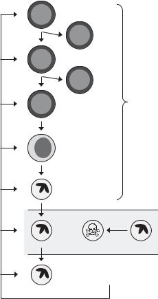
Figure 30-3. Regulation of granulopoiesis by G-CSF. G-CSF regulates granulopoiesis at multiple steps: progenitor and precursor cell survival in bone marrow, release from the bone marrow into the circulation, and neutrophil survival in tissues. Unstimulated neutrophils in circulation undergo constitutive apoptosis. MCL1 is required for progenitor survival and also specifically for terminal granulocytic di erentiation. CFU-GM, bipotential granulocytemacrophage progenitor; CFU-MegE, bipotential erythroid-megakaryocytic progenitor; CFUMix, multipotential progenitor; G-CSF, granulocyte-colony stimulating factor; Myelocyte, neutrophil precursor.
development.103,104 Finally, cytokine levels are markedly elevated in Mcl1fl/fl;Tg(LysM-Cre) and Gcsfr–/– mice,100 consistent with feedback regulation of neutrophil production.
In addition to promoting neutrophil precursor cell survival, cytokines also regulate the release of neutrophils from the bone marrow. Infection causes a marked increase in the systemic levels of G-CSF, macrophage colony-stimulating factor (M-CSF), and granulocyte-macrophage colony-stimulating factor (GM-CSF).105 Additionally, infection or pharmacological G-CSF causes the release of neutrophils and neutrophil precursors from the bone marrow into circulation and neutrophilia.106,107,108,109 Still, although cytokines have these properties, Gcsf–/–, Gcsfr–/–, Gmcsf–/–, Mcsf–/– and
mice generate neutrophils, indicating
a nonessential role, or redundancy, of these factors in neutrophil development.110,111,112,113,114
Neutrophils undergo constitutive apoptosis and have a short lifespan in circulation (Figure 30-3).115,116 In vitro, in the absence of cytokines or factors, most neutrophils undergo apoptosis within 24 hours. Akt signaling is important for neutrophil survival. In the absence of cytokine signaling, dissociation of heat shock protein 27 and MAPK-activated protein kinase- 2 from Akt-containing complexes prevents Akt activation and causes neutrophil commitment to apoptosis.117,118 Several lines of evidence suggest that the intrinsic death pathway also has a role in constitutive apoptosis. First, proapoptotic BCL2-related proteins are expressed in mature neutrophils, including BAX, BAK, BIK, BAD, and BID,119 but the antiapoptotic proteins BCL2 and BCL-XL are not expressed, and MCL1 and A1 are expressed, but downregulated. Additionally, MCL1 protein is downregulated through proteasomal degradation.120 Second, targeted disruption of A1a, one of three functioning and highly related A1 genes in mice,121 hastens the onset of constitutive neutrophil apoptosis, and enforced expression of BCL2, or targeted disruption of Bim, retards it.122,123,124 Third, mitochondrial depolarization precedes phosphatidylserine exposure and other signs of neutrophil apoptosis.125 Fourth, although the ability of broad-spectrum caspase inhibitors to prevent constitutive neutrophil apoptosis is controversial, caspase-3 is cleaved and activated during neutrophil apoptosis.126,127,128 Finally, despite expression of FAS by neutrophils129,130 and evidence for a mitochondria-independent mechanism,127 constitutive neutrophil apoptosis does not appear to be regulated by
FAS.123,128,131
Circulating neutrophils are recruited to sites of infection by chemotactic factors and cross the endothelium by diapedesis.132 In tissues, neutrophils are exposed to local cytokines and inflammatory mediators, such as G-CSF, GM-CSF, lipopolysaccharide (LPS), C5a, N- formyl-methionyl-leucyl-phenylalanine (fMLP), adenosine triphosphate, leukotriene B4, interleukin (IL)- 1β, IL-2, IL-3, IL-6, IL-15, and interferon-γ.133 These factors activate survival and death pathways, which both prolong neutrophil survival and limit the duration of the immune response. For example, as noted above, Akt activity declines in unstimulated neutrophils leading to apoptosis117; however, G-CSF, GM-CSF, interferon-γ, and leukotriene B4 activate Akt and inhibit apoptosis.117,134 Also, bacterial products and mimetics, such as LPS, peptidoglycan, and unmethylated CpG-DNA, inhibit apoptosis by activating Toll-like receptors, nuclear factor-kappa B (NF-κB), and
356 |
PAUL A. NEY |
Akt.135 Finally, G-CSF, GM-CSF, IL-1β, and LPS signaling increase levels of MCL1, A1, and phosphorylated BAD, suggesting that one or more of these are targets of their antiapoptotic activity.135,136 Other factors such as hypoxia and endothelial cell transmigration may also delay apoptosis and contribute to prolongation of neutrophil lifespan in tissues.137,138 In contrast, the death receptor ligands FASL and TNF-α, secreted by nearby macrophages, induce neutrophil apoptosis, which limits the duration of the immune response.131,139 In this regard, it should be noted that TNF-α can also inhibit apoptosis by activating NF-κB.140,141
Whereas the net effect of cytokines and inflammatory mediators in tissues is to prolong neutrophil survival, once neutrophils encounter bacteria, a series of events is initiated that ultimately leads to neutrophil clearance.142 When neutrophils encounter bacteria in tissues, the bacteria are phagocytosed and degraded. This is accomplished by the formation of phagosomes around bacteria and fusion with neutrophil granules and lysosomes to form a phagolysosomes. Within phagolysosomes, hydrogen peroxide, generated by the membrane-bound NADPH oxidase complex, is converted to hypochlorous acid and ROS.143,144 Neutrophils rely on glycolysis for energy,145 but oxygen is consumed to generate ROS; consequently, this event is known as the respiratory burst. The importance of the respiratory burst for bactericidal activity is illustrated by the susceptibility of patients with NADPH oxidase mutations to infection, a condition known as chronic granulomatous disease.146,147 Once neutrophils ingest and kill bacteria, they are removed by macrophages. This serves the dual functions of eliminating bacteria and limiting the immune response.
Once bacteria are ingested, it is suggested that ROS, generated during the respiratory burst, may trigger neutrophil apoptosis. In support of this view, ingestion of heat-inactivated Escherichia coli induces neutrophil apoptosis, which can be inhibited by antioxidants.142 In addition, NADPH oxidase-mutant neutrophils, which cannot generate a respiratory burst or ROS, exhibit diminished neutrophil apoptosis148 and accumulate in the tissues of patients with chronic granulomatous disease. On the other hand, ROS may target proapoptotic proteins for degradation, such as caspases, and therefore have an antiapoptotic effect,128 and bacterial ingestion can actually inhibit neutrophil apoptosis.149 Interpretation of these studies is complicated by the exceedingly short lifespan of unstimulated neutrophils in vitro; indeed, the demise of activated neutrophils in vivo is mediated at least in part by oxidant-induced phosphatidylserine exposure and pha-
gocytosis by macrophages, not caspase-dependent apoptosis per se.128,150
7. MONOPOIESIS
Monocytes, which differentiate into tissue macrophages, are an important component of the innate immune response. Like granulopoiesis, monopoiesis is regulated by the effect of cytokines, including GM-CSF and M-CSF, on monocyte progenitor survival. Also, similar to the effect of G-CSF deficiency on neutrophils, M-CSF deficiency is associated with a significant decrease in macrophages.113 Still, Gmcsf–/–, Mcsf–/–, and Gmcsf–/–;Mcsf–/– mice generate macrophages, indicating a nonessential role, or redundancy, of these factors in monocyte development.112,113,114
Similar to neutrophils, monocytes exhibit a high rate of constitutive apoptosis and spend a short time in circulation, about 32 hours151,152; however, once monocytes differentiate into macrophages, they are long-lived (days to weeks) and relatively resistant to apoptosis.152 Compared with neutrophils, monocytes are relatively resistant to FAS-induced death. This is attributed to the presence of BCL2129 and an inhibitor of FAS signaling, FADD-like interleukin-1 beta-converting enzyme– inhibitory protein, which is upregulated during monocyte differentiation.152 Also, Akt is constitutively active in macrophages and associated with upregulation of MCL1.153
The role of macrophages in the innate immune response is to phagocytose and kill bacteria and other cells, present antigen to lymphocytes, secrete cytokines that recruit neutrophils and prolong their survival, and secrete death receptor ligands that limit the duration of the immune response. To facilitate the destruction of phagocytosed bacteria, especially facultative intracellular pathogens, monocytes and macrophages undergo caspase-dependent apoptosis.149,154 Macrophages that have ingested bacteria undergo apoptosis, after an initial delay, which is attributed to a transient rise in MCL1, followed by expression of a proapoptotic dominant-negative MCL1 isoform.155 Macrophage apoptosis after ingestion of bacteria is also attributed to Toll-like receptor–dependent upregulation of the proapoptotic BH3-only protein BIM156 and to FASL secretion.131 Finally, it is suggested that ingestion of bacteria by macrophages may cause cathepsin D–dependent activation of caspase-3 and -7 and mitochondria-independent apoptosis.157 Apart from its bactericidal role, macrophage apoptosis and uptake by antigen-presenting dendritic cells also serves to stimulate the adaptive immune response.158,159
CELL DEATH REGULATION IN THE HEMATOPOIETIC SYSTEM |
357 |
Given the importance of macrophage apoptosis in the innate immune response, it is not surprising that pathogens have evolved mechanisms to evade and manipulate this host defense. For example, some bacteria secrete pore-forming exotoxins that cause early macrophage death, others induce apoptosis after ingestion through inhibition of NF-κB or MAPK signaling, and still others induce a form of programmed cell death, called pyroptosis, through caspase-1 activation.160 By inducing premature apoptosis, bacteria diminish the killing activity of macrophages. Another, and opposite, strategy employed by facultative intracellular pathogens, such as Mycobacterium tuberculosis, is to inhibit macrophage apoptosis. M. tuberculosis infection of macrophages is associated with upregulation of MCL1 and resistance to apoptosis,161 which, combined with its ability to interfere with phagosome-lysosome fusion,162 permits its growth and spread inside host cells.
There is also an emerging appreciation of the role of autophagy in the innate immune response. Intracellular viruses, bacteria, and protozoa are all targeted by the autophagy machinery.163 Autophagy is especially important in the defense against facultative intracellular pathogens. Stimulation of autophagy improves M. tuberculosis clearance by directing mycobacterial phagosomes to the autophagy pathway for degradation.164 Even without formation of typical double-membraned autophagosomes, phagocytosis of bacteria and Toll-like receptor signaling promotes recruitment of autophagy proteins to phagosomes, phagosome fusion with lysosomes, and phagosome maturation.165,166 As is the case for apoptosis, pathogens have evolved adaptations to subvert autophagy-dependent defenses.163
8. CONCLUSION
An overview has been provided of the physiologic roles of cell death pathways in hematopoiesis. The studies covered here illustrate several points about these pathways. First, proper regulation of apoptosis is essential for homeostasis in the hematopoietic system and presumably other tissue types as well. Second, although the effect of death pathways is often represented as a simple scale with only two possible outcomes (death or survival) these pathways are used in vivo in entirely different ways to achieve specific outcomes. Thus cell division and apoptosis are repressed to prevent depletion of HSCs, apoptosis regulates the exponential expansion of hematopoietic progenitors under normal and stress conditions, and apoptosis in neutrophils and macrophages is bactericidal. These pathways are highly regulated and respond to extracellular signals from the
local environment and distant sites. Third, although it remains somewhat speculative, death pathways appear to be involved in the regulation of other cell fates, such as autophagy. One important line of investigation in the future will be to understand how the differentiation program controls the transition between different states; for example, from a state where apoptosis is repressed to one where it is favored, or from a state where apoptosis is favored to one where autophagy is favored. Elucidation of the mechanisms underlying these state changes is important for our understanding of cellular differentiation, and the hematopoietic system is an ideal model in which to work.
ACKNOWLEDGMENTS
The author thanks Joseph Opferman, Janet Partridge, and Ji Zhang for review of the manuscript. This work was supported by grants to P.A.N. from the National Institutes of Health (CA084214 and DK074519), and by the American, Lebanese, and Syrian Associated Charities (ALSAC).
REFERENCES
1.Testa U. Apoptotic mechanisms in the control of erythropoiesis. Leukemia. 2004;18:1176–99.
2.Secchiero P, Zauli G. Tumor-necrosis-factor-related apo- ptosis-inducing ligand and the regulation of hematopoiesis. Curr Opin Hematol. 2008;15:42–8.
3.Ware CF. The TNF superfamily. Cytokine Growth Factor Rev. 2003;14:181–4.
4.Wolf BB, Green DR. Suicidal tendencies: apoptotic cell death by caspase family proteinases. J Biol Chem. 1999; 274:20049–52.
5.Youle RJ, Strasser A. The BCL-2 protein family: opposing activities that mediate cell death. Nat Rev Mol Cell Biol. 2008;9:47–59.
6.Danial NN, Korsmeyer SJ. Cell death: critical control points. Cell. 2004;116:205–19.
7.Certo M, Del Gaizo Moore V, Nishino M et al. Mitochondria primed by death signals determine cellular addiction to antiapoptotic BCL-2 family members. Cancer Cell. 2006;9:351–65.
8.Willis SN, Fletcher JI, Kaufmann T et al. Apoptosis initiated when BH3 ligands engage multiple Bcl-2 homologs, not Bax or Bak. Science. 2007;315:856–9.
9.Jurgensmeier JM, Xie Z, Deveraux Q et al. Bax directly induces release of cytochrome c from isolated mitochondria. Proc Natl Acad Sci U S A. 1998;95:4997–5002.
10.Rosse T, Olivier R, Monney L et al. Bcl-2 prolongs cell survival after Bax-induced release of cytochrome c. Nature. 1998;391:496–9.
11.Li H, Zhu H, Xu CJ, Yuan J. Cleavage of BID by caspase 8 mediates the mitochondrial damage in the Fas pathway of apoptosis. Cell. 1998;94:491–501.
358 |
PAUL A. NEY |
12.Luo X, Budihardjo I, Zou H, Slaughter C, Wang X. Bid, a Bcl2 interacting protein, mediates cytochrome c release from mitochondria in response to activation of cell surface death receptors. Cell. 1998;94:481–90.
13.Cheshier SH, Morrison SJ, Liao X, Weissman IL. In vivo proliferation and cell cycle kinetics of long-term selfrenewing hematopoietic stem cells. Proc Natl Acad Sci U S A. 1999;96:3120–5.
14.Nijnik A, Woodbine L, Marchetti C et al. DNA repair is limiting for haematopoietic stem cells during ageing. Nature. 2007;447:686–90.
15.Rossi DJ, Bryder D, Seita J et al. Deficiencies in DNA damage repair limit the function of haematopoietic stem cells with age. Nature. 2007;447:725–9.
16.Rossi DJ, Seita J, Czechowicz A et al. Hematopoietic stem cell quiescence attenuates DNA damage response and permits DNA damage accumulation during aging. Cell Cycle. 2007;6:2371–6.
17.Ivanova NB, Dimos JT, Schaniel C et al. A stem cell molecular signature. Science. 2002;298:6014.
18.Ramalho-Santos M, Yoon S, Matsuzaki Y, Mulligan RC, Melton DA. “Stemness”: transcriptional profiling of embryonic and adult stem cells. Science. 2002;298:597– 600.
19.Bruno L, Hoffmann R, McBlane F et al. Molecular signatures of self-renewal, differentiation, and lineage choice in multipotential hemopoietic progenitor cells in vitro. Mol Cell Biol. 2004;24:741–56.
20.Lessard J, Sauvageau G. Bmi-1 determines the proliferative capacity of normal and leukaemic stem cells. Nature. 2003;423:255–60.
21.Park IK, Qian D, Kiel M et al. Bmi-1 is required for maintenance of adult selfrenewing haematopoietic stem cells.
Nature. 2003;423:302–5.
22.Galan-Caridad JM, Harel S, Arenzana TL et al. Zfx controls the self-renewal of embryonic and hematopoietic stem cells. Cell. 2007;129:345–57.
23.Yoshida T, Hazan I, Zhang J et al. The role of the chromatin remodeler Mi-2beta in hematopoietic stem cell self-renewal and multilineage differentiation. Genes Dev. 2008;22:1174–89.
24.Wilson A, Trumpp A. Bone-marrow haematopoietic-stem- cell niches. Nat Rev Immunol. 2006;6:93–106.
25.Zhang CC, Lodish HF. Cytokines regulating hematopoietic stem cell function. Curr Opin Hematol. 2008;15: 307–11.
26.Opferman JT, Iwasaki H, Ong CC et al. Obligate role of antiapoptotic MCL-1 in the survival of hematopoietic stem cells. Science. 2005;307:1101–4.
27.Kikushige Y, Yoshimoto G, Miyamoto T et al. Human Flt3 is expressed at the hematopoietic stem cell and the granulocyte/macrophage progenitor stages to maintain cell survival. J Immunol. 2008;180:7358–67.
28.Matsuzaki Y, Nakayama K, Nakayama K et al. Role of bcl-2 in the development of lymphoid cells from the hematopoietic stem cell. Blood. 1997;89:853–62.
29.Motoyama N, Wang F, Roth KA et al. Massive cell death of immature hematopoietic cells and neurons in Bcl-x- deficient mice. Science 1995;267:1506–10.
30.Domen J, Cheshier SH, Weissman IL. The role of apoptosis in the regulation of hematopoietic stem cells: overexpression of Bcl-2 increases both their number and repopulation potential. J Exp Med. 2000;191:253–64.
31.Zhang J, Grindley JC, Yin T et al. PTEN maintains haematopoietic stem cells and acts in lineage choice and leukaemia prevention. Nature. 2006;441:518–522.
32.Tothova Z, Kollipara R, Huntly BJ et al. FoxOs are critical mediators of hematopoietic stem cell resistance to physiologic oxidative stress. Cell. 2007;128:325–39.
33.Necas E, Sefc L, Sulc K, Barthel E, Seidel HJ. Estimation of extent of cell death in different stages of normal murine hematopoiesis. Stem Cells. 1998;16:107–11.
34.Ogilvy S, Metcalf D, Print CG et al. Constitutive Bcl-2 expression throughout the hematopoietic compartment affects multiple lineages and enhances progenitor cell survival. Proc Natl Acad Sci U S A. 1999;96:14943–8.
35.Bouillet P, Metcalf D, Huang DC et al. Proapoptotic Bcl-2 relative Bim required for certain apoptotic responses, leukocyte homeostasis, and to preclude autoimmunity.
Science. 1999;286:1735–8.
36.Lindsten T, Ross AJ, King A et al. The combined functions of proapoptotic Bcl-2 family members bak and bax are essential for normal development of multiple tissues. Mol Cell. 2000;6:1389–99.
37.Schneider E, Moreau G, Arnould A et al. Increased fetal and extramedullary hematopoiesis in Fas-deficient C57BL/6- lpr/lpr mice. Blood. 1999;94:2613–21.
38.Suda T, Suda J, Ogawa M. Single-cell origin of mouse hemopoietic colonies expressing multiple lineages in variable combinations. Proc Natl Acad Sci U S A. 1983;80:6689– 93.
39.Leary AG, Strauss LC, Civin CI, Ogawa M. Disparate differentiation in hemopoietic colonies derived from human paired progenitors. Blood. 1985;66:327–32.
40.Akashi K, He X, Chen J et al. Transcriptional accessibility for genes of multiple tissues and hematopoietic lineages is hierarchically controlled during early hematopoiesis.
Blood. 2003;101:383–9.
41.Terskikh AV, Miyamoto T, Chang C, Diatchenko L, Weissman IL. Gene expression analysis of purified hematopoietic stem cells and committed progenitors. Blood.
2003;102:94–101.
42. Lagasse E, Weissman IL. Enforced expression of Bcl- 2 in monocytes rescues macrophages and partially reverses osteopetrosis in op/op mice. Cell. 1997;89:1021– 31.
43.Akashi K, Kondo M, von Freeden-Jeffry U, Murray R, Weissman IL. Bcl-2 rescues T lymphopoiesis in interleukin-7 receptor-deficient mice. Cell. 1997;89:1033–41.
44.Kondo M, Scherer DC, Miyamoto T et al. Cell-fate conversion of lymphoidcommitted progenitors by instructive actions of cytokines. Nature. 2000;407:383–6.
CELL DEATH REGULATION IN THE HEMATOPOIETIC SYSTEM |
359 |
45.Huang S, Guo YP, May G, Enver T. Bifurcation dynamics in lineage-commitment in bipotent progenitor cells. Dev Biol. 2007;305:695–713.
46.Sawada K, Krantz SB, Dai CH et al. Purification of human blood burst-forming units-erythroid and demonstration of the evolution of erythropoietin receptors. J Cell Physiol. 1990;142:219–30.
47.Wickrema A, Krantz SB, Winkelmann JC, Bondurant MC. Differentiation and erythropoietin receptor gene expression in human erythroid progenitor cells. Blood. 1992;80:1940–9.
48.Wu H, Liu X, Jaenisch R, Lodish HF. Generation of committed erythroid BFU-E and CFU-E progenitors does not require erythropoietin or the erythropoietin receptor. Cell. 1995;83:59–67.
49.Witthuhn BA, Quelle FW, Silvennoinen O et al. Jak2 associates with the erythropoietin receptor and is tyrosinephosphorylated and activated following stimulation with erythropoietin. Cell. 1993;74:227–36.
50.Parganas E, Wang D, Stravopodis D et al. Jak2 is essential for signaling through a variety of cytokine receptors. Cell. 1998;93:385–95.
51.Gregoli PA, Bondurant MC. The roles of Bcl-X(L) and apopain in the control of erythropoiesis by erythropoietin.
Blood. 1997;90:630–40.
52.Silva M, Grillot D, Benito A et al. Erythropoietin can promote erythroid progenitor survival by repressing apoptosis through Bcl-XL and Bcl-2. Blood. 1996;88:1576–82.
53.Socolovsky M, Fallon AE, Wang S, Brugnara C, Lodish HF. Fetal anemia and apoptosis of red cell progenitors in Stat5a-/-5b-/- mice: a direct role for Stat5 in Bcl-X(L) induction. Cell. 1999;98:181–91.
54.Dolznig H, Habermann B, Stangl K et al. Apoptosis protection by the Epo target Bcl-X(L) allows factorindependent differentiation of primary erythroblasts. Curr Biol. 2002;12:1076–85.
55.Rhodes MM, Kopsombut P, Bondurant MC, Price JO, Koury MJ. Bcl-x(L) prevents apoptosis of late-stage erythroblasts but does not mediate the antiapoptotic effect of erythropoietin. Blood. 2005;106:1857–63.
56.Motoyama N, Kimura T, Takahashi T, Watanabe T, Nakano T. Bcl-X prevents apoptotic cell death of both primitive and definitive erythrocytes at the end of maturation. J Exp Med. 1999;189:1691–8.
57.Koury MJ, Bondurant MC. Erythropoietin retards DNA breakdown and prevents programmed death in erythroid progenitor cells. Science. 1990;248:378–81.
58.Gregoli PA, Bondurant MC. Function of caspases in regulating apoptosis caused by erythropoietin deprivation in erythroid progenitors. J Cell Physiol. 1999;178:133– 43.
59.De Maria R, Testa U, Luchetti L et al. Apoptotic role of Fas/Fas ligand system in the regulation of erythropoiesis.
Blood. 1999;93:796–803.
60.Mohandas N, Prenant M. Three-dimensional model of bone marrow. Blood. 1978;51:633–43.
61.Liu Y, Pop R, Sadegh C, et al. Suppression of FasFasL coexpression by erythropoietin mediates erythroblast expansion during the erythropoietic stress response in vivo. Blood. 2006;108:123–133.
62.De Maria R., Zeuner A, Eramo A et al. Negative regulation of erythropoiesis by caspase-mediated cleavage of GATA-1.
Nature. 1999;401:489–93.
63.Varfolomeev EE, Schuchmann M, Luria V et al. Targeted disruption of the mouse Caspase 8 gene ablates cell death induction by the TNF receptors, Fas/Apo1, and DR3 and is lethal prenatally. Immunity. 1998;9:267–76.
64.Yeh WC, Pompa JL, McCurrach ME et al. FADD: essential for embryo development and signaling from some, but not all, inducers of apoptosis. Science. 1998;279:1954–8.
65.Dolznig H, Bartunek P, Nasmyth K, Mullner EW, Beug H. Terminal differentiation of normal chicken erythroid progenitors: shortening of G1 correlates with loss of D- cyclin/cdk4 expression and altered cell size control. Cell Growth Differ. 1995;6:1341–52.
66.Kelley LL, Green WF, Hicks GG et al. Apoptosis in erythroid progenitors deprived of erythropoietin occurs during the G1 and S phases of the cell cycle without growth arrest or stabilization of wild-type p53. Mol Cell Biol. 1994;14: 4183–92.
67.Dolznig H, Boulme F, Stangl K et al. Establishment of normal, terminally differentiating mouse erythroid progenitors: molecular characterization by cDNA arrays. FASEB J. 2001;15:1442–4.
68.Hou VC, Conboy JG. Regulation of alternative pre-mRNA splicing during erythroid differentiation. Curr Opin Hematol. 2001;8:74–9.
69.Groudine M, Weintraub H. Activation of globin genes during chicken development. Cell. 1981;24:393–401.
70.Zermati Y, Garrido C, Amsellem S et al. Caspase activation is required for terminal erythroid differentiation. J Exp Med. 2001;193:247–54.
71.Carlile GW, Smith DH, Wiedmann M. Caspase-3 has a nonapoptotic function in erythroid maturation. Blood. 2004;103:4310–16.
72.Krauss SW, Lo AJ, Short SA et al. Nuclear substructure reorganization during late-stage erythropoiesis is selective and does not involve caspase cleavage of major nuclear substructural proteins. Blood. 2005;106:2200–205.
73.Tarbutt RG. Cell population kinetics of the erythroid system in the rat. The response to protracted anaemia and to continuous gamma-irradiation. Br J Haematol. 1969;16: 9–24.
74.Ganzoni A, Hillman RS, Finch CA. Maturation of the macroreticulocyte. Br J Haematol. 1969;16:119–35.
75.Mel HC, Prenant M, Mohandas N. Reticulocyte motility and form: studies on maturation and classification. Blood. 1977;49:1001–9.
76.Waugh RE, McKenney JB, Bauserman RG et al. Surface area and volume changes during maturation of reticulocytes in the circulation of the baboon. J Lab Clin Med. 1997;129:527–35.
360 |
PAUL A. NEY |
77.Chasis JA, Prenant M, Leung A, Mohandas N. Membrane assembly and remodeling during reticulocyte maturation.
Blood. 1989;74:1112–20.
78.Gronowicz G, Swift H, Steck TL. Maturation of the reticulocyte in vitro. J Cell Sci. 1984;71:177–97.
79.Koury MJ, Koury ST, Kopsombut P, Bondurant MC. In vitro maturation of nascent reticulocytes to erythrocytes. Blood. 2005;105:2168–74.
80.Yorimitsu T, Klionsky DJ. Autophagy: molecular machinery for self-eating. Cell Death Differ. 2005;12 Suppl 2:1542–52.
81.Heynen MJ, Tricot G, Verwilghen RL. Autophagy of mitochondria in rat bone marrow erythroid cells. Relation to nuclear extrusion. Cell Tissue Res. 1985;239:235–9.
82.Kundu M, Lindsten T, Yang C et al. Ulk1 plays a critical role in the autophagic clearance of mitochondria and ribosomes during reticulocyte maturation. Blood. 2008;112:1493–502.
83.Schweers RL, Zhang J, Randall MS et al. NIX is required for programmed mitochondrial clearance during reticulocyte maturation. Proc Natl Acad Sci U S A. 2007;104:19500–5.
84.Sandoval H, Thiagarajan P, Dasgupta SK et al. Essential role for Nix in autophagic maturation of erythroid cells. Nature. 2008;454:232–5.
85.Aerbajinai W, Giattina M, Lee YT, Raffeld M, Miller JL. The proapoptotic factor Nix is coexpressed with Bcl-xL during terminal erythroid differentiation. Blood. 2003;102:712–17.
86.Diwan A, Koesters AG, Odley AM et al. Unrestrained erythroblast development in Nix−/− mice reveals a mechanism for apoptotic modulation of erythropoiesis. Proc Natl Acad Sci U S A. 2007;104:6794–9.
87.Maiuri MC, Le TG, Criollo A et al. Functional and physical interaction between Bcl-X(L) and a BH3-like domain in Beclin-1. EMBO J. 2007;26:2527–39.
88.Bratosin D, Estaquier J, Petit F et al. Programmed cell death in mature erythrocytes: a model for investigating death effector pathways operating in the absence of mitochondria. Cell Death Differ. 2001;8:1143–56.
89.Yoshida H, Kawane K, Koike M et al. Phosphatidylserinedependent engulfment by macrophages of nuclei from erythroid precursor cells. Nature. 2005;437:754–8.
90.Berg CP, Engels IH, Rothbart A et al. Human mature red blood cells express caspase-3 and caspase-8, but are devoid of mitochondrial regulators of apoptosis. Cell Death Differ. 2001;8:1197–206.
91.Patel SR, Hartwig JH, Italiano JE, Jr. The biogenesis of platelets from megakaryocyte proplatelets. J Clin Invest. 2005;115:3348–54.
92.Gurney AL, Carver-Moore K, de Sauvage FJ, Moore MW. Thrombocytopenia in c-mpl-deficient mice. Science. 1994;265:1445–7.
93.De BS, Sabri S, Daugas E et al. Platelet formation is the consequence of caspase activation within megakaryocytes.
Blood. 2002;100:1310–17.
94.Clarke MC, Savill J, Jones DB, Noble BS, Brown SB. Compartmentalized megakaryocyte death generates functional
platelets committed to caspaseindependent death. J Cell Biol. 2003;160:577–87.
95.Zauli G, Vitale M, Falcieri E et al. In vitro senescence and apoptotic cell death of human megakaryocytes. Blood. 1997;90:2234–43.
96.Kaluzhny Y, Yu G, Sun S et al. BclxL overexpression in megakaryocytes leads to impaired platelet fragmentation.
Blood. 2002;100:1670–8.
97.Wolf BB, Goldstein JC, Stennicke HR et al. Calpain functions in a caspaseindependent manner to promote apoptosis-like events during platelet activation. Blood. 1999;94:1683–92.
98.Sanz C, Benet I, Richard C et al. Antiapoptotic protein Bcl-x(L) is up-regulated during megakaryocytic differentiation of CD34(+) progenitors but is absent from senescent megakaryocytes. Exp Hematol. 2001;29:728–35.
99.Mason KD, Carpinelli MR, Fletcher JI et al. Programmed anuclear cell death delimits platelet life span. Cell. 2007;128:1173–86.
100.Richards MK, Liu F, Iwasaki H, Akashi K, Link DC. Pivotal role of granulocyte colony-stimulating factor in the development of progenitors in the common myeloid pathway.
Blood. 2003;102:3562–8.
101.Basu S, Hodgson G, Katz M, Dunn AR. Evaluation of role of G-CSF in the production, survival, and release of neutrophils from bone marrow into circulation. Blood. 2002;100:854–61.
102.Zhou P, Qian L, Bieszczad CK et al. Mcl-1 in transgenic mice promotes survival in a spectrum of hematopoietic cell types and immortalization in the myeloid lineage.
Blood. 1998;92:3226–39.
103.Dzhagalov I, St JA, He YW. The antiapoptotic protein Mcl-1 is essential for the survival of neutrophils but not macrophages. Blood. 2007;109:1620–6.
104.Steimer DA, Boyd K, Takeuchi O et al. Selective roles for antiapoptotic MCL-1 during granulocyte development and macrophage effector function. Blood. 2009;113:2805–15.
105.Cheers C, Haigh AM, Kelso A et al. Production of colonystimulating factors (CSFs) during infection: separate determinations of macrophage-, granulocyte-, granulocyte- macrophage-, and multi-CSFs. Infect Immun. 1988;56: 247–51.
106.Tamura M, Hattori K, Nomura H et al. Induction of neutrophilic granulocytosis in mice by administration of purified human native granulocyte colony-stimulating factor (G-CSF). Biochem Biophys Res Commun. 1987;142:454–60.
107.Cohen AM, Zsebo KM, Inoue H et al. In vivo stimulation of granulopoiesis by recombinant human granulocyte colony-stimulating factor. Proc Natl Acad Sci U S A. 1987;84:2484–8.
108.Metcalf D, Robb L, Dunn AR, Mifsud S, Di RL. Role of granulocyte-macrophage colony-stimulating factor and granulocyte colony-stimulating factor in the development of an acute neutrophil inflammatory response in mice.
Blood. 1996;88:3755–64.
CELL DEATH REGULATION IN THE HEMATOPOIETIC SYSTEM |
361 |
109.Gregory AD, Hogue LA, Ferkol TW, Link DC. Regulation of systemic and local neutrophil responses by G-CSF during pulmonary Pseudomonas aeruginosa infection. Blood. 2007;109:3235–43.
110.Lieschke GJ, Grail D, Hodgson G et al. Mice lacking granulocyte colony-stimulating factor have chronic neutropenia, granulocyte and macrophage progenitor cell deficiency, and impaired neutrophil mobilization. Blood. 1994;84:1737–46.
111.Liu F, Wu HY, Wesselschmidt R, Kornaga T, Link DC. Impaired production and increased apoptosis of neutrophils in granulocyte colony-stimulating factor receptordeficient mice. Immunity. 1996;5:491–501.
112.Stanley E, Lieschke GJ, Grail D et al. Granulocyte/ macrophage colony-stimulating factor-deficient mice show no major perturbation of hematopoiesis but develop a characteristic pulmonary pathology. Proc Natl Acad Sci U S A. 1994;91:5592–6.
113.Yoshida H, Hayashi S, Kunisada T et al. The murine mutation osteopetrosis is in the coding region of the macrophage colony stimulating factor gene. Nature. 1990; 345:442–4.
114.Lieschke GJ, Stanley E, Grail D et al. Mice lacking both macrophageand granulocyte-macrophage colonystimulating factor have macrophages and coexistent osteopetrosis and severe lung disease. Blood. 1994;84:27– 35.
115.Cartwright GE, Athens JW, Wintrobe MM. The kinetics of granulopoiesis in normal man. Blood. 1964;24:780–803.
116.Luo HR, Loison F. Constitutive neutrophil apoptosis: mechanisms and regulation. Am J Hematol. 2008;83: 288–95.
117.Zhu D, Hattori H, Jo H et al. Deactivation of phosphatidylinositol 3,4,5- trisphosphate/Akt signaling mediates neutrophil spontaneous death. Proc Natl Acad Sci U S A. 2006;103:14836–41.
118.Wu R, Kausar H, Johnson P et al. Hsp27 regulates Akt activation and polymorphonuclear leukocyte apoptosis by scaffolding MK2 to Akt signal complex. J Biol Chem. 2007;282:21598–608.
119.Moulding DA, Akgul C, Derouet M, White MR, Edwards SW. BCL-2 family expression in human neutrophils during delayed and accelerated apoptosis. J Leukoc Biol. 2001;70:783–92.
120.Derouet M, Thomas L, Cross A, Moots RJ, Edwards SW. Granulocyte macrophage colony-stimulating factor signaling and proteasome inhibition delay neutrophil apoptosis by increasing the stability of Mcl-1. J Biol Chem. 2004;279:26915–21.
121.Hatakeyama S, Hamasaki A, Negishi I et al. Multiple gene duplication and expression of mouse bcl-2-related genes, A1. Int Immunol. 1998;10:631–7.
122.Hamasaki A, Sendo F, Nakayama K et al. Accelerated neutrophil apoptosis in mice lacking A1-a, a subtype of the bcl- 2-related A1 gene. J Exp Med. 1998;188:1985–92.
123.Villunger A, O’Reilly LA, Holler N, Adams J, Strasser A. Fas ligand, Bcl-2, granulocyte colony-stimulating factor, and p38 mitogen-activated protein kinase: Regulators of distinct cell death and survival pathways in granulocytes.
J Exp Med. 2000;192:647–58.
124.Villunger A, Scott C, Bouillet P, Strasser A. Essential role for the BH3-only protein Bim but redundant roles for Bax, Bcl- 2, and Bcl-w in the control of granulocyte survival. Blood.
2003;101:2393–400.
125.Fossati G, Moulding DA, Spiller DG et al. The mitochondrial network of human neutrophils: role in chemotaxis, phagocytosis, respiratory burst activation, and commitment to apoptosis. J Immunol. 2003;170:1964–72.
126.Harter L, Keel M, Hentze H et al. Spontaneous in contrast to CD95-induced neutrophil apoptosis is independent of caspase activity. J Trauma. 2001;50:982–8.
127.Maianski NA, Mul FP, van Buul JD, Roos D, Kuijpers TW. Granulocyte colonystimulating factor inhibits the mitochondria-dependent activation of caspase-3 in neutrophils. Blood. 2002;99:672–9.
128.Fadeel B, Ahlin A, Henter JI, Orrenius S, Hampton MB. Involvement of caspases in neutrophil apoptosis: regulation by reactive oxygen species. Blood. 1998;92:4808–18.
129.Iwai K, Miyawaki T, Takizawa T et al. Differential expression of bcl-2 and susceptibility to anti-Fas-mediated cell death in peripheral blood lymphocytes, monocytes, and neutrophils. Blood. 1994;84:1201–8.
130.Liles WC, Kiener PA, Ledbetter JA, Aruffo A, Klebanoff SJ. Differential expression of Fas (CD95) and Fas ligand on normal human phagocytes: implications for the regulation of apoptosis in neutrophils. J Exp Med. 1996;184:429–40.
131.Brown SB, Savill J. Phagocytosis triggers macrophage release of Fas ligand and induces apoptosis of bystander leukocytes. J Immunol. 1999;162:480–5.
132.Dale DC, Boxer L, Liles WC. The phagocytes: neutrophils and monocytes. Blood. 2008;112:935–45.
133.Simon HU. Neutrophil apoptosis pathways and their modifications in inflammation. Immunol Rev. 2003;193: 101–10.
134.Klein JB, Buridi A, Coxon PY et al. Role of extracellular signal-regulated kinase and phosphatidylinositol-3 kinase in chemoattractant and LPS delay of constitutive neutrophil apoptosis. Cell Signal. 2001;13:335–43.
135.Francois S, El BJ, Dang PM et al. Inhibition of neutrophil apoptosis by TLR agonists in whole blood: involvement of the phosphoinositide 3-kinase/Akt and NF-kappaB signaling pathways, leading to increased levels of Mcl-1, A1, and phosphorylated Bad. J Immunol. 2005;174:3633–42.
136.Moulding DA, Quayle JA, Hart CA, Edwards SW. Mcl-1 expression in human neutrophils: regulation by cytokines and correlation with cell survival. Blood. 1998;92:2495– 502.
137.Hannah S, Mecklenburgh K, Rahman I et al. Hypoxia prolongs neutrophil survival in vitro. FEBS Lett. 1995;372: 233–7.
362 |
PAUL A. NEY |
138.Watson RW, Rotstein OD, Nathens AB, Parodo J, Marshall JC. Neutrophil apoptosis is modulated by endothelial transmigration and adhesion molecule engagement.
J Immunol. 1997;158:945–53.
139.Cowburn AS, White JF, Deighton J, Walmsley SR, Chilvers ER. z-VAD-fmk augmentation of TNF alpha-stimulated neutrophil apoptosis is compound specific and does not involve the generation of reactive oxygen species. Blood. 2005;105:2970–2.
140.Beg AA, Baltimore D. An essential role for NF-kappaB in preventing TNF-alpha-induced cell death. Science. 1996;274:782–4.
141.Van Antwerp DJ, Martin SJ, Kafri T, Green DR, Verma IM. Suppression of TNF-alpha-induced apoptosis by NFkappaB. Science. 1996;274:787–9.
142.Watson RW, Redmond HP, Wang JH, Condron C, BouchierHayes D. Neutrophils undergo apoptosis following ingestion of Escherichia coli. J Immunol. 1996;156:3986– 92.
143.Babior BM, Curnutte JT, McMurrich BJ. The particulate superoxide-forming system from human neutrophils. Properties of the system and further evidence supporting its participation in the respiratory burst. J Clin Invest
1976;58:989–96.
144.Klebanoff SJ. Myeloperoxidase: friend and foe. J Leukoc Biol. 2005;77:598–625.
145.Karnovsky ML. The metabolism of leukocytes. Semin Hematol. 1968;5:156–65.
146.Nunoi H, Rotrosen D, Gallin JI, Malech HL. Two forms of autosomal chronic granulomatous disease lack distinct neutrophil cytosol factors. Science. 1988;242:1298–301.
147.Volpp BD, Nauseef WM, Clark RA. Two cytosolic neutrophil oxidase components absent in autosomal chronic granulomatous disease. Science. 1988;242:1295–7.
148.Kasahara Y, Iwai K, Yachie A et al. Involvement of reactive oxygen intermediates in spontaneous and CD95 (Fas/APO-1)-mediated apoptosis of neutrophils. Blood. 1997;89:1748–53.
149.Baran J, Guzik K, Hryniewicz W et al. Apoptosis of monocytes and prolonged survival of granulocytes as a result of phagocytosis of bacteria. Infect Immun. 1996;64:4242– 8.
150.Lagasse E, Weissman IL. bcl-2 inhibits apoptosis of neutrophils but not their engulfment by macrophages. J Exp Med. 1994;179:1047–52.
151.van FR, Cohn ZA. The origin and kinetics of mononuclear phagocytes. J Exp Med. 1968;128:415–35.
152.Perlman H, Pagliari LJ, Georganas C et al. FLICE-inhibitory protein expression during macrophage differentiation confers resistance to fas-mediated apoptosis. J Exp Med. 1999;190:1679–88.
153.Liu H, Perlman H, Pagliari LJ, Pope RM. Constitutively activated Akt-1 is vital for the survival of human monocytedifferentiated macrophages. Role of Mcl-1, independent of nuclear factor (NF)-kappaB, Bad, or caspase activation.
J Exp Med. 2001;194:113–26.
154.Hacker H, Furmann C, Wagner H, Hacker G. Caspase- 9/-3 activation and apoptosis are induced in mouse macrophages upon ingestion and digestion of Escherichia coli bacteria. J Immunol. 2002;169:3172–9.
155.Marriott HM, Bingle CD, Read RC et al. Dynamic changes in Mcl-1 expression regulate macrophage viability or commitment to apoptosis during bacterial clearance. J Clin Invest. 2005;115:359–68.
156.Kirschnek S, Ying S, Fischer SF et al. Phagocytosis-induced apoptosis in macrophages is mediated by up-regulation and activation of the Bcl-2 homology domain 3-only protein Bim. J Immunol. 2005;174:671–9.
157.Albee L, Shi B, Perlman H. Aspartic protease and caspase 3/7 activation are central for macrophage apoptosis following infection with Escherichia coli. J Leukoc Biol. 2007;81:229–37.
158.Yrlid U, Wick MJ. Salmonella-induced apoptosis of infected macrophages results in presentation of a bacteriaencoded antigen after uptake by bystander dendritic cells.
J Exp Med. 2000;191:613–24.
159.Schaible UE, Winau F, Sieling PA et al. Apoptosis facilitates antigen presentation to T lymphocytes through MHC-I and CD1 in tuberculosis. Nat Med. 2003;9:1039–46.
160.Labbe K, Saleh M. Cell death in the host response to infection. Cell Death Differ. 2008;15:1339–49.
161.Sly LM, Hingley-Wilson SM, Reiner NE, McMaster WR. Survival of Mycobacterium tuberculosis in host macrophages involves resistance to apoptosis dependent upon induction of antiapoptotic Bcl-2 family member Mcl-1. J Immunol. 2003;170:430–7.
162.Armstrong JA, Hart PD. Response of cultured macrophages to Mycobacterium tuberculosis, with observations on fusion of lysosomes with phagosomes. J Exp Med. 1971; 134:713–40.
163.Orvedahl A, Levine B. Eating the enemy within: autophagy in infectious diseases. Cell Death Differ. 2008;16:57–69.
164. Gutierrez MG, Master SS, Singh SB et al. Autophagy is a defense mechanism inhibiting BCG and Mycobacterium tuberculosis survival in infected macrophages. Cell. 2004;119:753–66.
165.Blander JM, Medzhitov R. Regulation of phagosome maturation by signals from toll-like receptors. Science. 2004;304:1014–18.
166.Sanjuan MA, Dillon CP, Tait SW et al. Toll-like receptor signalling in macrophages links the autophagy pathway to phagocytosis. Nature. 2007;450:1253–7.