Добавил:
Upload Опубликованный материал нарушает ваши авторские права? Сообщите нам.
Вуз: Предмет: Файл:
Apoptosis_Physiology_and_Pathology.pdf
Скачиваний:
31
Добавлен:
12.02.2015
Размер:
10.82 Mб
Скачать

226

CHRISTIAN TAUBE AND MARTIN SCHULER

in tumor biopsies correlates with improved outcome of NSCLC patients undergoing chemotherapy. Hence the functionality of apoptotic signal transduction pathways appears to determine – at least in part – the preclinical and clinical efficacy of cytotoxic and molecularly targeted lung cancer therapies.

Although most, if not all, current anticancer drugs primarily induce apoptosis via the intrinsic pathway of caspase activation, the extrinsic pathway also provides targets for lung cancer therapy. The TNF-related apoptosis-inducing ligand (TRAIL) and its receptors are of particular interest, as recombinant TRAIL exhibits antitumor activity in preclinical models, including lung cancer. Whereas TRAIL monotherapy has modest activity, it highly sensitizes xenografted lung cancers to cytotoxic drug-induced apoptosis in vivo. In agreement, the anti-TRAIL receptor antibody mapatumumab has no apparent antitumor activity in patients with advanced lung cancers; however, due to its favorable tolerability, further development in combination therapies seems warranted. In summary, further understanding of the role of apoptosis in lung cancer will likely improve therapeutic options and outcome of patients suffering from this devastating disease.

SUGGESTED READINGS

ATS/ERS (2002). American Thoracic Society/European Respiratory Society International Multidisciplinary Consensus Classification of the Idiopathic Interstitial Pneumonias. This joint statement of the American Thoracic Society (ATS), and the European Respiratory Society (ERS) was adopted by the ATS board of directors, June 2001 and by the ERS Executive Committee, June 2001. Am J Respir Crit Care Med. 165, 277–304.

Albertine, K.H., Soulier, M.F., Wang, Z., Ishizaka, A., Hashimoto, S., Zimmerman, G.A., Matthay, M.A., and Ware, L.B. (2002). Fas and fas ligand are up-regulated in pulmonary edema fluid and lung tissue of patients with acute lung injury and the acute respiratory distress syndrome. Am J Pathol. 161, 1783– 96.

Aoshiba, K., Yokohori, N., and Nagai, A. (2003). Alveolar wall apoptosis causes lung destruction and emphysematous changes. Am J Respir Cell Mol Biol. 28, 555–62.

Apolinario, R.M., Van Der Valk, P., de Jong, J.S., Deville, W., van Ark-Otte, J., Dingemans, A.-M.C., van Mourik, J.C., Postmus, P.E., Pinedo, H.M., and Giaccone, G. (1997). Prognostic value of the expression of p53, bcl-2, and bax oncoproteins, and neovascularization in patients with radically resected nonsmall cell lung cancer. J Clin Oncol. 15, 2456–66.

Ashkenazi, A., Pai, R.C., Fong, S., Leung, S., Lawrence, D.A., Marsters, S.A., Blackie, C., Chang, L., McMurtrey, A.E., Hebert, A., DeForge, L., Koumenis, I.L., Lewis, D., Harris, L., Koeppen, H., Shahrokh, Z., and Schwall, R.H. (1999). Safety and antitu-

mor activity of recombinant soluble Apo2 ligand. J Clin Invest. 104, 155–62.

Barbas-Filho, J.V., Ferreira, M.A., Sesso, A., Kairalla, R.A., Carvalho, C.R., and Capelozzi, V.L. (2001). Evidence of type II pneumocyte apoptosis in the pathogenesis of idiopathic pulmonary fibrosis (IFP)/usual interstitial pneumonia (UIP).

J Clin Pathol. 54, 132–8.

Barnes, P.J., Shapiro, S.D., and Pauwels, R.A. (2003). Chronic obstructive pulmonary disease: molecular and cellular mechanisms. Eur Respir J. 22, 672–88.

Beer, D.G., Kardia, S.L.R., Huang, C.-C., Giordano, T.J., Levin, A.M., Misek, D.E., Lin, L., Gharib, T.G., Thomas, D.G., Lizyness, M.L., Kuick, R., Hayasaka, S., Taylor, J.M.G., Iannettoni, M.D., Orringer, M.B., and Hanash, S. (2002). Gene-expression profiles predict survial of patients with lung adenocarcinoma.

Nat Med. 8, 816–824.

Besse, B., Cande,´ C., Spano, J.P., Martin, A., Khayat, D., Le Chevalier, T., Tursz, T., Sabatier, L., Soria, J.C., and Kroemer, G. (2004). Nuclear localization of apoptosis protease activating factor-1 predicts survival after tumor resection in earlystage non-small cell lung cancer. Clin Cancer Res. 10, 5665–9.

Bruce, M.C., Honaker, C.E., and Cross, R.J. (1999). Lung fibroblasts undergo apoptosis following alveolarization. Am J Respir Cell Mol Biol. 20, 228–36.

Bull, T.M., Coldren, C.D., Geraci, M.W., and Voelkel, N.F. (2007). Gene expression profiling in pulmonary hypertension. Proc Am Thorac Soc. 4, 117–20.

Bykov, V.J.N., Issaeva, N., Shilov, A., Multcrantz, M., Pugacheva, E., Chumakov, P., Bergman, J., Wiman, K.G., and Selivanova, G. (2002). Restoration of the tumor suppressor function to mutant p53 by a low-molecular-weight compound. Nat Med. 8, 282–8.

Calabrese, F., Giacometti, C., Beghe, B., Rea, F., Loy, M., Zuin, R., Marulli, G., Baraldo, S., Saetta, M., and Valente, M. (2005). Marked alveolar apoptosis/proliferation imbalance in endstage emphysema. Respir Res. 6:14., 14.

Checinska, A., Hoogeland, B.S., Rodriguez, J.A., Giaccone, G., and Kruyt, F.A. (2007). Role of XIAP in inhibiting cisplatininduced caspase activation in non-small cell lung cancer cells: a small molecule Smac mimic sensitizes for chemotherapy-induced apoptosis by enhancing caspase-3 activation. Exp Cell Res. 313, 1215–24.

Chipuk, J.E., Maurer, U., Green, D.R., and Schuler, M. (2003). Pharmacologic activation of p53 elicits Bax-dependent apoptosis in the absence of transcription. Cancer Cell 4, 371–381.

Cvetanovic, M., Mitchell, J.E., Patel, V., Avner, B.S., Su, Y., Van Der Saag, P.T., Witte, P.L., Fiore, S., Levine, J.S., and Ucker, D.S. (2006). Specific recognition of apoptotic cells reveals a ubiquitous and unconventional innate immunity. J Biol Chem. 281, 20055–67.

De Paepe, M.E., Johnson, B.D., Papadakis, K., and Luks, F.I. (1999a). Lung growth response after tracheal occlusion in fetal rabbits is gestational age-dependent. Am J Respir Cell Mol Biol. 21, 65–76.

De Paepe, M.E., Johnson, B.D., Papadakis, K., Sueishi, K., and Luks, F.I. (1998). Temporal pattern of accelerated lung growth

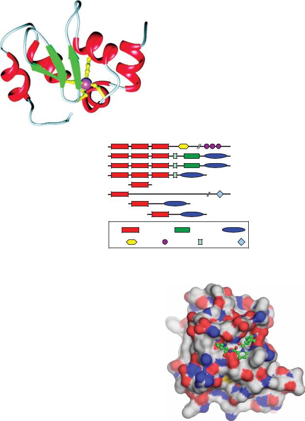
Plate 1. 3D structure of XIAP BIR3. See Figure 2-1 for details.

NAIP/BIRC1


1403

c-IAP1/BIRC2


604

c-IAP2/BIRC3


612

XIAP/BIRC4


497

Survivin/BIRC5


142

Apollon/Bruce/BIRC6


4830

Livin/ML-IAP/BIRC7


298

Ts-IAP/ILP-2/BIRC8


237

BIR

CARD


RING

NBD

LRR

UBA

UBC

Plate 2. Domain organization of the human IAP protein family. See Figure 2-2 for details.

Plate 3. Structure of the XIAP BIR3 domain complexed with SMAC tetrapeptide. SMAC peptide bound to BIR3 of XIAP. The BIR3 domain of XIAP (shown as a space-filling model) complexed with the SMAC tetrapeptide, AVPI. See Figure 2-4 for details.

CD95 and TRAIL signaling complex


FADD

Bax


c-Flip


Bak


mitochondria

tBid

Bid

caspase-8/10


Cytochrome C


ac ve caspase-8/10

Apaf-1


apoptosome

ac ve caspase-9

ac ve caspase-3

Smac/DIABLO

XIAP

Apoptosis

Plate 4. Schematic representation of apoptotic signaling by the CD95 and TRAIL systems. See Figure 3-2 for details.

TNF-R1 signaling complex

RIP1

TRADD


TRAF2/5

TAB2


TAK1


TAB1


NEMO IKKβ

cIAP1/2


IKKα


NF-κB JNK p38

Gene induction

Plate 5. Schematic representation of immunostimulatory, pro-inflammatory signaling by the TNF-R and DR3 systems. Binding of TNF and TL1A to their respective receptor leads to receptor trimerisation and formation of a receptor signaling complex. See Figure 3-3 for details.

Primarily apoptotic signaling systems

Primarily immunostimulatory, proinflammatory

(CD95 and TRAIL systems)

signaling systems (TNF and DR3 systems)

Complex I


Complex I


FADD

RIP1

TRADD


Complex II


TRAF2/5

Complex II

Caspase- 8

TAB2

TAK1


TAB1


NEMO

NEMOIKKβ


cIAP1/2


IKKα


NF-κB

MAPK

APOPTOSIS

Gene induction

APOPTOSIS

Plate 6. Complex I and complex II: spatial dissociation between proapoptotic and proinflammatory signaling in death receptor signal transduction. See Figure 3-4 for details.

- BIM or BID

- sensitizer BH3-only proteins - cytochrome c

- BCL-2 protein

- BAX/BAK protein

Normal cell

“Idealized” cancer cell

Plate 7. Cartoon representation of an unprimed mitochondrion versus a primed mitochondrion. See Figure 5-3 for details.

Plasma membrane


se

nCDase


Ma


aS


Cer

Sph


SM


L


Cer

Sph


M

GS


S


SM


e


Mas


nS


S1P

SK

SM

SM

GSL

Golgi

SM

L S G

GSL

Lysosomes

Cer

aSMase

SM

S


CS


MS

G


r


e

C


C


er


CERT

Serine

+

palmitoyl-CoA

h p S H d

rS e C


r

Des


e


C


dH


ER

Cer

Sph

CerS

SK

SPP

hpS

aCDase

GSL

C


r


e

GCase


e


r


C


lc


S1P


G


ia


r


d


n

h


o


p


h


?


S


c


to


r


i


e


M


?

C


S


M


P

S


L


S

P

T


Nucleus

M A

M s

Plate 8. Compartmentalization of sphingolipid metabolism. See Figure 9-2 for details.

extracellular ligand e.g., CD95L

aSMase

SM Cer

?

(a)

ExogenousCer

UV, IR,

Sph

DNA-damaging

agents

Receptor clustering


CDase

aSMase


Cer SM

flip-flop?


?

?

promotion of


apoptosis


Sphingomyelin

Ceramide

Sphingosine

Glycerophospholipid

extracellular ligands

cellular stresses


salvage

(e.g., cannabinoids)

(e.g., DNA damage)


pathway


promotion of


apoptosis


p53


SK


?


Sph


PP1, PP2A,


Bcl-2-like


SR proteins, p8, ???

proteins

acyl-CoA


acyl-CoA


dhSph

dhCer

Cer

SPT

CerS

Des

CerS

Myriocin

FB1


FB1

pro-survival pathways


ethano-

S1P

lamine


phosphate


+


hexa-


decenal

SPL

(b)

Plate 9. Summary of ceramide-mediated pathways. See Figure 9-8 for details.

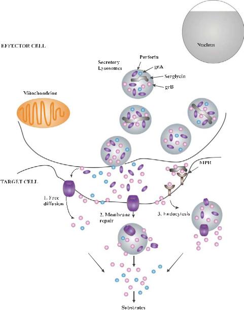
Plate 10. Several hypotheses have been proposed to explain how granzymes enter the target cell to mediate their cell death functions. See Figure 10-1 for details.

TARGET CELL

grA

grB

Human grB

Bid

Mouse grB

Bcl2

tBid

ROS

Procaspase-3

Bax / Bak

ER

Caspase-3

Caspase-9

SET complex

Cytochrome c

CAD

ICAD

IAP


SMAC/DIABLO

DNAse

Procaspase-9

Apaf-1

APOPTOSOME

Plate 11. GrA and grB show di erent substrate specificities within the target cell. GrA induces the release of ROS from the mitochondrial inner membrane, which mediates the translocation of the SET complex from the ER to the nucleus. See Figure 10-2 for details.

Plate 12. The unfolded protein response (UPR), a coordinated regulated response involving three sensor proteins: PERK (PKR-like ER kinase), ATF6 (activating transcription factor 6), and IRE1 (inositol requiring transmembrane kinase/endoribonuclease). See Figure 12-1 for details.

Plate 13. Proteins implicated in ER stress-pcd pathways. See Figure 12-2 for details.

Isolated intact mito

subcellular

Purified intact nuclei

fractionation

Intact DNA

Ca2+ overload

supernatant Supernatant containing AIF

~50 kb DNA fragments

pellet

Swollen, permeabilized

mito Condensed, fragmented nuclei

Plate 14. Identification of a caspase-independent mitochondrial apoptosis-inducing factor using an in vitro reconstitution assay with subcellular fractions. See Figure 14-4 for details.

Plate 15. Extrinsic and intrinsic signals of cell death and survival after spinal cord injury. See Figure 15-1 for details.

Plate 16. Retinal neovascularization in age-related macular degeneration (AMD). See Figure 16-2 for details.

Plate 17. The ear. See Figure 17-1 for details.

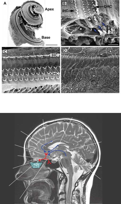
Plate 18. The cochlea. See Figure 17-2 for details.

corpus callosum

medial olfactory stria

olfactory tract

olfactory bulb

olfactory epithelium

lateral olfactory stria

striae medullares

longitudinal striae

medial forebrain bundle

to amygdala

brainstem

and prepyriform cortex

cerebellum

Plate 19. Gross anatomy. See Figure 18-1 for details.

olf. bulb

RMS

SVZ


fila


lateral ventricle


olfactoria

olf. epithelium

brainstem

spinal


cord


olf. bulb

RMS


fila olf.


basal cell


newborn


granule cell

developing

olf. tract

newly integrating

ORN


granule cell

supporting


apoptotic


granule cell

cell


mitral cell

ORN


tufted cell


apoptotic

ORN axon


ORN


mucus


glomerulum

periglomerular cell mature granule cell

Plate 20. Olfactory life and death on a microscopic level. See Figure 18-2 for details.

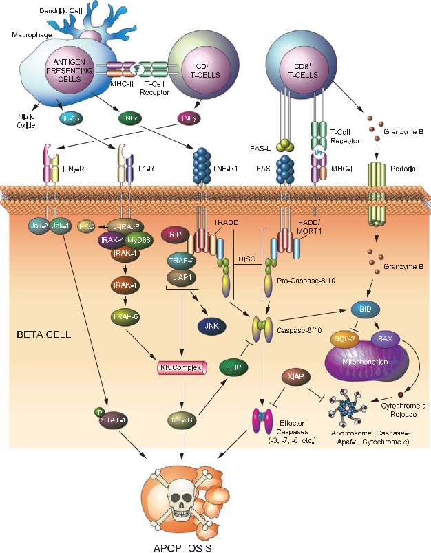
Plate 21. Signaling pathways leading to beta cell destruction in type 1 diabetes. See Figure 19-1 for details.

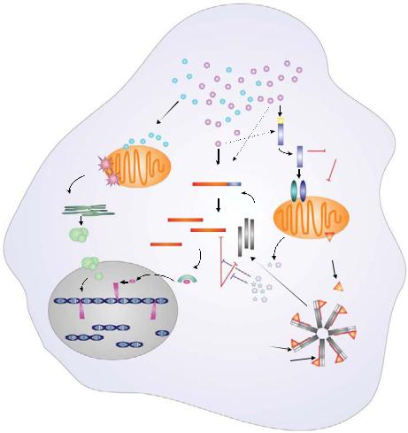
Plate 22. Shift in lipid partitioning associated with apoptosis in diabetic beta cells. See Figure 19-2 for details.

(a)

(b)

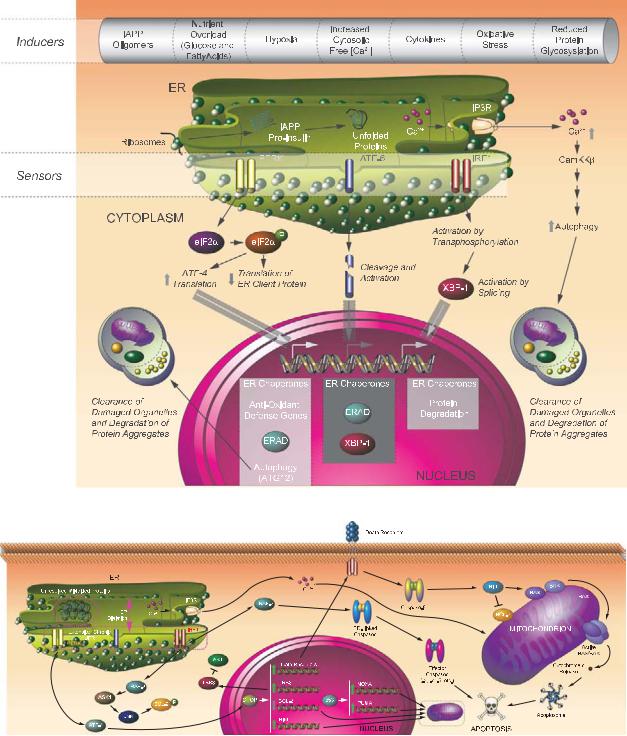
Plate 23. Signaling pathways in response to unfolded proteins. See Figure 19-3 for details.

A) Alteration in the BAX/BCL-2 ratio

C) Activation of caspase-9


CON

2d

5d

14d


M

M

M

M


BCL-2


BAX


D) Activation of caspase-3


COX IV


B) Release of cytochrome c and DIABLO


CON

2d

5d

14d


DIABLO


TUNEL

Caspase-3

Merged


E) PARP cleavage


Cyt.C


CON 2d

5d 14d


89 kDa

Actin

Actin

Plate 24. Hormonal deprivation results in activation of the intrinsic pathway signaling. See Figure 25-3 for details.

A)

CON 2d

5d 14d

p-ATF-2

Total ATF-2

B)

I

II

III

IV

V

IV

TUNEL

p-p38 MAPK

Merged

Plate 25. Activation of p38 MAPK in rat testes after GnRH-A treatment. See Figure 25-4 for details.

Plate 26. Testicular hyperthermia results in serine phosphorylation of BCL-2 in germ cells. See Figure 25-5 for details.

A B C

XII

XII

XII

Plate 27. Activation of ERK in the Sertoli cells. See Figure 25-6 for details.

Tunica adventitia connective tissue

Plate 28. Normal human artery consists of three layers. See Figure 26-1 for details.

Tunica intima

endothelium

internal elastic lamina

external elastic lamina

smooth muscle cells

Tunica media

a

b

Control Apoe-/-

SM22α-hDTR Apoe-/-

Control Apoe-/-

SM22α-hDTR Apoe-/-

Plate 29. Vascular smooth muscle cell apoptosis accelerates atherosclerotic plaque progression and induces plaque vulnerability. See Figure 26-3 for details.

a

b

Percent survival

d

LVEDP (mmHg)

100


C360A (n = 19)

80


WT (n = 197)

60


Line 169 (n = 34)


40


20


0

50 100 150 200 250 300

0


Time (d)


30


(mmHg/s)

20000


15000


20


10000


+dP/dt

10


5000


0

WT

Tg


0


n

7

6


n

IVS

LV-cavity

PW

– + – + WT Tg

7

5

6

3

e

f

WT

Tg

–dP/dt (mmHg/s)

IVS

LVcavity PW

10000


7500


5000


2500


0

+

+


n

WT


Tg


7

5

6

3


TUNEL-positive myocytes per

WT


Tg


c

4


(mm)

3


EDD

2


1


0

WT

Line 7

Line 169

C360A


n


9

9

5

3


100


(%)

75


50


FS


25


0

WT

Line 7

Line 169

C360A


n


9

9

5

3

nuclei

30


20


cardiac


10


5


10


0


WT

Line 7 C360A


n

5

6

4


Plate 30. Modest, but elevated, rates of cardiac myocyte apoptosis are su cient over time to induce lethal heart failure. See Figure 26-4 for details.


Bone

Skeletal Muscle


Vein

Muscle

A


Fascicle


Myofiber


Tendon

Capillary Myonucleus


Sarcomere


A-band I-band


M-line Z-line

Myofibril


ENDURANCE


TRAINING


T-tubule


Sarcoplasmic


reticulum


Myonucleus

Subsarcolemmal


(SS) mitochondria


Intermyofibrillar (IMF) mitochondria

B

OMM

Holoenzyme

MITOCHONDRION


assembly


IMM


Incorporation


into ETC


Electron transport

mtDNA

chain (ETC)

ATP


Nuclear pore

Import machinery

Translation

+1

Transcription

NUGEMPS


NUCLEUS

mRNA

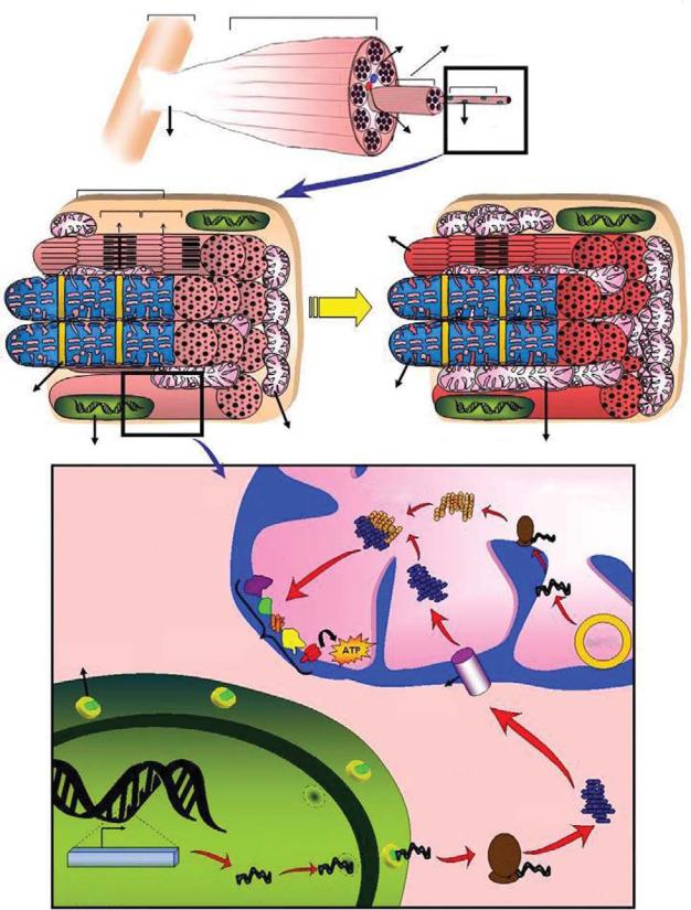
Plate 31. Unique morphology of skeletal muscle and exercise-induced mitochondrial biogenesis. See Figure 27-1 for details.

Loss of Myonuclei +

Satellite cells

Myonuclear

domain

Myofiber

Myonuclear

Domains

Satellite cell activation, proliferation

Atrophy

Hypertrophy

Fusion of satellite cells, myonuclear addition and resultant fiber hypertrophy

Myonuclear Domain is constant

Plate 32. Myonuclear domains during muscle hypertrophy and atrophy. See Figure 27-2 for details.

Plate 33. UVB signaling in keratinocytes. UVB can lead to di erent e ects in keratinocytes, ranging from cell cycle arrest, apoptosis, and inflammasome activation. UVB radiation primarily damages nuclear DNA as a result of direct absorption and generates ROS that can induce oxidative damage to DNA and cellular proteins. See Figure 28-3 for details.

Extrinsic pathway

Ligand


Death


receptors


FADD

FLIP

DISC


Procaspases


-8 and -10


Active caspases-8


and -10

Pro-caspases-3, -6, -7

Intrinsic pathway

Cytokine withdrawal, chemotherapeutic drugs, radiations…

BH3-only

proteins

Anti-apoptotic Bcl-2 family members

Bid

Bax/Bak

tBid

Cytochrome C

Apaf-1

Active effector


caspases-3, -6 and -7

Active caspase-9

Pro-caspase-9


Apoptosome

Plate 34. Two signaling pathways leading to apoptosis. See Figure 29-1 for details.

Plate 35. Lymphocyte apoptosis in spleen of septic patient. Hematoxylin and eosin staining of spleen of septic patient (400 × magnification). See Figure 31-1 for details.

Plate 36. Colonic epithelial apoptosis in a septic patient. See Figure 31-2 for details.

Gram-negative bacteria

Porin

Gram-positive bacteria

Lipopolysaccharide

Outer membrane

lipoprotein

Peptidoglycan

Plasma membrane

Plate 37. A representation of the cell wall from Gram-negative and Gram-positive bacteria. See Figure 32-2 for details.

teichoic acid

Peptidoglycan

Lipoteichoic acid

Plasma membrane

Mammalian

Insect


TNF TNF

TNF


TNFRTNFRTNFR


DD

DD

DD

DD

DD


DD


DD


Complex II


Complex I


DISC


TRADD


TRADD


RIP1


RIP1

DED

DED

DD

DD

DD

TRADD RIP1

TRAF2

RING


DED


TRAF2

TRAF2


RING


RING


p20


DED

FADD

DD

DD

TRADD RIP1

TRAF2

RING


DEDDED

DD

BIR BIR BIRCARD RING

p10


cIAP1/2


TAB2


TAK1


p20


Pro-caspase-8/10


IKK

IKK


p10


NEMO


Caspase-3/6/7


I B


p10p10


p20


p20


p50


RelA


Substrate processing

Apoptosis

DAP-type

Peptidoglycan

derivatives

PGRP

-LC

IMD

DD

BIR BIR BIRCARD RING

DIAP2

TAB2

TAK1

IKK Ird5

Kenny

Rel

Gram-negative

bacteria

dFADD


DED

DD

DED

DED


p20

Dredd

p10

Relish

ANK

Rel


NF B


NF B

p50

RelA


Rel

Rel

Induction of survival and

DNA fragmentation

Induction of Diptericin and other

inflammatory genes

Chromatin condensation

antimicrobial genes

Plate 38. Schematic representation of the mammalian TNF and insect Imd pathways. See Figure 32-6 for details.

Mammalian

Gram-positive and Gram-negative

bacteria

Insect


Gram-positive


bacteria

-

Lysine-type

PGRP

S


Peptidoglycan

derivatives


Flagellin


LPS


Bacterial

TIR

TIR

TLR4TIR

TIR

TIR

TIRTLR5

TIR


TLR1/6

TLR2


lipoproteins


TIR

TIR

TIR TIR


Mal

TRIF


MyD88


MyD88


TRAM


MyD88


IRAK1

IRAK4


Mal


TRAF6


FADD


DD

DD


TAB2


Apoptosis


DED


TAK1


TRIF RIP1


TAB1


TRAF6


TIR

TIR

TLR3


TRAF3


Nucleic


MKK3

MKK7


IKK

IKK


TBK1

TIR

TLR7

acids


TIR

TLR8


MKK6


NEMO


TIR

TLR9


p38

JNK


Endosome


IRF3

IRF7


Fungi

Spätzle

Yeast

Toll

TIR


dMyD88

TIR


DD


DD


DD


Pelle

Tube


Cactus


ANK


Dif

Rel

Rel

Dorsal

Rel


Induction of the interferon


response

NF B

NF B

p50 RelA

Induction of survival and inflammatory genes

Rel Rel

Induction of Drosomycin and other antimicrobial genes

Plate 39. A schematic representation of the mammalian TLR and insect Toll pathways. See Figure 32-7 for details.

NOD1


CARD

NBD


NOD2


CARD CARD

NBD


NOD3/9/27


X

NBD


(NLRC3/X1/5)


NALP1


PYD

NBD

FIIND CARD

(NLRP1)


NALP2-14


PYD

NBD


(NLRP2-14)


IPAF


CARD

NBD


(NLRC4)


NAIP

BIR

BIR BIR

CARD

NBD


(NLRB)


CIITA


AD

NBD


APAF1


CARD

NBD


Ced4


CARD

NBD


ASC


PYD

CARD


CARDINAL


FIIND

CARD


Caspase-


p20

p10

3/6/7


Caspase-8/10


DED DED

p20

p10


cIAP1/2

BIR

BIR

BIR

CARD

RING


XIAP

BIR

BIR

BIR


RING


TRAF2


RING


TZ

TZ


CC

MATH


TRAF6


RING


TZ

TZ


CC

MATH


RIP1


Kinase domain


DD


RIP2


Kinase domain


CARD


IKK /


Kinase domain

LZ

HLH

Nemo BD

IKK


CC1


CC2

LZ

ZF

FADD


DED

DD


TRADD


DD


TNFR1


EC


EC

EC

EC

TM

DD


Caspase-1


CARD


p20

p12


Caspase-9


CARD


p20

p10


Ced3


CARD


p20

p10


Plate 40. E ectors of inflammation. The domain structures are shown. See Figure 32-8 for details.

Cytochrome C PAMPs

Ced9

Ced4

Apaf1

NLRs

Bcl-2,


Bcl-xl


?


?

Apoptosome


Inflammasome


Active

Active

Active

Ced3

Caspase-9

Caspase-1

APOPTOSIS

APOPTOSIS

PYROPTOSIS &


INFLAMMATION

Plate 41. A parallel between mitochondrial apoptosis and NLR innate immunity pathways. See Figure 32-9 for details.


FasL FasL FasL


H. pylori

Fas Fas Fas


P. aeruginosa


DD

DD

DD

DD

DD


DD


DD


TRADD


TRADD

RIP1


RIP1

Ub

TRAF2 TRAF2

Ub

b


Ub

U


b


Ub

U


b

RING


RING

Ub

U


b


Ub


U


S. pneumoniae

BH3

BH3

IKK

IKK

B. Anthracis

Chlamydia spp.

Cytochrome c

PAMPs

Nigericin


(LT)

MKK


Maitotoxin

NEMO


Aerolysin


F. tularensis

P

P


Apaf1

NLRs

S. flexneri

I B


p50

RelA


Apoptosome

Inflammasome

B. Anthracis


S. typhimurium

MAPK


(LT)

R. rickettsii


(AvrA)


L. monocytogenes


Y. pestis


(LLO)


(YopJ)


NF B


S. typhimurium

p50

RelA


(TTSS)


L. pneumophila


Active

Active

P. aeruginosa


Caspase-9

Caspase-1


( Exo U)


SURVIVAL


APOPTOSIS

PYROPTOSIS &


INFLAMMATION


Plate 42. Modulation of cell survival/death pathways by microbial e ectors. See Figure 32-10 for details.

Pyroptosis


Apoptosis

Oncosis


Autophagy

Unconventional


Apoptotic


protein secretion


Membrane


bodies


rupture


Membrane


Membrane

Atg7


blebbing


blebbing


Caspase-1


Organelle

Atg8/LC3


Caspase-3, 7

swelling


Atg8/LC3


Autophagosome


Immature

Other

Glycolysis


Cyto C


Autophago-


Substrates release


Membrane


lysosome

cytokines

substrates

enzymes


vesicles


Nuclear


m


swelling


Nuclear


Nuclear


condensation


condensation


DNA


DNA


fragmentation


fragmentation


Cell


Membrane


Mature


Membrane


shrinkage


permeability


cytokines


swelling


Plate 43. Pathogen-induced host cell death. See Figure 32-11 for details.

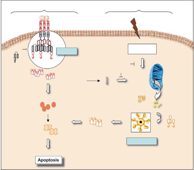
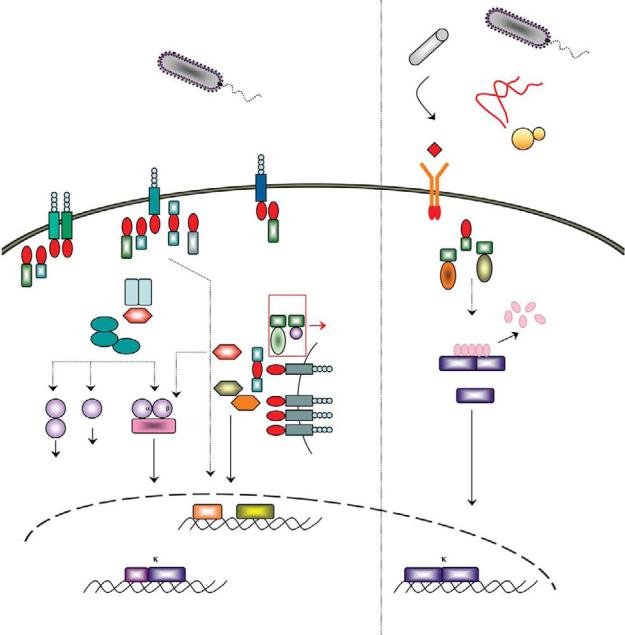
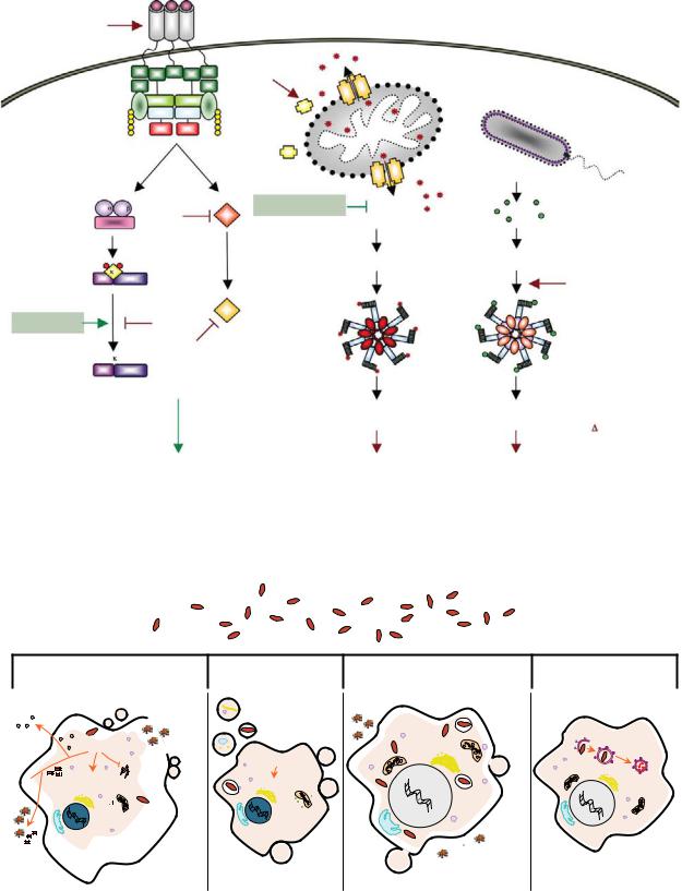
Plate 44. The four male-specific chemosensory (CEM) neurons located in the cephalic region of the animal undergo programmed cell death in hermaphrodites. See Figure 34-3 for details.

12.5 Gy

acridine orange

TUNEL

p53 +/+

non-injected

chk1 MO

p53e7/e7

non-injected

chk1 MO

Plate 45. A rapid morpholino loss-of-function screen identifies chk1 knockdown as a caspase-3–independent radiation sensitizer in p53 mutant embryos. See Figure 36-3 for details.

Regions of developmental cell death

Brain

Spinal cord

Ear

Eye

Plate 46. Cell death zones in developing zebrafish embryos. See Figure 36-5 for details.

Germ cells

Excretory system

Tail bud

A


B


C


D

E


F


G


H

Plate 47. Images of microglia consuming dying neurons by phagocytosis in wild-type and atpv0a1 morphant larvae. See Figure 36-6 for details.

APOPTOSIS IN THE PHYSIOLOGY AND DISEASES OF THE RESPIRATORY TRACT

227

after tracheal occlusion in the fetal rabbit. Am J Pathol. 152, 179–90.

De Paepe, M.E., Mao, Q., Chao, Y., Powell, J.L., Rubin, L.P., and Sharma, S. (2005). Hyperoxia-induced apoptosis and Fas/FasL expression in lung epithelial cells. Am J Physiol Lung Cell Mol Physiol. 289, L647–59.

De Paepe, M.E., Sardesai, M.P., Johnson, B.D., Lesieur-Brooks, A.M., Papadakis, K., and Luks, F.I. (1999b). The role of apoptosis in normal and accelerated lung development in fetal rabbits. J Pediatr Surg. 34, 863–70.

Del Riccio, V., van Tuyl, M., and Post, M. (2004). Apoptosis in lung development and neonatal lung injury. Pediatr Res. 55, 183–9.

Droemann, D., Aries, S.P., Hansen, F., Moellers, M., Braun, J., Katus, H.A., and Dalhoff, K. (2000). Decreased apoptosis and increased activation of alveolar neutrophils in bacterial pneumonia. Chest. 117, 1679–84.

Fadok, V.A., Bratton, D.L., Konowal, A., Freed, P.W., Westcott, J.Y., and Henson, P.M. (1998). Macrophages that have ingested apoptotic cells in vitro inhibit proinflammatory cytokine production through autocrine/paracrine mechanisms involving TGF-beta, PGE2, and PAF. J Clin Invest. 101, 890–8.

Fedorov, L.M., Tyrsin, O.Y., Papadopoulos, T., Camarero, G., Gotz,¨ R., and Rapp, U.R. (2002). Bcl-2 determines susceptibility to induction of lung cancer by oncogenic C-Raf. Cancer Res. 62, 6297–303.

Fujita, M., Kuwano, K., Kunitake, R., Hagimoto, N., Miyazaki, H., Kaneko, Y., Kawasaki, M., Maeyama, T., and Hara, N. (1998). Endothelial cell apoptosis in lipopolysaccharideinduced lung injury in mice. Int Arch Allergy Immunol. 117, 202–8.

Galie, N., Hoeper, M.M., Humbert, M., Torbicki, A., Vachiery, J-L., Barbera, J.A., Beghetti, M., Corris, P., Gaine, S., Gibbs, J.S., Gomez-Sanchez, M.A., Jondeau, G., Klepetko, W., Opitz, C., Peacock, A., Rubin, L., Zellweger, M., and Simonneau, G. (2009). Guidelines for the diagnosis and treatment of pulmonary hypertension. Eur Respir J; 34: 1219–1263.

Greco, F.A., Bonomi, P., Crawford, J., Kelly, K., Oh, Y., Halpern, W., Lo, L., Gallant, G., and Klein, J. (2008). Phase 2 study of mapatumumab, a fully human agonistic monoclonal antibody which targets and activates the TRAIL receptor-1, in patients with advanced non-small cell lung cancer. Lung Cancer. 61, 82–90.

Hagimoto, N., Kuwano, K., Miyazaki, H., Kunitake, R., Fujita, M., Kawasaki, M., Kaneko, Y., and Hara, N. (1997). Induction of apoptosis and pulmonary fibrosis in mice in response to ligation of Fas antigen. Am J Respir Cell Mol Biol. 17, 272–8.

Hargitai, B., Szabo, V., Hajdu, J., Harmath, A., Pataki, M., Farid, P., Papp, Z., and Szende, B. (2001). Apoptosis in various organs of preterm infants: histopathologic study of lung, kidney, liver, and brain of ventilated infants. Pediatr Res. 50, 110– 14.

Hodge, S., Hodge, G., Ahern, J., Jersmann, H., Holmes, M., and Reynolds, P.N. (2007). Smoking alters alveolar macrophage recognition and phagocytic ability: implications in chronic

obstructive pulmonary disease. Am J Respir Cell Mol Biol. 37, 748–55.

Hodge, S., Hodge, G., Holmes, M., and Reynolds, P.N. (2005). Increased airway epithelial and T-cell apoptosis in COPD remains despite smoking cessation. Eur Respir J. 25, 447–54.

Hodge, S., Hodge, G., Nairn, J., Holmes, M., and Reynolds, P.N. (2006). Increased airway granzyme B and perforin in current and ex-smoking COPD subjects. COPD. 3, 179–87.

Hoffarth, S., Zitzer, A., Wiewrodt, R., Hahnel,¨ P.S., Beyer, V., Kreft, A., Biesterfeld, S., and Schuler, M. (2008). pp32/PHAPI determines the apoptosis response of non-small-cell lung cancer. Cell Death Differ. 15, 170.

Holopainen, R., Aho, H., Laine, J., Peuravuori, H., Soukka, H., and Kaapa, P. (1999). Human meconium has high phospholipase A2 activity and induces cellular injury and apoptosis in piglet lungs. Pediatr Res. 46, 626–32.

Huynh, M.L., Fadok, V.A., and Henson, P.M. (2002). Phos- phatidylserine-dependent ingestion of apoptotic cells promotes TGF-beta1 secretion and the resolution of inflammation. J Clin Invest. 109, 41–50.

Imai, K., Mercer, B.A., Schulman, L.L., Sonett, J.R., and D’Armiento, J.M. (2005). Correlation of lung surface area to apoptosis and proliferation in human emphysema. Eur Respir J. 25, 250–8.

Jiang, X., Kim, H.-E., Shu, H., Zhao, Y., Zhang, H., Kofron, J., Donnelly, J., Burns, D., Ng, S.-C., Rosenberg, S., and Wang, X. (2003). Distinctive roles of PHAP proteins and prothymosin-a in a death regulatory pathway. Science. 299, 223–6.

Jin, H., Yang, R., Fong, S., Totpal, K., Lawrence, D., Zheng, Z., Ross, J., Koeppen, H., Schwall, R., and Ashkenazi, A. (2004). Apo2 Ligand/tumor necrosis factor-related apoptosisinducing ligand cooperates with chemotherapy to inhibit orthopic lung tumor growth and improve survival. Cancer Res. 64, 4900–5.

Johnson, L., Mercer, K., Greenbaum, D., Bronson, R.T., Crowley, D., Tuveson, D.A., and Jacks, T. (2001). Somatic activation of the K-ras oncogene causes early onset lung cancer in mice.

Nature. 410, 1111–16.

Johnstone, R.W., Ruefli, A.A., and Lowe, S.W. (2002). Apoptosis: a link between cancer genetics and chemotherapy. Cell. 108, 153–64.

Karrasch, S., Holz, O., and Jorres, R.A. (2008). Aging and induced senescence as factors in the pathogenesis of lung emphysema. Respir Med. 102, 1215–30.

Kasahara, Y., Tuder, R.M., Cool, C.D., Lynch, D.A., Flores, S.C., and Voelkel, N.F. (2001a). Endothelial cell death and decreased expression of vascular endothelial growth factor and vascular endothelial growth factor receptor 2 in emphysema. Am J Respir Crit Care Med. 163, 737–44.

Kasahara, Y., Tuder, R.M., Cool, C.D., Lynch, D.A., Flores, S.C., and Voelkel, N.F. (2001b). Endothelial cell death and decreased expression of vascular endothelial growth factor and vascular endothelial growth factor receptor 2 in emphysema. Am J Respir Crit Care Med. 163, 737–44.

Kasahara, Y., Tuder, R.M., Taraseviciene-Stewart, L., Le Cras, T.D., Abman, S., Hirth, P.K., Waltenberger, J., and Voelkel, N.F.

228

CHRISTIAN TAUBE AND MARTIN SCHULER

(2000). Inhibition of VEGF receptors causes lung cell apoptosis and emphysema. J Clin Invest. 106, 1311–19.

Kawasaki, M., Kuwano, K., Hagimoto, N., Matsuba, T., Kunitake, R., Tanaka, T., Maeyama, T., and Hara, N. (2000). Protection from lethal apoptosis in lipopolysaccharide-induced acute lung injury in mice by a caspase inhibitor. Am J Pathol. 157, 597–603.

Kitamura, Y., Hashimoto, S., Mizuta, N., Kobayashi, A., Kooguchi, K., Fujiwara, I., and Nakajima, H. (2001). Fas/ FasL-dependent apoptosis of alveolar cells after lipo- polysaccharide-induced lung injury in mice. Am J Respir Crit Care Med. 163, 762–9.

Kresch, M.J., Christian, C., Wu, F., and Hussain, N. (1998). Ontogeny of apoptosis during lung development. Pediatr Res. 43, 426–31.

Kuwano, K., Hagimoto, N., Kawasaki, M., Yatomi, T., Nakamura, N., Nagata, S., Suda, T., Kunitake, R., Maeyama, T., Miyazaki, H., and Hara, N. (1999). Essential roles of the Fas-Fas ligand pathway in the development of pulmonary fibrosis. J Clin Invest. 104, 13–19.

Kuwano, K., Kunitake, R., Kawasaki, M., Nomoto, Y., Hagimoto, N., Nakanishi, Y., and Hara, N. (1996). P21Waf1/Cip1/Sdi1 and p53 expression in association with DNA strand breaks in idiopathic pulmonary fibrosis. Am J Respir Crit Care Med. 154, 477–83.

Kuwano, K., Kunitake, R., Maeyama, T., Hagimoto, N., Kawasaki, M., Matsuba, T., Yoshimi, M., Inoshima, I., Yoshida, K., and Hara, N. (2001). Attenuation of bleomycin-induced pneumopathy in mice by a caspase inhibitor. Am J Physiol Lung Cell Mol Physiol. 280, L316–25.

Lane, K.B., Machado, R.D., Pauciulo, M.W., Thomson, J.R., Phillips, J.A., III, Loyd, J.E., Nichols, W.C., and Trembath, R.C. (2000). Heterozygous germline mutations in BMPR2, encoding a TGF-beta receptor, cause familial primary pulmonary hypertension. The International PPH Consortium. Nat Genet. 26, 81–4.

Lee, C.G., Cho, S.J., Kang, M.J., Chapoval, S.P., Lee, P.J., Noble, P.W., Yehualaeshet, T., Lu, B., Flavell, R.A., Milbrandt, J., Homer, R.J., and Elias, J.A. (2004). Early growth response gene 1-mediated apoptosis is essential for transforming growth factor beta1-induced pulmonary fibrosis. J Exp Med. 200, 377–89.

Levesque, B.M., Vosatka, R.J., and Nielsen, H.C. (2000). Dihydrotestosterone stimulates branching morphogenesis, cell proliferation, and programmed cell death in mouse embryonic lung explants. Pediatr Res. 47, 481–91.

Lu, Q., Xu, D.Z., Davidson, M.T., Hasko, G., and Deitch, E.A. (2004). Hemorrhagic shock induces endothelial cell apoptosis, which is mediated by factors contained in mesenteric lymph. Crit Care Med. 32, 2464–70.

Lukkarinen, H.P., Laine, J., and Kaapa, P.O. (2003). Lung epithelial cells undergo apoptosis in neonatal respiratory distress syndrome. Pediatr Res. 53, 254–9.

Mantell, L.L., Horowitz, S., Davis, J.M., and Kazzaz, J.A. (1999). Hyperoxia-induced cell death in the lung–the correlation of

apoptosis, necrosis, and inflammation. Ann N Y Acad Sci. 887, 171–80.

Martin, T.R., Hagimoto, N., Nakamura, M., and Matute-Bello, G. (2005). Apoptosis and epithelial injury in the lungs. Proc Am Thorac Soc. 2, 214–20.

Matute-Bello, G., Liles, W.C., Frevert, C.W., Nakamura, M., Ballman, K., Vathanaprida, C., Kiener, P.A., and Martin, T.R. (2001). Recombinant human Fas ligand induces alveolar epithelial cell apoptosis and lung injury in rabbits. Am J Physiol Lung Cell Mol Physiol. 281, L328–35.

Matute-Bello, G., Liles, W.C., Radella, F., Steinberg, K.P., Ruzinski, J.T., Jonas, M., Chi, E.Y., Hudson, L.D., and Martin, T.R. (1997). Neutrophil apoptosis in the acute respiratory distress syndrome. Am J Respir Crit Care Med. 156, 1969–77.

Matute-Bello, G., Liles, W.C., Steinberg, K.P., Kiener, P.A., Mongovin, S., Chi, E.Y., Jonas, M., and Martin, T.R. (1999). Soluble Fas ligand induces epithelial cell apoptosis in humans with acute lung injury (ARDS). J Immunol. 163, 2217–25.

May, M., Strobel, P., Preisshofen, T., Seidenspinner, S., Marx, A., and Speer, C.P. (2004). Apoptosis and proliferation in lungs of ventilated and oxygen-treated preterm infants. Eur Respir J. 23, 113–21.

McDonnell, T.J., Fang, B., Yu, R., Kagawa, S., Hunt, K.K., McDonnell, T.J., Roth, J.A., and Swisher, S.G. (2000). Adenoviral Bak overexpression mediates caspase-dependent tumor killing.

Cancer Res. 60, 788–92.

McMurtry, M.S., Archer, S.L., Altieri, D.C., Bonnet, S., Haromy, A., Harry, G., Bonnet, S., Puttagunta, L., and Michelakis, E.D. (2005). Gene therapy targeting survivin selectively induces pulmonary vascular apoptosis and reverses pulmonary arterial hypertension. J Clin Invest. 115, 1479–91.

Metzger, R.J., Klein, O.D., Martin, G.R., and Krasnow, M.A. (2008). The branching programme of mouse lung development. Nature. 453, 745–50.

Morrell, N.W., Yang, X., Upton, P.D., Jourdan, K.B., Morgan, N., Sheares, K.K., and Trembath, R.C. (2001). Altered growth responses of pulmonary artery smooth muscle cells from patients with primary pulmonary hypertension to transforming growth factor-beta(1) and bone morphogenetic proteins.

Circulation. 104, 790–5.

Nakamura, M., Matute-Bello, G., Liles, W.C., Hayashi, S., Kajikawa, O., Lin, S.M., Frevert, C.W., and Martin, T.R. (2004). Differential response of human lung epithelial cells to fasinduced apoptosis. Am J Pathol. 164, 1949–58.

Oltersdorf, T., Elmore, S.W., Shoemaker, A.R., Armstrong, R.C., Augeri, D.J., Belli, B.A., Bruncko, M., Deckwerth, T.L., Dinges, J., Hajduk, P.J., Joseph, M.K., Kitada, S., Korsmeyer, S.J., Kunzer, A.R., Letai, A., Li, C., Mitten, M.J., Nettesheim, D.G., Ng, S., Nimmer, P.M., O’Connor, J.M., Oleksijew, A., Petros, A.M., Reed, J.C., Shen, W., Tahir, S.K., Thompson, C.B., Tomaselli, K.J., Wang, B., Wendt, M.D., Zhang, H., Fesik, S.W., and Rosenberg, S.H. (2005). An inhibitor of Bcl-2 family proteins induces regression of solid tumours. Nature. 435, 677–81.

Petrache, I., Natarajan, V., Zhen, L., Medler, T.R., Richter, A.T., Cho, C., Hubbard, W.C., Berdyshev, E.V., and Tuder, R.M.

APOPTOSIS IN THE PHYSIOLOGY AND DISEASES OF THE RESPIRATORY TRACT

229

(2005). Ceramide upregulation causes pulmonary cell apoptosis and emphysema-like disease in mice. Nat Med. 11, 491–8.

Plataki, M., Koutsopoulos, A.V., Darivianaki, K., Delides, G., Siafakas, N.M., and Bouros, D. (2005). Expression of apoptotic and antiapoptotic markers in epithelial cells in idiopathic pulmonary fibrosis. Chest. 127, 266–74.

Potti, A., Mukherjee, S., Petersen, R., Dressman, H.K., Bild, A., Koontz, J., Kratzke, R., Watson, M.A., Kelley, M., Ginsburg, G.S., West, M., Harpole, D.H., and Nevins, J.R. (2006). A genomic strategy to refine prognosis in early-stage non- small-cell lung cancer. N Engl J Med. 355, 570–80.

Rabe, K.F., Hurd, S., Anzueto, A., Barnes, P.J., Buist, S.A., Calverley, P., Fukuchi, Y., Jenkins, C., Rodriguez-Roisin, R., van, W.C., and Zielinski, J. (2007). Global strategy for the diagnosis, management, and prevention of chronic obstructive pulmonary disease: GOLD executive summary. Am J Respir Crit Care Med. 176, 532–55.

Rabinovitch, M. (2008). Molecular pathogenesis of pulmonary arterial hypertension. J Clin Invest. 118, 2372–9.

Radetzki, S., Kohne,¨ C.H., von Haefen, C., Gillisen, B., Sturm, I., Dorken,¨ B., and Daniel, P.T. (2002). The apoptosis promoting Bcl-2 homologues Bak and Nbk/Bik overcome drug resistance in Mdr-1-negative and Mdr-1-overexpressing breast cancer cell lines. Oncogene. 21, 227–38.

Ranger, A.M., Malynn, B.A., and Korsmeyer, S.J. (2001). Mouse models of cell death. Nat Genet. 28, 113–18.

Roth, J.A., Nguyen, D., Lawrence, D.D., Kemp, B.L., Carrasco, C.H., Ferson, D.Z., Hong, W.K., Komaki, R., Lee, J.J., Nesbitt, J.C., Pisters, K.M.W., Putnam, J.B., Schea, R., Shin, D.M., Walsh, G.L., Dolormente, M.M., Han, C.-I., Martin, F.D., Yen, N., Xu, K., Stephens, L.C., McDonnell, T.J., Mukhopadhyay, T., and Cai, D. (1996). Retrovirus-mediated wild-type p53 gene transfer to tumors of patients with lung cancer. Nat Med. 2, 985–91.

Scavo, L.M., Ertsey, R., Chapin, C.J., Allen, L., and Kitterman, J.A. (1998). Apoptosis in the development of rat and human fetal lungs. Am J Respir Cell Mol Biol. 18, 21–31.

Schittny, J.C., Djonov, V., Fine, A., and Burri, P.H. (1998). Programmed cell death contributes to postnatal lung development. Am J Respir Cell Mol Biol. 18, 786–93.

Segura-Valdez, L., Pardo, A., Gaxiola, M., Uhal, B.D., Becerril, C., and Selman, M. (2000). Upregulation of gelatinases A and B, collagenases 1 and 2, and increased parenchymal cell death in COPD. Chest. 117, 684–94.

Shapiro, S.D. (2003). Proteolysis in the lung. Eur Respir J Suppl. 44, 30s–2s.

Sordella, R., Bell, D.W., Haber, D.A., and Settleman, J. (2004). Gefitinib-sensitizing EGFR mutations in lung cancer activate anti-apoptotic pathways. Science 305, 1163–7.

Sweet-Cordereo, A., Mukherjee, S., Subramanian, A., You, H., Roix, J.J., Ladd-Acosta, C., Mesirov, J., Golub, T.R., and Jacks, T. (2005). An oncogenic KRAS2 expression signature identified by cross-species gene-expression analysis. Nat Genet. 37, 48–55.

Takata, T., Tanaka, F., Yamada, T., Yanagihara, K., Otake, Y., Kawano, Y., Nakagawa, T., Miyahara, R., Oyanagi, H., Inui, K., and Wada, H. (2001). Clinical significance of caspase-3 expression in pathologic-stage I, nonsmall-cell lung cancer.

Int J Cancer. 96, 54–60.

Tang, K., Rossiter, H.B., Wagner, P.D., and Breen, E.C. (2004). Lung-targeted VEGF inactivation leads to an emphysema phenotype in mice. J Appl Physiol. 97, 1559–66.

Thannickal, V.J. and Horowitz, J.C. (2006). Evolving concepts of apoptosis in idiopathic pulmonary fibrosis. Proc Am Thorac Soc. 3, 350–6.

Tse, C., Shoemaker, A.R., Adickes, J., Anderson, M.G., Chen, J., Jin, S., Johnson, E.F., Marsh, K.C., Mitten, M.J., Nimmer, P., Roberts, L., Tahir, S.K., Xiao, Y., Yang, X., Zhang, H., Fesik, S., Rosenberg, S.H., and Elmore, S.W. (2008). ABT-263: a potent and orally bioavailable Bcl-2 family inhibitor. Cancer Res. 68, 3421–8.

Tuder, R.M., Zhen, L., Cho, C.Y., Taraseviciene-Stewart, L., Kasahara, Y., Salvemini, D., Voelkel, N.F., and Flores, S.C. (2003). Oxidative stress and apoptosis interact and cause emphysema due to vascular endothelial growth factor receptor blockade. Am J Respir Cell Mol Biol. 29, 88–97.

Uhal, B.D., Joshi, I., Hughes, W.F., Ramos, C., Pardo, A., and Selman, M. (1998). Alveolar epithelial cell death adjacent to underlying myofibroblasts in advanced fibrotic human lung.

Am J Physiol. 275, L1192–9.

Vandivier, R.W., Fadok, V.A., Hoffmann, P.R., Bratton, D.L., Penvari, C., Brown, K.K., Brain, J.D., Accurso, F.J., and Henson, P.M. (2002). Elastase-mediated phosphatidylserine receptor cleavage impairs apoptotic cell clearance in cystic fibrosis and bronchiectasis. J Clin Invest. 109, 661–70.

Varfolomeev, E., Blankenship, J.W., Wayson, S.M., Fedorova, A.V., Kayagaki, N., Garg, P., Zobel, K., Dynek, J.N., Elliott, L.O., Wallweber, H.J., Flygare, J.A., Fairbrother, W.J., Deshayes, K., Dixit, V.M., and Vucic, D. (2007). IAP antagonists induce autoubiquitination of c-IAPs, NF-kappaB activation, and TNFalpha-dependent apoptosis. Cell, 131, 669–81.

Ventura, A., Young, A.F., Winslow, M.M., Lintault, L., Meissner, A., Erkeland, S.J., Newman, J., Bronson, R.T., Crowley, D., Stone, J.R., Jaenisch, R., Sharp, P.A., and Jacks, T. (2008). Targeted deletion reveals essential and overlapping functions of the miR-17 through 92 family of miRNA clusters. Cell. 132, 875–86.

Walczak, H., Miller, R.E., Ariail, K., Gliniak, B., Griffith, T.S., Kubin, M., Chin, W., Jones, J., Woodward, A., Le, T., Smith, C., Smolak, P., Goodwin, R.G., Rauch, C.T., Schuh, J.C.L., and Lynch, D.H. (1999). Tumoricidal activity of tumor necrosis factor related apoptosis inducing ligand in vivo. Nat Med. 5, 157–63.

Wallach-Dayan, S.B., Izbicki, G., Cohen, P.Y., Gerstl-Golan, R., Fine, A., and Breuer, R. (2006). Bleomycin initiates apoptosis of lung epithelial cells by ROS but not by Fas/FasL pathway.

Am J Physiol Lung Cell Mol Physiol. 290, L790–6.

Wang, H.C., Shun, C.T., Hsu, S.M., Kuo, S.H., Luh, K.T., and Yang, P.C. (2002). Fas/Fas ligand pathway is involved in the

230

CHRISTIAN TAUBE AND MARTIN SCHULER

resolution of type II pneumocyte hyperplasia after acute lung injury: evidence from a rat model. Crit Care Med. 30, 1528–34.

Wang, R., Ibarra-Sunga, O., Verlinski, L., Pick, R., and Uhal, B.D. (2000). Abrogation of bleomycin-induced epithelial apoptosis and lung fibrosis by captopril or by a caspase inhibitor. Am J Physiol Lung Cell Mol Physiol. 279, L143–51.

Wang, R., Ramos, C., Joshi, I., Zagariya, A., Pardo, A., Selman, M., and Uhal, B.D. (1999). Human lung myofibroblastderived inducers of alveolar epithelial apoptosis identified as angiotensin peptides. Am J Physiol. 277, L1158– 64.

Wesarg, E., Hoffarth, S., Wiewrodt, R., Kroll,¨ M., Biesterfeld, S., Huber, C., and Schuler, M. (2007). Targeting BCL-2 family proteins to overcome drug resistance in non-small cell lung cancer. Int J Cancer. 121, 2387–94.

Yang, L., Mashima, T., Sato, S., Mochizuki, M., Sakamoto, H., Yamori, T., Oh-Hara, T., and Tsuruo, T. (2003). Predominant suppression of apoptosome by inhibitor of apoptosis protein in non-small cell lung cancer H460 cells: therapeutic effect

of a novel polyarginine-conjugated Smac peptide. Cancer Res. 63, 831–7.

Yokohori, N., Aoshiba, K., and Nagai, A. (2004). Increased levels of cell death and proliferation in alveolar wall cells in patients with pulmonary emphysema. Chest. 125, 626–32.

Zagariya, A., Bhat, R., Chari, G., Uhal, B., Navale, S., and Vidyasagar, D. (2005). Apoptosis of airway epithelial cells in response to meconium. Life Sci. 76, 1849–58.

Zhang, W.W., Fang, X., Mazur, W., French, B.A., Georges, R.N., and Roth, J.A. (1994). High-efficiency gene transfer and highlevel expression of wildtype p53 in human lung cancer cells mediated by recombinant adenovirus. Cancer Gene Ther. 1, 5–13.

Zheng, T., Kang, M.J., Crothers, K., Zhu, Z., Liu, W., Lee, C.G., Rabach, L.A., Chapman, H.A., Homer, R.J., Aldous, D., De Sanctis, G.T., Underwood, S., Graupe, M., Flavell, R.A., Schmidt, J.A., and Elias, J.A. (2005). Role of cathepsin S- dependent epithelial cell apoptosis in IFN-gamma-induced alveolar remodeling and pulmonary emphysema. J Immunol. 174, 8106–15.

Соседние файлы в предмете [НЕСОРТИРОВАННОЕ]