Добавил:
Опубликованный материал нарушает ваши авторские права? Сообщите нам.
Вуз: Предмет: Файл:

Intermediate Physics for Medicine and Biology - Russell K. Hobbie & Bradley J. Roth

.pdf
Скачиваний:
92
Добавлен:
10.08.2013
Размер:
20.85 Mб
Скачать

472 16. Medical Use of X Rays

ExcessEffect

Supralinear

LNT


Threshold

Hormesis


0


0

Dose


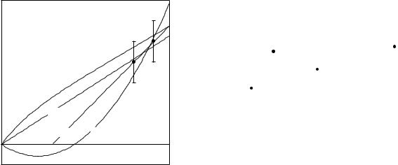
FIGURE 16.55. Possible responses to various doses. The two lowest-dose measurements are shown. With zero dose there is no excess e ect. The curves are discussed in the text.

Other Models

Figure 16.55 plots the excess e ect vs. dose, showing four possibilities for how some e ect might depend on dose. By definition, there is no excess e ect when the dose is zero. The two data points represent the lowest doses at which the e ect has been measured. The LNT line is a linear interpolation to zero from these points. Lines are also shown for three other possibilities: (1) a threshold below which there is no excess e ect, (2) a supralinear response, which is higher than predicted by the LNT model, and (3) hormesis. In hormesis there is a limited range in which the excess e ect is negative—a reduction in the e ect. Hormesis has been seen in the response of some organisms to chemicals and in some cases to radiation. Two issues of

Critical Reviews in Toxicology, Vol. 31 No. 4–5 and Vol. 33, No. 3–4, have been devoted to reviews of hormetic e ects in all fields.

Some investigators feel that there is evidence for a threshold dose, and that the LNT model overestimates the risk [Kathren (1996); Kondo (1993); Cohen (2002)]. Mossman (2001) argues against hormesis but agrees that the LNT model has led to “enormous problems in radiation protection practice” and unwarranted fears about radiation.

On the other hand, annual screening CTs [Brenner and Elliston (2004)] and CTs to children [Hall (2002)], lead to doses that are large enough so that there is a measured excess risk of developing cancer in an individual; no LNT extrapolation to lower doses is needed. This is shown in Fig. 16.56. A pediatric CT study can lead to a dose in the range of 5–100 mSv. This can be compared with data from the extensive study of 35,000 atomic bomb survivors who have been followed for 55 years. There is a


0.10


Dose range for pediatric CT


Risk

0.08


Relative

0.06


Excess

0.04


0.02


0.00


25

50

75

100

125

150


0

Dose (mSv)

FIGURE 16.56. Excess relative risk for atomic bomb survivors who were exposed to a dose of 150 mSv or less and followed for 55 years show a small, but statistically significant increase in cancer incidence. The range of doses from pediatric CT is also shown. Redrawn from Hall, E. J. Helical CT and cancer risk: Introduction to session I. Pediatr. Radiol. 32: 225–227 (2002). With kind permission of Springer Science and Business Media.

small but statistically significant excess risk of developing cancer.

16.13.4 Radon

The question of a hormetic e ect or a threshold e ect has received a great deal of attention for the case of radon, where remediation at fairly low radon levels has been proposed. Radon is produced naturally in many types of rock. It is a noble gas, but its radioactive decay products can become lodged in the lung. An excess of lung cancer has been well documented in uranium miners, who have been exposed to fairly high radon concentrations as well as high dust levels and tobacco smoke. Radon at lower concentrations seeps from the soil into buildings and contributes up to 55% of the exposure to the general population. Radon concentrations in the air are measured in the number of radioactive decays per second per cubic meter of air. One becquerel (Bq) is one decay per second.

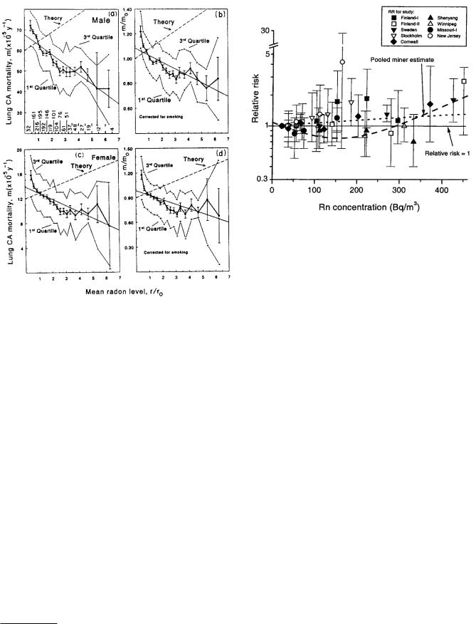
Figure 16.57 shows a study by B. L. Cohen (1995) that plots annual age-adjusted lung-cancer mortality rates in 1601 counties in the United States vs the average radon concentration measured in that county. The radon concentration is expressed as r/r0, where r0 is 37 Bq m3 (1.0 pCi l1 in old units, which will be discussed in the next chapter). The upper two panels are for males, and the lower two are for females. The two panels on the right are corrected for the e ects of smoking, using the radon-and- smoking model from BEIR Report V (1990). The dashed lines labeled Theory are based on the LNT model. The mortality rate falls with increasing radon concentration, though other studies have shown that it rises at radon concentrations higher than shown here.

Symbols Used

473

FIGURE 16.57. Lung cancer mortality rates vs mean radon level in 1601 U.S. counties. Graphs (a) and (b) are for males;

(c) and (d) are for females. Graphs (b) and (d) have been corrected for smoking levels. Error bars show the standard deviation of the mean. The meaning of radon level is discussed in the text. From B. L. Cohen. Test of the linear-no threshold theory of radiation carcinogenesis for inhaled radon decay products. Health Physics 68(2): 157–174, 1995. Used by permission of the Health Physics Society.

FIGURE 16.58. Relative risk (on a log scale) vs. radon concentration. The data points are for several studies, not including Cohen’s. The horizontal line shows a relative risk of 1. The dotted line is a linear extrapolation from the miner study. The dashed quadratic line is Cohen’s model. From Lubin, J. H. (1999). Response to Cohen’s comments on the Lubin rejoinder. Health Phys. 77(3): 330–332. Used by permission.

radon in Qu´ebec. They predicted a total of 109 deaths from lung cancer in a population of 60,000. Mitigating radon in all residences with concentrations of 200 Bq m3 or more would reduce this number from 109 to 105. The same number of lives would be saved by reducing smoking by 0.04%.

Epidemiological studies are di cult and can only be suggestive. A number of authors have criticized Cohen’s study for dealing with county-wide averages, and Cohen has defended his results.30 Cohen argues that his data are valid below about 150 Bq m3. Lubin (1999) compares an LNT fit and Cohen’s model to several other radon studies, shown in Fig. 16.58. The error bars are much larger than in Cohen’s figure because the populations are smaller. Lubin argues that this is irrelevant because Cohen has systematic errors. Cohen’s data points are not inconsistent with those shown by Lubin. Recall from Table 16.6 that the average annual dose from radon is about 2 mSv, and note where this lies on Fig. 16.56.

Even if the LNT model for radon is correct, some of our remediation e orts are misdirected. Ayotte et al. (1998) used the LNT model to assess the lung cancer risk from

30For example, see Lubin (1998); Cohen (1998); Lubin (1998b); Cohen (1999); Lubin (1999) and BEIR VI (1999).

Symbols Used in Chapter 16

Symbol

Use

Units

First


used on


page

b

Thickness of slice being

m

456


scanned


c

Velocity of light

m s1

443

e

Electron charge

C

440

f

Fraction of photons


451


that interact or


detective quantum


e ciency


f

General function to be


455


represented


g

Incremental signal

kg C1

442


transfer function


h

Planck’s constant

J s

438

j

Total angular


438


momentum quantum


number


474

16. Medical Use of X Rays


Symbol

Use

Units


First

Q

Quality factor


468


used on

R

Resistance

Ohm (Ω)

444


page

S

Area

m2

452

k

Minimum


452

S

Surviving fraction

J m1

462


signal-to-noise ratio


Se (or Sc)

Collision stopping

467

kB

Boltzmann factor

J K1


445


power


l

Orbital angular


438

T

Kinetic energy

J

438


momentum quantum


T0

Initial kinetic energy

J

439


number


T

Optical transmission


441

m

Mass

kg


440

T

Temperature

K

445

me

Mass of electron

kg


443

W

Mean energy expended

J or eV

439

m

Mean number


451


per ion pair formed


m

Number of scans


457

WR

Radiation weight factor


468

n

Principal quantum


438

WT

Tissue weighting factor

C kg1

469


number


X

Exposure

440

n

Number of slices in a


456

Z

Atomic number


437


scan


α

Integral of attenuation

Gy1

454

n

Number of moles of a

mol


439

α

Dose proportionality

460


substance


constant

Gy1


n

Number of fractions


462

α

Excess risk

471

p

Probability


463


proportionality


q

Charge

C


440


constant


r

Distance

m


447

β

Coe cient

Gy2

457

r

Risk or probability

varies


470

β

Squared dose

460

r, r0

Radon concentrations

Bq m3

472


proportionality


v

Voltage di erence

V


444


constant


w

Width of picture

m


456

γ

Fraction


457


element


γ

Film contrast

m1

441

wi

Mass fraction of ith


447

µ, µatten

Attenuation coe cient

447


constituent


µen

Energy absorption

m1

440

x

Photon


443


coe cient


energy/electron rest


ν, ν0

Frequency

Hz

438


mass energy


ρ

Density

kg m3

440

x, y, z

Coordinates

m


441

σ

Standard deviation

m2

471

A

Proportionality

C m2 kg1

451

Φ

Particle fluence

438


constant


ΦT

Particle fluence per

m2 J1 or

467

C

Constant

J1 m2

438


unit energy interval

m2 eV1


C

Capacitance

F


444

Ψ

Energy fluence

J m2

438

Cin

Exposure contrast


451


Cout

Brightness contrast


451

Problems


D

Absorbed dose

Gy (J kg1)

440


D

Absorbed dose in one

Gy


457

Section 16.1


scan


D0

Reciprocal of

Gy


460

Problem 1 Use Eqs. 15.3 and 16.2 to answer the follow-


proportionality


constant α


ing questions. Then compare your answers to values given

DR,T

Absorbed dose of

Gy


468

in tables, such as those in the Handbook of Chemistry and


radiation type R to


Physics. What is the minimum energy of electrons strik-


target organ T


ing a copper target that will cause the K x-ray lines to

E

Energy

J or eV

438

appear? What is the approximate energy of the Kα line?

E

E ective dose to an or-

Sv (J kg1)

468


gan


Repeat for iodine, molybdenum, and tungsten.


F

Projection (integral) of


455

Problem 2 When tungsten is used for the anode of an x-


f along some direction


G

Radiation chemical

mol J1

439

ray tube, the characteristic tungsten Kα line has a wave-


yield

kg C

1


length of 2.1 × 10¹¹ m. Yet a voltage of 69, 525 V must

G

Large signal transfer


442

be applied to the tube before the line appears. Explain the


factor


discrepancy in terms of an energy-level diagram for tung-

H

Hounsfield CT unit


456


sten.


H

Dose equivalent

Sv (J kg1)

468


HE

E ective dose

Sv


469

Problem 3 Henry Moseley first assigned atomic num-


equivalent


bers to elements by discovering that the square root of the

HT

Equivalent dose

Sv (J kg1)

468

Kc

Collision kerma

J kg1

440

frequency of the Kα photon is linearly related to Z. Solve

L

Length of object

m


452

Eq. 16.2 for Z and show that this is true. Plot Z vs the

N

Number


439

square root of the frequency and compare it to data you

OD

Optical density


441


look up.


P

Probability


462


Problem 4 Compare the shapes of the curves of Eqs. 16.3a and 16.3b for hν0 = 100 keV, over the range 40– 100 keV. Are the characteristics of Eq. 16.3b reflected in the theoretical points of Fig. 16.5?

Problem 5 Express the formula for the thick-target x- ray energy fluence rate [Eq. 16.3a] as dΨ/dλ and plot it.

Problem 6 Starting from Eq. 16.3b, written as dΦ/dE = (1/hν)dΨ/d(), use the chain rule to verify that dΨ/dλ = (h2c23)dΦ/dE.(Recall that λν = c.)

Section 16.2

Problem 7 A beam of 0.08-MeV photons passes through a body of thickness L. Assume that the body is all muscle

with ρ = 1.0×10³ kg m3. The energy fluence of the beam is Ψ J m2.

(a)What is the skin dose where the beam enters the

body?

(b)Assume the beam is attenuated by an amount e−µL as it passes through the body. Calculate the average dose as a function of the fluence, the body thickness, and µ.

(c)What is the limiting value of the average dose as

µL → 0?

(d)What is the limiting value of the average dose as µL → ∞? Does the result make sense? Is it useful?

Problem 8 The obsolete unit, the roentgen (R), is defined as 2.08 × 10⁹ ion pairs produced in 0.001 293 g of dry air. (This is 1 cm3 of dry air at standard temperature and pressure.) Show that if the average energy required to produce an ion pair in air is 33.7 eV (an old value), then 1 R corresponds to an absorbed dose of 8.69 × 10³ Gy and that 1 R is equivalent to 2.58 × 10⁴ C kg1.

Problem 9 During the 1930s and 1940s it was popular to have an x-ray fluoroscope unit in shoe stores to show children and their parents that shoes were properly fit. These marvellous units were operated by people who had no concept of radiation safety and aimed a beam of x rays upward through the feet and right at the reproductive organs of the children! A typical unit had an x-ray tube operating at 50 kVp with a current of 5 mA.

(a)What is the radiation yield for 50-keV electrons on tungsten? How much photon energy is produced with a 5-mA beam in a 30-s exposure?

(b)Assume that the x rays are radiated uniformly in all directions (this is not a good assumption) and that the x rays are all at an energy of 30 keV. (This is a very poor assumption.) Use the appropriate values for striated muscle to estimate the dose to the gonads if they are at a distance of 50 cm from the x-ray tube. Your answer will be an overestimate. Actual doses to the feet were typically 0.014–0.16 Gy. Doses to the gonads would be less because of 1/r2. Two of the early articles pointing out the danger are Hempelmann, L. H. (1949), Potential dangers in the uncontrolled use of shoe fitting fluoroscopes, New Engl. J.

Problems 475

Med. 241: 335–337, and Williams, C. R. (1949), Radiation exposures from the use of shoe-fitting fluoroscopes, New Engl. J. Med. 241: 333–335.

Section 16.3

Problem 10 Rewrite Eq. 16.9 in terms of exponential decay of the viewing light and relate the optical density to the attenuation coe cient and thickness of the emulsion.

Problem 11 Derive the useful rule of thumb ∆(OD) = 0.43γ X/X.

Problem 12 The atomic cross sections for the materials in a gadolinium oxysulfide screen for 50-keV photons are

Element

Cross section per atom (m2)

A

Gd

1.00 × 1025

157

S

3.11 × 1027

32

O

5.66 × 1028

16

(a)What is the cross section per target molecule of GdO2S?

(b)How many target molecules per unit area are there in a thickness ρdx of material?

(c)What is the probability that a photon interacts in traversing 1.2 kg m2 of GdO2S?

Problem 13 The film speed is often defined as the reciprocal of the exposure (in roentgens) required to give an optical density that is 1 greater than the base density. Assume that in Fig. 16.6 a relative exposure of 1 corresponds to 105 C kg1. Calculate the film speed.

Problem 14 A dose of 1.74 × 10⁴ Gy was estimated for part of the body just in front of an unscreened x-ray film. Suppose that a screen permits the dose to be reduced by a factor of 20. Calculate the skin dose on the other side of the body (the entrance skin dose) assuming 50-keV photons and a body thickness of 0.2 m. Ignore buildup, and assume that only unattenuated photons are detected.

Problem 15 Find an expression for photon fluence per unit absorbed dose in a beam of monoenergetic photons. Then find the photon fluence for 50-keV photons that causes a dose of 105 Gy in muscle.

Problem 16 A dose of 100 Gy might cause noticeable radiation damage in a sodium iodide crystal. How long would a beam of 100-keV photons have to continuously and uniformly strike a crystal of 1-cm2 area at the rate of 104 photon s1, in order to produce this absorbed dose? For NaI µen= 0.1158 m2 kg1.

Problem 17 Another method to measure the absorbed dose is by calorimetry. Show that if all the energy imparted warms the sample, the temperature rise is 2.39 × 10⁴ C per Gy.

476 16. Medical Use of X Rays

Section 16.4

Problem 18 Plot µ for lead, iodine, and barium from 10 to 200 keV.

Problem 19 Use a spreadsheet to make the following calculations. Consider a photon beam with 100 kVp.

(a)Use Eq. 16.3b to calculate the photon fluence from a thick target at 1, 10, 20, 30, 40, 50, 60, 70, 80, 90, and 100 keV.

(b)The specific gravity of aluminum is 2.7. Make a table of the photon fluence at these energies emerging from 2 and 3 mm of aluminum. Compare the features of this table to Fig. 16.16.

(c)Use trapezoidal integration to show that the average photon energy is 44 keV after 2-mm filtration and 47 keV after 3-mm filtration.

(d)Repeat for 120 kVp and show that the average energies after the same filtrations are 52 and 55 keV.

Problem 20 X-ray beams have a spectrum of photon energies. It would be very laborious to measure the spectrum every time we want to check the quality of the beam. In addition to kVp, one simple measurement that is used to check beam quality (related to the energy spectrum) is the “half-value layer” HVL–the thickness of a specified absorber (often Cu or Al) that reduces the intensity of the beam to one-half.

(a)For a monoenergetic beam, relate HVL to the at-

tenuation coe cient. What is the HVL if the attenuation coe cient is 0.46 mm1?

(b)For a monoenergetic beam, how does the quartervalue layer QVL relate to HVL?

(c)Suppose a beam has equal numbers of photons at two

di erent energies. The attenuation coe cients at these energies are 0.46 mm1 and 0.6 mm1. Find the HVL and QVL for this beam. You may need to plot a graph or use a computer algebra program.

Problem 21 The “half-value layer” (HVL) is often used to characterize an x-ray beam. It is the thickness of a specified absorber that attenuates the beam to one-half the original value. Figure 16.45 refers to a beam with a 3.0 mm Cu HVL. What is the value of the attenuation coefficient? What monoenergetic x-ray beam does this correspond to?

Problem 22 Assume an antiscatter grid is made of lead sheets 3 mm long with a spacing between sheets of 0.3 mm. Ignore the thickness of the sheets. If all photons hitting the sheets are absorbed, what is the largest angle from the incident beam direction that a photon can be scattered and still emerge?

Section 16.5

Problem 23 Suppose that two measurements are made: one of the combination of signal and noise, y = s + n,

and one of just the noise n. One wishes to determine s = y − n.

(a)Find s − s in terms of y, y, n, and n.

(b)Show that if y and n are “uncorrelated,” (s − s)2 = (y − y)2 + (n − n)2 and state the mathematical condition for being “uncorrelated.”

(c)If y and n are Poisson distributed, under what con-

ditions is the 2 factor of Footnote 13 needed?

Section 16.7

Problem 24 A molybdenum target is used in special x- ray tubes for mammography. The electron energy levels in Mo are as follows:

K 20, 000 eV LI

2886 eV

MI

505 eV

LII

2625 eV

MII

410 eV

LIII

2520 eV

MIII

392 eV


MIV

230 eV


MV

227 eV

What is the energy of the Kα line(s)? The Kβ line(s) (defined in Fig. 16.2)?

Problem 25 As a simple model for mammography, consider two di erent tissues: a mixture of 2/3 fat and 1/3 water, with a composition by weight of 12% hydrogen, 52% carbon and 36% oxygen; and glandular tissue, composed of 11% hydrogen, 33% carbon, and 56% oxygen. The density of the fat and water combination is 940 kg m3, and the density of glandular tissue is 1020 kg m3. What is the attenuation in 1 mm of fat and in 1 mm of glandular tissue for 50-keV photons? For 30-keV photons?

Section 16.9

Problem 26 For a first-generation CT scanner with

µ = 18 m1, µ/µ = 0.005, L = 30 cm, w = 0.1 cm, and b = 1 cm, calculate, for a scan time of 15 s,

(a)N ,

(b)m,

(c)N , and

(d)The dose.

Problem 27 It is often said that the number of photons that must be detected in order to measure a di erence in fluence with a certain resolution can be calculated from N = (∆Φ/Φ)2. (For example, if we want to detect a change in Φ of 1% we would need to count 104 photons.) Use Eq. 16.20 to make this statement more quantitative. Discuss the accuracy of the statement.

Problem 28 Spiral CT uses interpolation to calculate the projections at a fixed value of z before reconstruction. This has an e ect on the noise. Let σ0 be the noise standard deviation in the raw projection data and σ be the noise in the interpolated data. The interpolated signal, α, is the weighted sum of two values: α = 1 + (1 − w)α2.

(a) Show that the variance in α is σ2 = w2σ02 + (1 − w)2σ02. Plot σ/σ0 vs w.

(b) Averaging over a 360 scan involves integrating uniformly over all weights:

1

σ2 = w2σ02 + (1 − w)2σ02 dw.

0

Find the ratio σ/σ0.

Problem 29 An experimental technique to measure cerebral blood perfusion is to have the patient inhale xenon, a noble gas with Z = 54, A = 131 [Suess et al. (1995)]. The solubility of xenon is di erent in red cells than in plasma. The equation used is

5.15θXe

Arterial enhancement = (µ/ρ)w /(µ/ρ)Xe CXe(t),

where the arterial enhancement is in Hounsfield units, CXe is the concentration of xenon in the lungs (end tidal volume), and

θXe = (0.011)(Hct) + 0.10.

Hct is the hematocrit: the fraction of the blood volume occupied by red cells. Discuss why the equation has this form.

Section 16.10

Problem 30 Use Equations 16.32 and 16.33 to obtain expressions for dose vs number of tumor cells for a probability of cure of 50%.

Section 16.11

Problem 31 Geiger’s rule is an approximation to the range-energy relationship:

R = AEp.

For protons in water A = 0.0022 when R is in cm and E is in MeV. The exponent p = 1.77. This is a good approximation for E < 200 MeV. Use Geiger’s approximation to find dE/dx as a function of R for 100 MeV protons. Make a plot to show the Bragg peak when straggling is ignored.

Section 16.12

Problem 32 Calculate (Se)wg in argon for 0.1-, 1.0- and 10-MeV electrons. The values of Se/ρ for argon at these energies are 2.918, 1.376, and 1.678 cm2 g1.

Problem 33 An ion chamber contains 10 cm3 of air at standard temperature and pressure. Find q vs D for 0.5- MeV electrons.

References 477

Section 16.13

Problem 34 Suppose that the probability p per year of some event (death, mutations, cancer, etc.) consists of a spontaneous component S and a component proportional to the dose of something else, D: p = S + AD. The dose may be radiation, chemicals, sunlight, etc. Investigations of women given mammograms showed that if p is the probability of acquiring breast cancer, S = 1.91 × 10³ and A = 4 × 10⁴ Gy1. How many women had to be studied to distinguish between A = 0 and the value above if D = 2 Gy? If D = 102 Gy?

References

AAPM Report 87 (2005). Diode in Vivo Dosimetry for Patients Receiving External Beam Radiation Therapy.

College Park, MD. American Association of Physicists in Medicine. Report of Task Group 62 of the Radiation Therapy Committee.

Attix, F. H. (1986). Introduction to Radiological Physics and Radiation Dosimetry. New York, Wiley.

Ayotte, P., B. L´evesque, D. Gauvin, R. G. McGregor, R. Martel, S. Gingras, W. B. Walker, and E. G. L´etourneau (1998). Indoor exposure to 222Rn: A public health perspective. Health Phys. 75(3): 297–302.

Barth, R. F. (2003). A critical assessment of boron neutron capture therapy: an overview. J. Neuro-Oncol. 62: 1–5. The entire issue of the journal is devoted to a review of BNCT.

BEIR Report V (1990). Health E ects of Exposure to Low Levels of Ionizing Radiation. Washington, DC, National Academy Press. Committee on the Biological Effects of Ionizing Radiation.

BEIR VI (1999). Committee on Health Risks of Exposure to Radon. Health E ects of Exposure to Radon.

Washington, D.C., National Academy Press.

BEIR Report VII (2005). Health Risks from Exposure to Low Levels of Ionizing Radiation. Washington, DC, National Academy Press. Committee to Assess Health Risks from Exposure to Low Levels of Ionizing Radiation.

Birch, R., and M. Marshall (1979). Computation of bremsstrahlung x-ray spectra and comparison with spectra measured with a Ge(Li) detector. Phys. Med. Biol. 24: 505–517.

Boice, J. D., Jr. (1996). Risk estimates for radiation exposures. In W. R. Hendee and F. M. Edwards, eds.

Health E ects of Exposure to Low-Level Ionizing Radiation. Bristol, Institute of Physics.

Brenner, D. J., and C. D. Elliston (2004). Estimated radiation risks potentially associated with full-body CT screening. Radiology 232: 735–738.

Broad, W. J. (1980). Riddle of the Nobel debate. Science 207: 37–38.

478 16. Medical Use of X Rays

Brooks, A. L. (2003). Developing a scientific basis for radiation risk estimates: Goal of the DOE low dose research program. Health Phys. 85(1): 85–93.

Brooks, R. A., and G. DiChiro (1976). Principles of computer-assisted tomography in radiographic and radioisotope imaging. Phys. Med. Biol. 21: 689–732; Statistical limitations in x-ray reconstructive tomography.

Med. Phys. 3: 237–240.

Cho, Z.-H., J. P. Jones, and M. Singh (1993). Foundations of Medical Imaging. New York, Wiley-Interscience.

Cohen, B. L. (1995). Test of the linear–no threshold theory of radiation carcinogenesis for inhaled radon decay products. Health Phys. 68(2): 157–174.

Cohen, B. L. (1998). Response to Lubin’s proposed explanations of the discrepancy. Health Phys. 75(1): 18–22.

Cohen, B. L. (1999). Response to the Lubin rejoinder. Health Phys. 76(4): 437–439.

Cohen, B. L. (2002). Cancer risk from low-level radiation.[see comment]. AJR. Amer. J. Roentgenology.

179(5):1137–43.

Cohen, L. (1993). History and future of empirical and cell kinetic models for risk assessment in radiation oncology. In B. Paliwal, et al., eds. Prediction of Response in Radiation Therapy: Radiosensitivity and Repopulation.

Woodbury, NY, AIP for the American Association of Physicists in Medicine.

Cormack, A. M. (1980). Nobel award address: Early two-dimensional reconstruction and recent topics stemming from it. Med. Phys. 7(4): 277–282.

Dainty, J. C., and R. Shaw (1974). Image Science. New York, Academic.

DeVita, V. T. (2003). Hodgkin’s disease—Clinical trials and travails. New Engl. J. Med. 348 (24): 2375–2376.

DiChiro, G., and R. A. Brooks (1979). The 1979 Nobel prize in physiology or medicine. Science. 206: 1060–1062.

Doi, K., H. K. Genant, and K. Rossman (1976). Comparison of image quality obtained with optical and radiographic magnification techniques in fine-detail skeletal radiography: E ect of object thickness. Radiology. 118: 189–195.

Douglas, J. G., W. J. Koh, M. Austin-Seymour, and G. E. Laramore (2003). Treatment of salivary gland neoplasms with fast neutron radiotherapy. Arch. Otolaryngol.–Head & Neck Surg. 129(9): 944–948.

Duncan, W. (1994). An evaluation of the results of neutron therapy trials. Acta Oncolog. 33(3): 299–306. This issue of the journal is devoted to fast-neutron therapy.

Evans, R. D. (1955). The Atomic Nucleus. New York, McGraw-Hill.

Goodsitt, M., E. Christodoulou, P. Strouse, A. Chien, J. Platt and E. Kazerooni (2002). Radiation doses for corresponding CT and radiographic/fluoroscopic exams. Med. Phys. 29(6): 1298. (Meeting abstract)

Habrand, J. L., P. Schlienger, L. Schwartz, D. Pontvert, C. Lenir-Cohen-Solal, S. Helfre, C. Haie, A. Mazal, and J. M. Cosset (1995). Clinical applications of proton therapy.

Experiences and ongoing studies. Radiat. Environment. Biophys. 34(1): 41–44.

Hall, E. J. (2000). Radiobiology for the Radiologist. 5th ed. Philadelphia, Lippincott Williams & Wilkins.

Hall, E. J. (2002). Helical CT and cancer risk: Introduction to session I. Pediatr. Radiol. 32: 225–227.

Hall, E. J. (2003). The bystander e ect. Health Phys. 85(1): 31–35.

Hendee, W. R., and R. Ritenour (2002). Medical Imaging Physics, 4th. ed. New York, Wiley-Liss.

Higson, D. J. (2004). The bell tolls for LNT. Health Phys. 87(Supplement 2): S47–S50.

Holmes, T. W., T. R. Mackie, and P. Reckwerdt (1995). An iterative filtered backprojection inverse treatment planning algorithm for tomotherapy. Int. J. Radiation Oncolog. Biol. Phys. 32(4): 1215–1225.

Hounsfield, G. N. (1980). Nobel award address: Com-

puted medical imaging. Med. Phys. 7(4): 283–290.


Hubbell, J. H., and S. M. Seltzer (1996).

Ta-

bles of X-Ray Mass Attenuation Coe cients

and

Mass Energy-Absorption Coe cients 1 keV to

20

MeV for Elements Z=1 to 92 and 48 Additional Substances of Dosimetric Interest. National Institute of Standards and Technology Report NISTIR 5632. physics.nist.gov/PhysRefData/XrayMassCoef/cover.html Huda, W. (2002). Adult and pediatric dose in CT. Med.

Phys. 29(6): 1363. Meeting abstract.

Hunt, D. C., S. S. Kirby, and J. A. Rowlands (2002). X-ray imaging with amorphous selenium: X-ray to charge conversion gain and avalanche multiplication gain. Med. Phys. 29(11): 2464–2471.

ICRP (1991). 1990 Recommendations of the International Commission on Radiological Protection. Annals of the ICRP. 21 no. 1–3.

ICRU Report 31 (1979). Average Energy to Produce an Ion Pair. Bethesda, MD, International Commission on Radiation Units and Measurements.

ICRU Report 33 (1980, Reprinted 1992). Radiation Quantities and Units. Bethesda, MD, International Commission on Radiation Units and Measurements.

ICRU Report 37 (1984). Stopping Powers for Electrons and Positrons. Bethesda, MD, International Commission on Radiation Units and Measurements.

ICRU Report 39 (1985). Determination of Dose Equivalents Resulting from External Radiation Sources.

Bethesda, MD, International Commission on Radiation Units and Measurements.

ICRU Report 41 (1986). Modulation Transfer Function of Screen-Film Systems. Bethesda, MD, International Commission on Radiation Units and Measurements.

ICRU Report 54 (1996). Medical Imaging—The Assessment of Image Quality. Bethesda, MD, International Commission on Radiation Units and Measurements.

Kalender, W. A. (2000). Computed Tomography: Fundamentals, System Technology, Image Quality and Applications. Munich, Publicis MCD.

Kassis, A. I. (2004).The amazing world of Auger electrons. Int. J. Rad. Biol., 80(11-12): 789–803.

Kathren, R. L. (1996). Pathway to a paradigm: The linear nonthreshold dose-response model in historical context: The American Academy of Health Physics 1995 Radiology Centennial Hartman Oration. Health Phys. 70(5): 621–635.

Khan, F. M. (2003). The Physics of Radiation Therapy.

3rd ed. Philadelphia, Lippincott Williams & Wilkins. Kondo, S. (1993). Health E ects of Low-Level Radia-

tion. Osaka, Japan, Kinki University Press. English translation: Madison, WI, Medical Physics.

Lubin, J. H. (1998). On the discrepancy between epidemiologic studies in individuals of lung cancer and residential radon and Cohen’s ecologic regression. Health. Phys. 75(1): 4–10.

Lubin, J. H. (1998b). Rejoinder: Cohen’s response to “On the discrepancy between epidemiologic studies in individuals of lung cancer and residential radon and Cohen’s ecologic regression.” Health Phys. 85(1): 29–30.

Lubin, J. H. (1999). Response to Cohen’s comments on the Lubin rejoinder. Health Phys. 77(3): 330–332.

Lutz, G. (1999). Semiconductor Radiation Detectors.

New York, Springer.

Macovski, A. (1983). Medical Imaging Systems. Englewood Cli s, NJ, Prentice-Hall.

McCollough, C. H. and B. A. Schueler (2000). Educational Treatise: Calculation of e ective dose. Med. Phys. 27(5): 828–837.

Metz, C. E., and K. Doi (1979). Transfer function analysis of radiographic imaging systems. Phys. Med. Biol. 24(6): 1079–1106.

Miralbell, R., A. Lomax, L. Cella, and U. Schneider (2002). Potential reduction of the incidence of radiationinduced second cancers by using proton beams in the treatment of pediatric tumors. Int. J. Radiation Oncology Biol. Phys. 54(3): 824–829.

Mossman, K. L. (2001). Deconstructing radiation hormesis. Health Phys. 80(3): 263–269.

Moulder, J. E., and J. D. Shadley (1996). Radiation interactions at the cellular and tissue levels. In W. R. Hendee and F. M. Edwards, eds. Health E ects of Exposure to Low-Level Ionizing Radiation. Bristol, Institute of Physics.

Moyers, M. (2003). Proton therapy refresher course, Part II. Practical challenges and opportunities for proton therapy. Med. Phys. 30(6): 1445–1446.

Nagel, H. D., M. Galanski, N. Hidajat, W. Maer and T. Schmidt (2000). Radiation Exposure in Computed Tomography: Fundamentals, Influencing Parameters, Dose Assessment, Optimisation, Scanner Data, Terminology.

COCIR, European Coordination Committee of the Radiological and Electromedical Industries. Hamburg, CTB Publications (ctb-publications@gmx.de)

NCRP Report 93 (1987). Ionizing Radiation Exposure of the Population of the United States. Bethesda, MD,

References 479

National Council on Radiation Protection and Measurements.

NCRP Report 94 (1987). Exposure of the Population in the United States and Canada from Natural Background Radiation. Bethesda, MD, National Council of Radiation Protection and Measurements.

NCRP Report 100 (1989). Exposure of the U.S. Population from Diagnostic Medical Radiation. Bethesda, MD, National Council of Radiation Protection and Measurements.

NCRP Report 136 (2001). Evaluation of the LinearNonthreshold Dose-Response Model for Ionizing Radiation. Bethesda, MD, National Council of Radiation Protection and Measurements.

Orton, C. (1997). Fractionation: Radiobiological principles and clinical practice. Chapter 11 in F. M. Khan and R. A. Potish, eds. Treatment Planning in Radiation Oncology. Baltimore, MD, Williams and Wilkins.

Platzman, R. L. (1961). Total ionization in gases by high-energy particles: An appraisal of our understanding.

Intl. J. Appl. Rad. Isotopes 10: 116–127.

Rowlands, J. A. (2002). The physics of computed radiography. Phys. Med. Biol. 47: R123–R126.

Schlesinger, T. E. and R. B. James, eds. (1995). Semiconductors for Room Temperature Nuclear Applications.

New York, Academic Press.

Schulz, R. J., and A. R. Kagan (2002). On the role of intensity-modulated radiation therapy in radiation oncology. Med. Phys. 29(7): 1473–1482.

Seibert, J. A. (2003). Digital fluoroscopic imaging: Acquisition, processing and display. Med. Phys. 30(6): 1413.

Shabashon, L. (1996). Radiation interactions: Physical and chemical e ects. In W. R. Hendee and F. M. Edwards, eds. Health E ects of Exposure to Low-Level Ionizing Radiation. Bristol, Institute of Physics.

Shani, G. (1991). Radiation Dosimetry: Instrumentation and Methods. Boca Raton, FL, CRC Press.

Shani, G. (2001). Radiation Dosimetry: Instrumentation and Methods. 2nd. ed. Boca Raton, FL, CRC Press.

Slater, J. D., C. J. Rossi, Jr., L. T. Yonemoto, D. A. Bush, B. R. Jabola, R. P. Levy, R. I. Grove, W. Preston, and J. M. Slater (2004). Proton therapy for prostate cancer: The initial Loma Linda University experience. Int. J. Radiat. Oncol. Biol. Phys. 59(2): 348–352.

Sobol, W. T. (2002). High frequency x-ray generator basics. Med. Phys. 29(2): 132–144.

Steel, G. G. (1996). From targets to genes: a brief history of radiosensitivity. Phys. Med. Biol. 41(2): 205–222.

Suess, C., A. Polacin, and W. A. Kalender (1995). Theory of xenon/computed tomography cerebral blood flow methodology. In M. Tomanaga, A. Tanaka, and H. Yonas, eds. Quantitative Cerebral Blood Flow Measurements Using Stable Xenon/CT: Clinical Applications. Armonk, NY, Futura.

Suit, H., and M. Urie (1992). Proton beams in radiation therapy. J. Natl. Cancer Inst. 84(3): 155–164.

480 16. Medical Use of X Rays

Upton, A. C. (2003). The state of the art in the 1990’s: NCRP Report No. 136 on the scientific bases for linearity in the dose-response relationship for ionizing radiation.

Health Phys. 85(1): 15–22.

van Eijk, C. W. E. (2002). Inorganic scintillators in medical imaging. Phys. Med. Biol. 47: R85–R106

Wagner, R. F. (1977). Toward a unified view of radiological imaging systems. Part II: Noisy images. Med. Phys. 4(4): 279–296.

Wagner, R. F. (1983). Low-contrast sensitivity of radiologic, CT, nuclear medicine, and ultrasound medical

imaging systems. IEEE Trans. Med. Imaging MI-12(3): 105–121.

Wagner, R. F., K. E. Weaver, E. W. Denny, and R. G. Bostrom (1974). Toward a unified view of radiological imaging systems. Part I: Noiseless images. Med. Phys. 1(1): 11–24.

Webb, S. (2001). Intensity-Modulated Radiation Therapy. Bristol and Philadelphia. Institute of Physics Publishing.

Winsor, R. (2003). CCD digital detectors. Med. Phys. 30(6): 1433. Meeting abstract.

17

Nuclear Physics and Nuclear Medicine

Each atom contains a nucleus about 100,000 times smaller than the atom. The nuclear charge determines the number of electrons in the neutral atom and hence its chemical properties. The nuclear mass determines the mass of the atom. For a given nuclear charge there can be a number of nuclei with di erent masses or isotopes. If an isotope is unstable, it transforms into another nucleus through radioactive decay.

In this chapter we will consider some of the properties of radioactive nuclei and their use for medical imaging and for treatment, primarily of cancer.

Four kinds of radioactivity measurements have proven useful in medicine. The first involves no administration of a radioactive substance to the patient. Rather, a sample from the patient (usually blood) is mixed with a radioactive substance in the laboratory, and the resulting chemical compounds are separated and counted. This is the basis of various competitive binding assays, such as those for measuring thyroid hormone and the availability of iron-binding sites. The most common competitive binding technique is called radioimmunoassay. A wide range of proteins are measured in this manner.

In the second kind of measurement, radioactive tracers are administered to the patient in a way that allows the volume of a compartment within the body to be measured. Examples of such compartments are total body water, plasma volume, and exchangeable sodium. Timedependent measurements include red-blood-cell survival and iron and calcium kinetics. Counting is of the whole body or of blood or urine samples drawn at di erent times after administration of the isotope.

For the third class of measurements, a gamma camera generates an image of an organ from radioactive decay of a drug that has been administered and taken up by the organ. These images are often made as a function of time.

The fourth class is an extension of these in which tomographic reconstructions of body slices are made. These include single-photon emission computed tomography and positron emission tomography.

Radioactive isotopes are also used for therapy. The patient is given a radiopharmaceutical that is selectively absorbed by a particular organ (for example, radioactive iodine for certain thyroid diseases). The isotope emits charged particles that lose their energy within a short distance, thereby giving a high dose to the target organ. Isotopes are also used in self-contained implants for brachytherapy.

The first four sections introduce some of the nuclear properties that are important: size, mass, and the modes of radioactive decay and the amount of energy released.

It is important to know the dose to the patient from a nuclear medicine procedure, and a standard technique for calculating it has been developed by the Medical Internal Radiation Dose (MIRD) Committee of the Society of Nuclear Medicine. Sections 17.5–17.8 show the steps in making these calculations. Section 17.9 describes some of the pharmacological considerations in selecting a suitable isotope, and Sec. 17.10 provides a sample dose calculation using this technique. Section 17.11 extends the dose discussion to the special considerations when an isotope emits Auger electrons and is bound to the DNA in the cell nucleus.

The next few sections describe various ways of forming images. Section 17.12 describes the gamma camera, and Sec. 17.13 extends this to single-photon emission tomography. Section 17.14 describes positron emission tomography, which can be done with special equipment when positron sources are available in the hospital.

Radiotherapy is described in Sec. 17.15, including both the relatively common brachytherapy and the less common injection of isotopes that target particular organs.