Добавил:
Опубликованный материал нарушает ваши авторские права? Сообщите нам.
Вуз: Предмет: Файл:

Intermediate Physics for Medicine and Biology - Russell K. Hobbie & Bradley J. Roth

.pdf
Скачиваний:
93
Добавлен:
10.08.2013
Размер:
20.85 Mб
Скачать

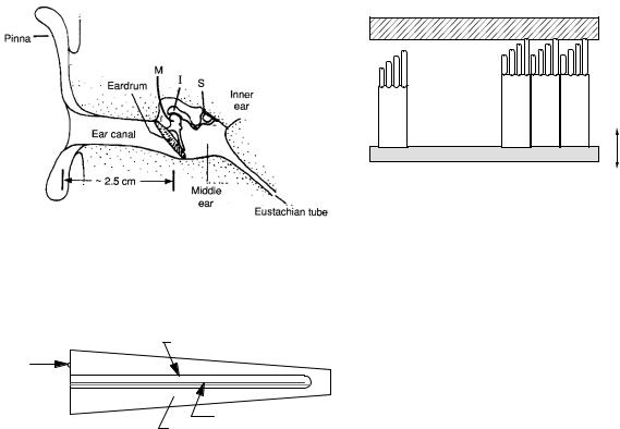
FIGURE 13.8. Details of the middle ear. From J. R. Cameron, J. G. Skofronick, and R. M. Grant. Physics of the Body. Madison, WI. Medical Physics Publishing. 1999. Used by permission.

Cochlear duct

Stapes and

Oval Vestibular Chamber

Window

Round Tympanic Chamber

Window

Tectorial Membrane

Basilar Membrane

FIGURE 13.9. A schematic representation of the cochlea.

The middle ear transforms the impedance by two mechanisms. The first is a simple area change. The ear drum vibrates in response to the pressure changes in the sound wave. If a sound wave with pressure amplitude pair impinges on the ear drum of area Sear drum, the total excess force on the ear drum is F = pairSear drum. For this simplest model, assume that the three bones behave like a single rigid rod and there are no e ects of the boundary at the circumference of the ear drum. Then the bones transmit this force to the membrane at the oval window, which has area Soval window. The pressure induced in the liquid in the inner ear is then pinner ear = F/Soval window = pairSear drum/Soval window. The area of the ear drum is about Sear drum = 64 mm2, while the area of the area of the base of the stapes is 3.2 mm2 [Newman (1957)]. Therefore pinner ear = 20pair. Actually, the ear drum and the membrane at the oval window are not connected by a simple rigid rod. The malleus, incus, and stapes are pivoted in such a way that they serve as a set of levers multiplying the force at the oval window by an additional factor of 1.3. Therefore the total pressure amplification by the middle ear is 26. This corresponds to a 28 dB change in sound intensity, which almost compensates for the 30 dB loss going from air to the liquid of the inner ear. The bones of the middle ear have muscles that change their sti ness, so they can reduce the amount of pressure amplification to protect the inner ear from very loud, low-frequency noises.

13.6 Attenuation

349

Tectorial Membrane

Cilia on Ends of Hair Cells

Hair Cells

Basilar Membrane

Movement

Basilar Membrane

FIGURE 13.10. A cross section of the cochlea. The hair cells are deformed as the basilar membrane moves.

The inner ear contains three semicircular canals, which help control our sense of balance, and the cochlea, which changes the sound to nerve impulses. All are filled with liquid. The cochlea is a small spiral about the size of the tip of your little finger. If it is unwound, it is about 3 cm long. Figure 13.9 shows it schematically. There are three chambers. The vestibular chamber connects to the stapes in the middle ear through the oval window. At the other end of the cochlea the vestibular chamber connects to the tympanic chamber. The round window opens onto the middle ear and allows the pressure to be equalized at low frequencies. The third chamber is the cochlear duct.

When the stapes moves the oval window, it generates a sound wave that travels through the liquid in the cochlea. This produces a displacement of the basilar membrane in the third chamber, the cochlear duct. Two types of hair cells sit on the basilar membrane: one row of inner hair cells and three rows of outer hair cells. The hair cells in turn have very fine “hairs” on them, called cilia. The cilia of the outer hair cells touch another membrane, the tectorial membrane, but the cilia of the inner hair cells do not. A cross section of this is shown schematically in Figure 13.10. When the basilar and tectorial membranes are displaced by the sound wave, the cilia on the inner hair cells move in the liquid that fills the region between the membranes. It is just as if you submerged your head in a swimming pool and shook your head back and forth. Your hair would move in the water, but the motion of your hair would be altered as the water “dragged” it. As a result of this motion of the cilia in the liquid, the inner hair cells generate nerve impulses that then travel to the brain and provide our sensation of sound. The mechanism was discussed briefly in Sec. 9.9

13.6 Attenuation

A plane wave of sound propagating through a medium is attenuated : there is a decrease in intensity because of dissipative factors such as viscosity and heat conduction, which we did not include in Sec. 13.1. The attenuation

13.7.1 Ultrasound Transducers
Ultrasound is typically produced using a piezoelectric transducer. A piezoelectric material converts a stress (or pressure) into an electric field, and vice versa. A highfrequency oscillating voltage applied across a piezoelectric material creates a sound wave at the same frequency.

350 13. Sound and Ultrasound

is exponential. The amplitude attenuation coe cient5 is


105


defined by


Lung


1 dp


(13.35)


α = p dx ,

-1)

104


where x is the distance the wave travels in the medium.

m


Skull


(dB

10

3


Muscle across fibers


The sound pressure amplitude decays exponentially:


coefficient


p(x) = p(0)e−αx.

(13.36)


Muscle along fibers


102


Liver


Since the intensity is proportional to p2,


Attenuation


Blood


101


I(x) = I(0)e2αx.

(13.37)


The intensity attenuation coe cient is µ = 2α. In

100


acoustics, the attenuation is usually expressed in deci-


Water


bels per meter, which is then independent of whether µ


10-1


or α is used.6


6

2

3

4

5

6 7

8 9


7


10


The wave equation for acoustics is an approximation,


10


Frequency (Hz)


because the basic equations of fluid dynamics are non-


linear. Therefore e ects that we have ignored, such as

FIGURE 13.11. Representative values of the attenuation co-

waveform distortion, the generation of harmonics, and in-

e cient for ultrasound.


creased attenuation may occur, particularly at high sound


intensities.


13.7 Medical Uses of Ultrasound


In air, the attenuation depends on the frequency of the


sound and the temperature and humidity of the air [Lind-

Ultrasound has several uses in medicine. The most com-

say and Beyer (1989); Denny (1993)]. Sound that we can

mon is to provide diagnostic images that complement

hear (in the frequency range of 20 Hz to 20 kHz) is atten-

those made with x-rays, nuclear medicine, and magnetic

uated by about 0.1–10 dB km1. Water transmits sound

resonance. Another is to heat tissue (diathermy). It is

better than air, but its attenuation is an even stronger

also used to

break up

gall stones

and

kidney

stones

function of frequency. It also depends on the salt con-

(lithotripsy), and, experimentally, to destroy tissue by in-

tent. At 1, 000 Hz, sound attenuates in fresh water by

tense heating.


about 4 ×10⁴ dB km1. The attenuation in sea water is


The

highest-frequency sounds


that

we

can

hear

about a factor of ten higher [Lindsay and Beyer (1989)].

(15 kHz) have a wavelength in water of 0.1 m. One

The low attenuation of sound in water (especially at low

frequencies) allows aquatic animals to communicate over

property of waves is that di raction limits our ability to

produce an image. Only objects larger than or approxi-

large distances [Denny (1993)].


mately equal to the wavelength can be imaged e ectively.

The attenuation of sound depends strongly

on fre-

This property is what limits light microscopes (using elec-

quency. Figure 13.11 shows some representative values.

tromagnetic waves to form an image) to resolutions equal

As a rule of thumb, at ultrasonic frequencies the atten-

to about the wavelength of visible light, 500 nm. If we

uation is proportional to frequency, with the constant

used audible sound to form images, our resolution would

of proportionality being 100 dB m1 MHz1. There are

be limited to about 0.07 m, which would be a poor im-

large variations in attenuation in tissue, depending on the

age indeed. To overcome this di culty, higher frequencies

age of the subject and other factors. Values can be found

(ultrasound) are used to form images. Typically, diag-

in Appendix A of ICRU 61 (1998).


nostic ultrasound uses frequencies on the order of 1 to

There can also be scattering of the sound from some

15 MHz, corresponding to wavelengths of 1.4 to 0.1 mm

object, just as there is for light. The total scattering cross

in tissue. Higher frequencies would result in even shorter

section for the object is defined by


wavelengths, but higher frequency sound has increased


Ws


attenuation, which ultimately sets an upper bound to the

σs = I0 ,

(13.38)

useful frequency.


where Ws is the total power scattered and I0 is the incident intensity. As in Chapter 14, the di erential scattering cross section can also be defined.

5ICRU 61 (1998).

6Sometimes the attenuation coe cient is expressed in nepers m1, in which case the natural logarithm of the intensity or pressure ratio is used.

13.7 Medical Uses of Ultrasound

351

r'

a

(r'2 + z2 )1/2

z

FIGURE 13.12. Coordinate system for calculating the intensity of sound radiated from a transducer of radius a. The z axis passes through the center of the transducer and is perpendicular to it.

Conversely, an oscillating pressure applied to a piezoelectric material creates an oscillating voltage across it. Measurement of this voltage provides a way to record ultrasonic waves. Thus, the same piezoelectric material can serve as both source and detector. One piezoelectric material often used in medical transducers is lead zirconate titanate (PZT). Its density is 7.5 ×10³ kg m3, the speed of sound in the material is 4,065 m s1, and the acoustic impedance is 30×106 Pa s m1. About half of the electrical energy is converted to sound energy, and vice versa.

There are some important features of the radiation pattern from a transducer which we review next. Consider a circular transducer, the surface of which is oscillating back and forth in a fluid. Both faces set up disturbances in the fluid; however, we consider the radiation from only one face, since the transducer is placed in a holder that prevents radiation from the rear surface. We can easily calculate the intensity along the z axis, which we set up perpendicular to the piston and passing through its center, as shown in Fig. 13.12.

The displacement of the face of the transducer, ξ, is the same as the displacement of the fluid in contact with it. The entire face of the piston, and therefore the fluid immediately in front of it, vibrates with a fluid velocity dξ/dt = v0 cos ωt.7 Each small element of the vibrating fluid creates a wave that travels radially outward, the points of constant phase being expanding hemispheres. The amplitude of each spherical wave decreases as 1/r, the intensity falling as 1/r2. We want the pressure at a point z on the axis of the transducer. It is obtained by summing up the e ect of all the spherical waves emanating from the face of the transducer. At time t the phase of the wave is the same as the phase of the wave leaving the annular ring r dr at the earlier time t − r/c:

p


(z, t)


a

2πr dr

cos[ω(t − r/c)]

dt

r


0


Near field

Far field


units


1/r2


on axis, arbitrary


Intensity


Frequency


= 2 MHz


Wavelength


= 0.077 cm


Piston radius

= 0.5 cm


a/λ


= 6.5


0.00

0.02

0.04


0.06

0.08

0.10


(a) (b)


(c)


Distance along axis, m

FIGURE 13.13. The sound intensity on the axis of a circular transducer. The sound frequency is 2 MHz, and the transducer radius is 0.5 cm. Both nearand far-field regions are shown. Labels (a), (b), and (c) show the positions of the transverse radial scans in Fig. 13.14.

FIGURE 13.14. Scans across the beam from the transducer shown in Fig. 13.13. (a) In the near field at an on-axis maximum 0.0102 4 m from the transducer. (b) In the near field at an on-axis minimum 0.0157 5 m from the transducer. (c) In the far field 0.060 m from the transducer.

This is easily evaluated by changing variables. Since r2 = r 2 + z2, 2rdr = 2r dr :


2


2


cos[ω(t − r/c)]


p


2π r= a

+z


rdr


r


r=z


2π


1


=


sin[ω(t −


#a2 + z2)] sin[ω(t − z/c)] .


k

c

To find the average intensity we square and average over one period. The result is

2

ω


#


2


2

I sin


z −

a


+ z


(13.39)

2c


The result is plotted in Fig. 13.13 for a fairly typical but

7We use dξ/dt because it is in phase with the excess pressure. small transducer (a = 0.5 cm, f = 2 MHz).

352 13. Sound and Ultrasound


ka = 10


ka = 20


Relative Intensity

ka = 40


-20

-10

0

10

20


Angle from central axis (degrees)


FIGURE 13.15. The far-field intensity as a function of angle, calculated from Eq. 13.40. The value ka = 10 corresponds to 1 MHz and transducer radius a = 0.25 cm. The value ka = 20 corresponds to f = 2 MHz and a = 0.25 cm or f = 1 MHz and a = 0.5 cm. Value ka = 40 corresponds to 4 MHz and a = 0.25 cm or f = 2 MHz and a = 0.5 cm, the case examined in Fig. 13.13.

There are several important features of Fig. 13.13. Close to the transducer there are large oscillations in intensity along the axis; there are corresponding oscillations perpendicular to the axis, as shown in Fig. 13.14. The maxima and minima form circular rings. This is called the near field or Fresnel zone. Further away the intensity falls as 1/r2, in the far field or Fraunho er zone. The depth of the Fresnel zone is approximately a2. For the example shown (2 MHz, transducer diameter 1 cm), the depth is about 3 cm; for a larger transducer or higherfrequency ultrasound, it would be greater.

In the far field, approximations can be made to simplify the calculation. The intensity is then given by

1

J1(ka sin θ)

2

(13.40)

I


.

r2

ka sin θ

Function J1(x) is the Bessel function of order 1. It is found in math tables and is available in many spreadsheets. The angular dependence of the far-field intensity is plotted in Fig. 13.15.

By shaping the face of the transducer, it is possible to bring the beam to a focus at some particular depth. This improves the spatial resolution and increases the strength of the returning echo. Ultrasound imaging may be done in the near field, the far field, or the transition region. Modern transducers typically consist of an array of transducers which may lie on a straight or curved line. They can be driven in such a way as to produce waves that come to a focus, or that travel in an o -axis direction [see Hendee and Ritenour (2002) or Fig. 13.17].

FIGURE 13.16. An A scan of the eye. From ICRU 61, p. 2. Used by permission.

The impedance of a typical transducer is about 30×106 Pa s m1, so it is necessary to have an impedancematching material between the transducer and the patient’s skin. See Problem 16.

13.7.2 Pulse Echo Techniques

Most ultrasonic imaging is based on a pulse-echo technique. A short pulse (typically 0.5 µs in duration with a central frequency of about 5 MHz) is applied to the tissue by a piezoelectric transducer. The pulse travels with a speed of about c = 1540 m s1 (or 1.54 mm µs1). Whenever it approaches a boundary between two tissues having di erent acoustic impedances, part of the incident pulse is reflected as an echo, which can be detected by the same piezoelectric transducer. The longer the time ∆t between the generation and detection of the pulse, the farther away the reflecting boundary. In general, the distance from the source to the boundary is ∆x = ct/2, where c is the speed of sound in the tissue. Multiple boundaries produce multiple echoes, with each echo corresponding to a di erent distance from the source to boundary. A plot of echo intensity vs. time is called an A scan. An A scan of the eye is shown in Fig. 13.16. Because the attenuation is high, it is customary to increase the gain of the receiving amplifier as the echo time increases.

To form a two-dimensional image, it is necessary to scan in many di erent directions. In a B scan the brightness of the screen corresponds to the intensity of the echo, plotted vs. position in the body in the plane of the scan. The B-scan transducer sends a narrow beam into the body. The direction of the beam is rapidly changed to cover a fan-shaped region. This can be done with an oscillating or rotating transducer head (often containing three transducers), with an array of transducers that are

(a)

(b))

(c)

FIGURE 13.17. This shows how a phased array or de- layed-pulse array works. Five transducers have been pulsed; the semi-circles show the propagating lines of constant phase from each one. The thick lines show the advancing wave front. In (a), all five transducers have been pulsed at the same time. The signals from each transducer add along the plane wave front traveling to the right. In (b), the top transducer was pulsed first. Each lower transducer was pulsed at successively later times, so the pulses have not traveled as far. This steers the beam downward. In (c), the outer transducers were pulsed first. As one goes inward, each transducer was pulsed later than the one before. This focuses the beam. The same technique can be used to steer or focus the scattered wave during detection.

pulsed sequentially, or with a phased array of transducers that are pulsed together. The operation of sequential pulsing or a phased array can be understood by referring to Fig. 13.17. The basic principle of using multiple onedimensional (x) echo scans along di erent lines through the body is explored in Problems 23 and 24.

Two-dimensional ultrasound is widely used in diagnostic medicine; for instance in monitoring the fetus during pregnancy. Figure 13.18 shows a typical ultrasound image of a fetus.

Other imaging methods include motion or M mode to observe the beating heart as a function of time, and detecting sound backscattered from structures in an organ that are smaller than a wavelength. Doppler-shift imaging is discussed below.

Because the tissue response to high-intensity ultrasound is nonlinear, harmonics of the original ultrasound pulse are generated in the tissue; the second-harmonic signals are used to form harmonic images. Several techniques are being developed to measure the elastic properties of tissue. For example, an A-mode signal is measured with and without a static force on the tissue; the slight changes in signal reflect changes in tissue density.

13.8 The Doppler E ect

353

FIGURE 13.18. A B scan of a 16-week fetus.

The skin intensities used in diagnostic ultrasound range from 0.1 W m2 for an obstetric examination to 25,000 W m2 for some procedures that image the heart or blood vessels. These intensities occur over a small area of the body and for a limited period of time. Many studies have been done to see if any harm results from these sound intensities. No harmful e ects have been found. The primary potential causes of harm are heating—direct warming of the tissue because of the energy deposited, and cavitation, a process in which very high intensity sound waves cause tiny bubbles of steam to form and then collapse violently. Cavitation requires intensities of 3.5× 10⁷ W m2 or more.

Another use of ultrasound is lithotripsy,8 the destruction of kidney stones using sharply focused ultrasound. Lithotripsy uses extremely intense, pulsed ultrasound waves. The peak intensity is about 3.8 ×10⁸ W m2. The sound is intense enough so that bubbles of steam form and then collapse. When they collapse near the surface of the stone they “hammer” on the stone. With repeated blows, the stone shatters. These smaller pieces may pass in the urine, avoiding surgery.

13.8 The Doppler E ect

When the source of an ultrasound wave is moving, the frequency of the wave observed by a stationary receiver is di erent than the frequency of the source. This phenomenon is called the Doppler e ect. When the source is moving toward the receiver, the frequency is higher, and when the source moves away from the receiver, the frequency is lower.

To see why this happens, consider the source moving to the right with speed vs in a fluid for which the speed

8Litho- means stone.

354 13. Sound and Ultrasound

of sound is c. At t = 0, the source emits the crest of

l

Length

m


344

a wave with period T (frequency f = 1/T ). The wave

p

Excess pressure

Pa


344

travels to the right. This crest takes a time t = L/c to

sn

Normal stress

Pa


344

reach a stationary receiver a distance L away. At t = T ,

r, r

Position

m


351

t

Time

s


344

one period later, another crest is emitted by the source.


v

Fluid or particle

m s1


345

This crest takes less time to reach the receiver because


velocity


the source has moved closer to the receiver. Specifically,


vs, vo

Velocity of

m s1


354

the distance from source to receiver is now L − vsT , so


source, observer


the crest reaches the receiver at t = T + (L − vsT )/c.

x, y, z

Position

m


343

The time T between crests reaching the receiver is T =

E

Young’s modulus

Pa


344

T + (L − vsT )/c − L/c = T (1 − vs/c). The frequency

F

Force

N


2

344


W m


observed by the receiver is


I

Intensity


347


J1

Bessel function of


352


f = 1/T =


f

(13.41)


order 1


.

L

Distance

m


354


1

− vs/c


M

Mass

kg


345

If the source is moving toward the receiver with a speed

P

Pressure

Pa


344

R

Reflection


347

equal to 10% of the speed of sound, then f

is about 11%


coe cient


higher than f . When the source is moving away from the


S

Area

m

2


344

receiver, f

= f /(1 + vs/c) (see Problem 32). It is not


T

Transmission


347

di cult to include the e ect of motion of the reflecting


coe cient


surface at an angle with the ultrasound beam.

T

Period

s


354

In medical ultrasound applications, the detected wave

V

Volume

m3


344

is usually a reflection from moving tissue, such as red

Ws

Power scattered

W


350

blood cells. In this case, the relationship between the fre-

Z

Acoustic

Pa s m1or kg

346

quency f produced by a stationary source and the fre-


impedance

m2s1


α

Amplitude atten-

m1


350

quency f received by the stationary receiver after reflec-


uation coe cient


tion from an object moving away from it at speed vo is


κ

Compressibililty

Pa1


344

(see Problem 33)


n

Normal strain


344


1 − vo/c


λ

Wavelength

m


345


f = f

.

(13.42)

µ

Intensity attenua-

m1


350


1 + vo/c


tion coe cient

kg m3


ρ

Density

344

The di erence in frequency between f and f contains

σ

Scattering cross

m2


350

information about the speed of the object (Problem 35).


section


Doppler ultrasound is often used in medicine to measure

θ

Angle

m


352

speed, such as the speed of moving blood cells. Often the

ξ

Displacement


344


from equilibrium


Doppler shift is measured for a pulse of ultrasound, so


ω

Angular

s1


345

that one can be sure of the depth at which the Doppler


frequency


shift occurred. A distribution of red cell velocities can


be measured by looking at the Doppler shift frequency

Problems


spectrum.


Symbols Used in Chapter 13


Section 13.1


Problem 1 Show that 1/


has units of speed.


ρ0κ

Symbol

Use

Units

First

Problem 2 Show that the pressure p satisfies the wave


used on

equation. Hint: Use Eqs. 13.13 and 13.14. Di erentiate

a

Transducer radius

m

page

to obtain ∂2p/∂x2 and ∂2p/∂t2. Also use the fact that

351

when multiple partial derivatives are taken, the order of

c

Speed of sound

m s1

344

di erentiation can be interchanged (Appendix N).

f, g

Arbitrary


345


functions


Problem 3 Show that v and

ρ also satisfy

the wave

f, f

Frequency

Hz

345

h

Arbitrary


345

equation.


function


k

Wave number

m1

345

Problem 4 Derive Eq. 13.15.


Section 13.2

Problem 5 Use the chain rule, with u = x −ct, to show that f (x − ct) obeys the wave equation for any function f . Show that g(x + ct) also obeys the wave equation.

Problem 6 Calculate the wavelength in air for the lowest audible frequency (20 Hz for most people) and the highest audible frequency (20 kHz for most young people).

Problem 7 The ear canal is about 2.5 cm long. It is open to the air at one end and closed by the ear drum at the other. This can cause a standing wave to form, which has a pressure node (zero amplitude) at the opening and pressure maximum at the ear drum. What is the longest wavelength of a standing wave that is set up? What frequency does this correspond to? Compare this to the most sensitive frequency of the ear (Fig. 13.6).

Problem 8 . Use the trigonometric identity sin(a ±b) = sin a cos b ±cos a sin b to show that a traveling wave can be written as the sum of two out-of phase standing waves, and that a standing wave can be written as the sum of two oppositely propagating traveling waves.

Section 13.3

Problem 9 Derive the relationships between p0, ξ0 and v0 (Eqs. 13.22 and 13.23), where p0, ξ0 and v0 are the amplitudes of a sinusoidally varying plane wave.

Problem 10 For the following five tissues, calculate the density and compressibility [data are from W. R. Hendee and E. R. Ritenour (2002)]:

Problems 355

of Z1 and Z2. Use the data in Problem 10 to calculate what fraction of the incident intensity is reflected at the boundary going from muscle to fat. Do the same for the boundary going from fat to muscle.

Problem 14 When an incident sound wave in fluid 1 encounters the boundary with fluid 2, the transmission coe cient, T , is defined as the fraction of the incident intensity that is transmitted. Derive an expression for T in terms of Z1 and Z2. Hint: recall that fluids 1 and 2 are di erent, so that the value of Z in Eq. 13.29 is di erent for the incident and transmitted waves.

Problem 15 Use the results of Problems 13 and 14 to show that R + T = 1.

Problem 16 (a) Show that when sound goes from a transducer with Ztransducer = 30 × 106 Pa s m1 to tissue with Ztissue = 1.5 × 10³ Pa s m1, the transmission coe cient is T = 2 × 10⁴.

(b) Show that a coupling medium between the trans-

ducer and tissue will maximize the overall transmission

if Zcoupling = ZtransducerZtissue. Show that in that case

the transmission is T = 8 × 10⁴. Ignore interference e ects (λ the thickness of the coupling medium).

Section 13.4

Problem 17 If the intensity of a sound wave falls to half its original value, what is the change in dB?


Tissue

Z (Pa s m1)

c (m s1)


Fat


6

1475


1.38 × 10⁶

. Brain

1.55

× 106

1560


Blood

1.61

× 106

1570


Muscle

1.65

× 106

1580


Bone

6.10

× 10

3360

Problem 11 Show that the intensity of a sound wave (Eq. 13.29) can be written as 12 ZV 2, as 12 P V , or as 12 PZ2 .

Problem 12 The threshold for audible sound is 1012 W m2. Use Eq. 13.29 to convert this to the amplitude of the pressure oscillation in air, using Zair = 400 Pa s m1. Compare this to 105 Pa (atmospheric pressure), and to 5×10⁶ Pa (which is on the order of the amplitude of random pressure variations in the air due to thermal motion). Are the pressure oscillations small? Perform the

same analysis for the threshold for pain, I = 104 W m2.

Problem 13 When an incident sound wave in fluid 1 encounters the boundary with fluid 2, the reflection coefficient, R, is defined as the fraction of the incident intensity that is reflected. Derive an expression for R in terms

Section 13.5

Problem 18 A sound wave with intensity of 1 × 10¹² W m2 is the threshold for hearing. Convert that to a pressure amplitude P . Convert the pressure amplitude to a displacement amplitude using Eq. 13.22, with f = 1 kHz, κair = 105 Pa1, and cair = 344 m s1. Compare your result with the size of an atom, which is on the order of 0.1 nm. Surprised?

Problem 19 The ear can just hear sound at about 1, 000 Hz at a level that corresponds to a pressure change of 2 ×10⁵ Pa. Atmospheric pressure is 105 Pa. Since atmospheric pressure is due to collisions of molecules with the eardrum, there are pressure fluctuations because of fluctuations in the number of collisions in time t. We can expect that p/p is about 1/(number of collisions)1/2. Suppose that the eardrum has area S and that when detecting a signal at 1, 000 Hz it averages over a time interval of 0.5 ms. The number of collisions per unit area per unit time is given by nv/4, where n is the number of air molecules per unit volume and v is an average velocity of 482 m s1. The radius of the eardrum is 4.5 mm. Find

p/p.

356 13. Sound and Ultrasound

Section 13.6

Problem 20 Find the conversion between α in dB m1 and m1 (as in I = I0e−αx).

Section 13.7

Problem 21 An ultrasound pulse used in medical imaging has a frequency of 5 MHz and a pulse width of 0.5 µs. Approximately how many oscillations of the sound wave occur in the pulse? The number of oscillations is sometimes called the quality, Q, of the pulse. A pulse with little damping has Q 1, whereas a heavily damped pulse has Q ≈ 1. Is the ultrasound pulse heavily damped?

Problem 22 A heavily damped pulse does not represent a single frequency. Consider a pulse p(t) having the shape

p(t) = e(t/τ )2 cos ω0t.

Using the techniques developed in Sec. 11.9, calculate the Fourier transform of this pulse. Determine the shape of the power spectrum. How is the parameter τ related to the width of the power spectrum? What is the central frequency of the power spectrum?

Problem 23 Suppose you send a short ultrasound pulse into the body at t = 0, and observe echoes at t = 31, 79, and 95 µs. How far from the source are the three tissue boundaries? Assume c = 1540 m s1 in each tissue, and ignore attenuation. Draw a line corresponding to the x axis (x = 0 is the source location), and draw a dot at the position corresponding to each boundary. You have just created a one-dimensional ultrasound image, using the B mode, where each dot (“bright” spot in the image, hence “B”) represents a boundary.

Problem 24 Suppose you emit an ultrasound pulse in the x direction from a source at each of eight di erent positions y. Each pulse receives a series of echoes, as shown in the table below (Echo times are in µs.):

y (mm):

0

10

20

30

40

50

60

70

Echo 1

35

37

39

40

45

47

48

49

Echo 2

97

98

58

56

57

96

91

90

Echo 3


71

73

71


Echo 4


99

99

98


Draw an x-y coordinate system (x = 0 is location of the source) and put a bright spot corresponding to each echo. Assume c = 1, 540 m s1 in each tissue, and ignore attenuation. You have just created a two-dimensional ultrasound image.

Problem 25 Assume the attenuation is proportional to frequency, and is given by 100 dB m1 MHz1. If you use a 5 MHz ultrasound wave to image a surface 30 mm below the surface of the skin, the measured echo is what fraction of the original intensity? Ignore impedance di erences at

the surface of the skin and assume that 100% of the wave is reflected by the surface, so that the reduction of the echo intensity is caused entirely by attenuation. Remember that you must consider the round-trip distance traveled by the wave. Express your answer in decibels.

Problem 26 The intensity of echoes depends on not only the nature of the boundary they reflect from, but also on the distance to the boundary. Consider a boundary that reflects 50% of the incident wave intensity. Compare the intensity of the echoes recorded by the detector for boundaries 10, 20, and 30 mm from the source. Assume an attenuation coe cient of 500 dB m1. Ignore any inverse-square fall-o . Clinical ultrasound imaging devices often use a technique called time gain compensation to selectively amplify later echoes, thereby correcting for the e ect of attenuation that you just calculated.

Problem 27 The depth resolution of an ultrasound image depends on the speed of sound and the width of the ultrasound pulse. A pulse having a width of 0.5 µs has what spatial width (assume c = 1, 540 m s1)? Structures smaller than the spatial pulse width are di cult to resolve using ultrasound imaging.

Problem 28 Ultrasound images are often generated using a series of ultrasound pulses, with echoes detected from each pulse. Images are obtained more quickly if the time between pulses is short. However, if this time is too short, echoes from consecutive pulses overlap, making the ultrasound signal di cult to interpret. Assume the deepest structure you wish to image is 80 mm from the source, and the speed of sound is 1, 540 m s1. What is the minimum time between pulses you can use without overlapping echoes? How many pulses per second does this correspond to? If you need to use 256 pulses in order to build up a two-dimensional image, how many images can you generate per second?

Problem 29 Suppose that an ultrasound wave is traveling to the right in muscle, which contains a 3 mm layer of fat (use the data in Problem 10 for the acoustic properties of these tissues). Part of the wave will reflect o the left surface of the fat, but part will be transmitted and then reflect o the right surface. This second echo is called a reverberation echo, and is one source of artifact in an ultrasound image. You can have more than one, since the wave can reflect back and forth between the left and right surfaces multiple times. Calculate the time between the first three reverberation echoes, and the relative intensities of each one (ignore attenuation).

Problem 30 Assume there is a fat-muscle boundary 50 mm below the tissue surface. Calculate the intensity of the reflected wave, ignoring attenuation, using the data in Problem 10. Now, assume there is a bone that lies in the region from 20 to 30 mm below the surface, with the fat-muscle boundary still 50 mm below the surface. Calculate the intensity of the wave reflected from the fat-muscle

boundary, accounting for the front and back bone surfaces, ignoring attenuation. If the minimum measurable intensity is −25 dB, will the fat-muscle boundary be observable in each case? In general, surfaces behind a bone do not appear in ultrasound images. The bone casts an acoustic shadow.

Problem 31 Verify Eq. 13.39.

Section 13.8

Problem 32 Show that when a source of sound waves is moving away from the receiver, the frequency of the source, f , and the frequency measured by the receiver, f , are related by f = f /(1 + vs/c).

Problem 33 Suppose a stationary source sends ultrasound waves to the right. They are reflected from an object moving to the right with speed vo, and they are recorded by the stationary receiver (the receiver and source are at the same location). Derive the relationship in Eq. 13.42 between the frequency of the source, f , and the frequency recorded by the receiver, f , using the following steps.

(a)Find the time t1 when the receiver records a signal that was emitted by the source at t = 0, traveled a distance L, was reflected, and then returned to the receiver.

(b)Find the time t2 when the receiver records a signal that was emitted by the source at t = T , traveled a distance L + ∆L, was reflected, and then returned to the receiver.

References 357

(c)Relate the distance L to the speed of the speed of the object.

(d)Solve for T = t2 − t1.

(e)Determine f in terms of f .

Problem 34 Show that if vo c, Eq. 13.42 reduces to f = f (1 2vo/c).

Problem 35 Solve Eq. 13.42 for vo as a function of f /f . This allows you to measure the emitted and received frequencies and determine the speed of the object.

References

Denny, M. W. (1993) Air and Water, Princeton, NJ, Princeton University Press.

Hendee, W. R. and E. R. Ritenour (2002) Medical Imaging Physics, 4th. ed. New York, Wiley-Liss.

ICRU (1998). Tissue Substitutes, Phantoms and Computational Modeling in Medical Ultrasound. International Commission on Radiation Units and Measurements Report 61. Bethesda, MD, ICRU.

Lindsay, R. B., and R. T. Beyer (1989). Acoustics. Ch. 2 in H. L. Anderson, editor in chief, A Physicist’s Desk Reference, New York, American Institute of Physics.

Morse, P. M., and K. U. Ingard (1968) Theoretical Acoustics, New York, McGraw-Hill.

Newman E. B. (1957). Speech and hearing. in D. E. Gray, coordinating editor, American Institute of Physics Handbook. New York, McGraw-Hill. p. 3-123.

14

Atoms and Light

This chapter describes some of the biologically important properties of infrared, visible, and ultraviolet light. X rays are discussed in Chapters 15 and 16. A brief discussion of geometrical optics accompanies the description of image formation in the eye and errors of refraction.

This chapter considers light to be photons (Sec. 14.1). Photons can be emitted or absorbed when single atoms change energy levels, and they have certain frequencies characteristic of the atom, as described in Sec. 14.2. Molecules have additional energy levels shown in Sec. 14.3. Biological examples include spectrophotometry, photodissociation, immunofluorescence, infrared spectroscopy, and Raman scattering. There is such an extensive literature about these that the discussion here is quite brief.

Section 14.4 describes the scattering and absorption of radiation, processes that are important in the rest of this chapter and in Chapters 15–17. The probability of scattering or absorption is measured by the cross section, which is also introduced here. Photons may scatter many times in a substance without being absorbed. This leads to the concept of turbid media such as milk or clouds. In some cases the process can be modeled accurately with the di usion approximation developed in Sec. 14.5. Biological examples of infrared scattering (including Raman scattering) are described in Sec. 14.6

Photons can be absorbed and emitted by some substances in a continuous range of frequencies or wavelengths. This happens when many atoms interact with one another and blur the energy levels, as in liquids and solids. This leads to the concept of thermal radiation described in Sec. 14.7. Examples of thermal radiation are infrared radiation by the skin and ultraviolet radiation by the sun. The former is discussed in Sec. 14.8.

Blue and ultraviolet light are used for therapy, as described in Sec. 14.9. They can also be harmful, particularly to skin and eyes.

Lasers are used to heat tissue, often rapidly enough to do surgery as water in the tissue suddenly boils. Models of this process include the bioheat equation that is developed in Sec. 14.10.

Section 14.11 describes the problem of radiometry: measuring radiation. All of the important quantities are defined, and the corresponding photometric and actinometric quantities are also introduced.

Section 14.12 describes how the eye focuses an image on the retina and the correction of simple errors of refraction. A final example of the photon nature of light is given in Sec. 14.13: the statistical limit to dark-adapted vision— shot noise—which is important when the eye is operating in its most sensitive mode.

14.1The Nature of Light: Waves versus Photons

Light travels in a vacuum with a velocity c = 3 × 10⁸ m s1 (to an accuracy of 0.07%). When light travels through matter, its speed is less than this and is given by

cn =

c

,

(14.1)

n


where n is the index of refraction of the substance. The value of the index of refraction depends on both the composition of the substance and the color of the light.

A controversy over the nature of light existed for centuries. In the seventeenth century, Sir Isaac Newton explained many properties of light with a particle model. In the early nineteenth century, Thomas Young performed some interference experiments that could be explained only by assuming that light is a wave. By the end of the nineteenth century, nearly all known properties of light, including many of its interactions with matter, could be