

Intermediate Physics for Medicine and Biology - Russell K. Hobbie & Bradley J. Roth
.pdf390 14. Atoms and Light
Retina
FIGURE 14.41. Depth of field is illustrated by this ray diagram. The retina is slightly behind the plane of focus. In dim light, the pupil of the eye is fully open and light from a point object is spread out over the larger circle on the retina. When the light is brighter and the pupil is smaller, light from the same point object is confined to the smaller circle defined by the dashed lines.
A concept important in both vision and photography is depth of field. The retina has a finite spatial resolution, so the image of a point still appears sharp, even if it is slightly out of focus. Consider Fig. 14.41. The retina is behind the plane in which the image is in focus. In dim light, the pupil of the eye is fully open and light from a point object is spread out over the larger circle on the retina defined by the solid rays. In brighter light the pupil is smaller, and light from the same point object is confined to the smaller circle defined by the dashed lines. As long as this circle is smaller than the spatial resolution, the image is sharp. This is why we can see better in brighter light. An older person whose accommodation is less and who is trying to avoid bifocals often finds that bright light makes it easier to see nearby objects.
Point-spread functions and modulation transfer functions can be used to describe the image. [See, for example, Charman (1995) or Grievenkamp et al. (1995).] A simpler model describes the image by a Gaussian with a certain standard deviation, equal to the square root of the sum of the variances due to various e ects. The maximum photopic (bright-light) resolution of the eye is limited by four e ects: di raction of the light passing through the circular aperture of the pupil (5–8 µm), spacing of the receptors (≈ 3 µm), chromatic and spherical aberrations (10–20 µm) and noise in eyeball aim (a few micrometers) [Stark and Theodoris (1973)]. The total standard deviation is (62 + 32 + 152 + 52)1/2 = 17 µm in the image on the retina. Since the diameter of the eyeball is about 2 cm, this corresponds to an angular size (α in Fig. 14.39) of (17 × 10−6)/(2 × 10−2) = 8.5 × 10⁻⁴ rad = 0.048 ◦ = 2.9 minutes of arc. [For further discussion, see Cornsweet (1970, Chapter 3).]
14.13Quantum E ects in Dark-Adapted Vision
The visual process involves two steps. First, the eye creates an image of an external object on the retina as described above. Then the photon stimulus is transduced into neurological signals that are interpreted by the cen-
FIGURE 14.42. An example of a 10-minute-of-arc field superimposed on the rods and cones in the retina in the region of greatest sensitivity.
tral nervous system. The discussion here is limited to a classic experiment on scotopic vision that shows the importance of quantum e ects (shot noise) in human vision in dim light.
The experiment was performed by Hecht et al. in 1942. It has been described in many places. A detailed nonmathematical description is that by Cornsweet (1970). A more mathematical review by Pirenne (1962) is also available.
The retina can be divided into two regions. The fovea, the area of greatest visual discrimination, is composed entirely of cones. The percentage of rods is highest a few millimeters away from the fovea, and this part of the retina is most sensitive to faint light. The dark-adapted eye increases sensitivity by a factor of about 5,000.
The experiment was done by having the subject look directly at a very dim red fixation point while a green light was flashed in such a place that its image fell on the most sensitive part of the retina. Experiments on the sensitivity of the dark-adapted eye to flashes of weak light have shown that if the flash duration is less than 100 ms and the light on the retina covers a receptor field less than 10 minutes of arc in size, the scotopic response of the eye depends on the total amount of energy or the total number of photons in the flash. Photons striking anywhere within the receptor field during this time have the same e ect; the eye must combine the e ects occurring in all receptors in the receptor field in a tenth of a second. A scotopic receptor field is shown in Fig. 14.42. This scotopic field size (10 minutes of arc) cannot be compared to the 2.9 minutes for maximum resolution, which is for photopic vision on a di erent part of the retina.
In the Hecht–Schlaer–Pirenne experiment the flashes were short enough and small enough so that only the total number of photons was important. The fraction of flashes that the subject recognized was measured as a function of the total flash energy. A typical response curve is shown
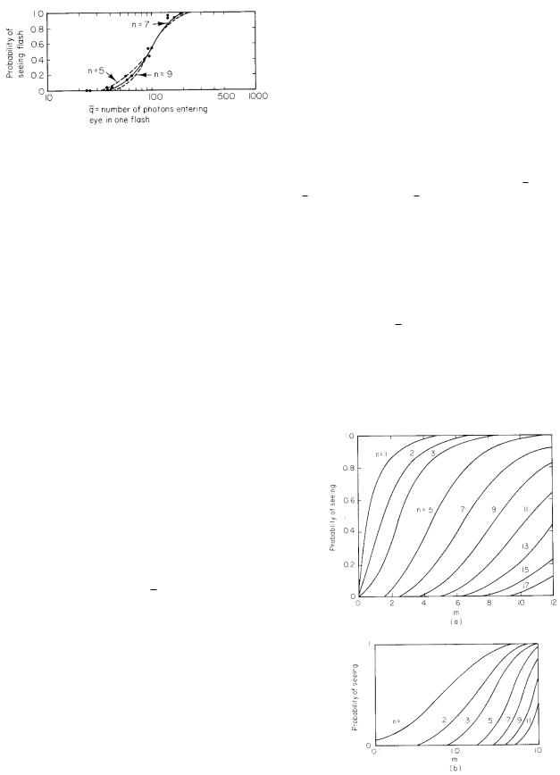
FIGURE 14.43. Typical response in the experiments of Hecht, Schlaer, and Pirenne. Curves are calculated using Eq. 14.68. Data are from S. Hecht, S. Schlaer, and M. H. Pirenne. The Journal of General Physiology 25: 819–840 (1942) by copyright permission of the Rockefeller University Press.
in Fig. 14.43. Let q be the number of photons striking the cornea in front of the pupil in each flash, which is the total energy in the flash divided by the energy of each photon. For the 510-nm green light used, the photon energy is hc/λ = 3.89 × 10⁻¹⁹ J. The number of photons striking the cornea can be determined as follows. Let Lt be the radiance times the duration of the flash. Consider Eq. 14.56 with both θs and θd nearly zero. Refer also to the lower half of Fig. 14.39. The energy striking the cornea over the pupil area is
(Lt) dSsdSd |
= |
(Lt) dS πa2 |
. |
r2 |
|
u2 |
|
Because h = h v/u, the area on the retina where photons from dS fall is dS = dS (v/u)2. The number of photons striking the cornea that would be in dS if there were no losses is
q = |
(Lt)(πa2)dS |
= |
(Lt)(πa2) dS |
. |
(14.66) |
hνu2 |
|
||||
|
|
hνv2 |
|
||
The number of photons fluctuates from flash to flash. Therefore we should speak of q, the average number of photons striking the cornea per flash. Of these, only some fraction f actually reach the retina and are absorbed by a visual pigment molecule. The average number absorbed is
m = f |
q. |
(14.67) |
Let us next postulate that some minimum number of quanta n must be absorbed during the flash in order for the subject to see it. If the average number absorbed per flash is m, there will sometimes be more and sometimes less than n photons absorbed per flash. The probability of absorbing x photons per flash is given by the Poisson distribution P (x; m) (Appendix J). The probability of seeing the flash is the probability that x is greater than
14.13 Quantum E ects in Dark-Adapted Vision |
391 |
or equal to n :
∞ |
n−1 |
|
|
|
|
|
||
|
|
|
P (x; m) |
|
|
|
|
|
P (seeing) = P (x; m) = 1 − |
|
|
|
|
|
|
|
|
x=n |
x=0 |
|
|
|
|
|
||
|
|
m2 |
+ · · · + |
mn−1 |
||||
= 1 − e−m 1 + m + |
|
|
|
|
|
|
. |
|
2! |
|
(n |
− |
1)! |
||||
|
|
|
|
|
|
|
|
|
(14.68)
This function is plotted in Fig. 14.44 as a function of m for various values of n, with both a linear and a logarithmic scale for m.
Hecht et al. used an ingenious method to determine n. They plotted their data vs. the logarithm of q. Since m = f q, log m = log f + log q; di erent values of f correspond to shifting the curve along the axis. They then compared the experimental data to the various theoretical curves for the probability of seeing a flash, plotted against log m. Sliding the paper containing the data along the log m axis is equivalent to trying di erent values of f . The data in Fig. 14.43 are shown along with the curves for n = 5, 7, and 9. For these data, n = 7 gives the best fit. From Fig. 14.43, a 55% chance of detecting the flash corresponds to 100 photons for q while being consistent with m = 7. Therefore, f = 0.07.
Hecht, Schlaer, and Pirenne deduced that about seven photons must be absorbed by the rods in the area of integration shown in Fig. 14.42 within 0.1 s in order for the brain to detect the flash of light. Their data were
FIGURE 14.44. The probability of seeing a flash, plotted vs
(a) m; (b) log m.
392 14. Atoms and Light
consistent with the hypothesis that the photons arrived |
n |
||||||
at random, with the actual number in each flash obey- |
n |
||||||
|
|
|
|||||
ing a Poisson distribution. Later work by Sakitt (1972) |
n |
|
|||||
|
|
|
|||||
is consistent with the rods counting individual photons, |
n |
||||||
with false positives produced by thermal noise within the |
|
|
|
||||
retina [Barlow (1956)]. |
|
|
p |
||||
The phototransduction mechanism |
is quite |
compli- |
q |
||||
q |
|||||||
cated. Rieke and Baylor (1998) reviewed the detection |
|||||||
|
|
|
|||||
q |
|||||||
of photons by rod cells. When stimulated with dim light |
|||||||
r |
|||||||
pulses, the rod cell responds to each flash consistent with |
r, r |
||||||
the absorption of 0, 1 or 2 photons. The rods have a |
s |
||||||
dark current that is reduced when light falls on them. In |
s |
||||||
|
|
|
|||||
other words, the light hyperpolarizes the cell. This low- |
t |
||||||
ers the rate of release of a neurotransmitter, cyclic GMP. |
|||||||
v |
|||||||
The review discusses what is known about the chemical |
v |
||||||
transduction process. |
|
|
u, v |
||||
If the light intensity is increased, m increases. There |
wtot |
||||||
x, z |
|||||||
will be shot-noise fluctuations with a standard deviation |
|||||||
z0 |
|||||||
equal to m1/2, and the eye should be unable to detect |
|||||||
A |
|||||||
brightness changes smaller than this. Measurements by |
A |
||||||
H. B. Barlow in 1957 showed that as long as short flashes |
B |
||||||
spanning only one visual field are used, the minimum |
C |
||||||
Cb, Ct |
|||||||
detectable intensity depends on the square root of the |
|||||||
|
|
|
|||||
light intensity. This statistical limit to detecting intensity |
D |
||||||
changes is a lower limit; for larger sources and longer ex- |
D |
||||||
posure times the minimum detectable brightness change |
D |
||||||
is larger and is more nearly proportional to the intensity |
E |
||||||
E |
|||||||
than to the square root of the intensity [Rose (1973)]. |
|||||||
Ep |
|||||||
|
|
|
|
||||
|
|
|
|
Er |
|||
|
|
|
|
Ev |
|||
Symbols Used in Chapter 14 |
|
E |
|||||
|
F, F |
||||||
|
|
|
|
F |
|||
Symbol |
Use |
Units |
First |
I |
|||
|
|
|
used on |
||||
|
|
|
K |
||||
|
|
|
page |
|
|
|
|
a |
Radius |
m |
367 |
Km |
|||
c |
Speed of light in a vacuum |
m s−1 |
359 |
||||
Km |
|||||||
cn |
Speed of light in a medium |
m s−1 |
359 |
||||
L |
|||||||
e |
Charge on an electron |
C |
361 |
||||
L |
|||||||
f |
Focal length |
m |
388 |
||||
|
|
|
|||||
f |
Fraction of photons reaching |
|
391 |
N |
|||
|
retina |
|
|
||||
|
|
|
Na |
||||
g |
Scattering anisotropy factor |
|
367 |
||||
|
Ns |
||||||
h |
Planck’s constant |
J s |
360 |
||||
NA |
|||||||
h, h |
Image height, object height |
m |
388 |
||||
NT |
|||||||
|
Planck’s constant divided by |
J s |
360 |
||||
|
|
|
|||||
|
2π |
|
|
P |
|||
i |
Label of energy level |
|
361 |
||||
|
P |
||||||
j |
Total angular momentum |
|
361 |
||||
|
|
|
|
||||
|
quantum number |
|
|
P |
|||
jH |
Energy transport in heat flow |
W m−2 |
382 |
||||
Pv |
|||||||
k |
Spring constant |
N m−1 |
363 |
||||
Q |
|||||||
kB |
Boltzmann constant |
J K−1 |
372 |
||||
R, R |
|||||||
l |
Orbital angular momentum |
|
361 |
||||
|
Rλ |
||||||
|
quantum number |
|
|
||||
|
|
|
|
|
|
||
m |
Mass |
kg |
362 |
R |
|||
m |
Average number |
|
391 |
||||
|
R |
||||||
me |
Mass of electron |
kg |
361 |
||||
S, S |
|||||||
mi |
Mass of ith particle |
kg |
363 |
T |
|||
mj , ml, ms |
z quantum number for |
|
361 |
||||
|
T |
||||||
|
|
|
|
||||
angular momentum
Index of refraction Principal quantum number Average number of photons that interact
Minimum number of photons to trigger a response Probability
Electric charge Number of photons Average value of q
Rotational quantum number Coordinate
Spin quantum number Source term in di usion equation
Time Velocity
Vibrational quantum number Object and image distances Net power radiated
Distance
Depth of first scattering Amplitude of wave Molar mass
Magnetic field Concentration
Heat capacity of blood, tissue
Di usion constant Photon di usion constant
Thermal di usion constant Electric field
Energy Potential energy
Rotational energy Vibrational energy Irradiance
Force
Converging power of a lens
Moment of inertia
Thermal conductivity
Luminous e ciency, photopic Luminous e ciency, scotopic Angular momentum Radiance
Number of photons Number absorbed Number scattered Avogadro’s number Number of target entities per unit area along beam Probability
Tissue perfusion
Radiant power Luminous flux Rate of production
Coordinate of atom, distance Radiant energy per unit wavelength interval Reflected fluence rate Radiant energy
Surface area Period Kinetic energy
359
361
365
391
366
C360
391
391
363
m362
361
m−3 s−1 |
367 |
s360
m s−1 |
360 |
|
364 |
m388
W376
m360
m369
370
kg |
366 |
T360
m−3 |
367 |
J kg−1 |
382 |
K−1 |
|
m2 s−1 |
367 |
m367
m2 s−1 |
382 |
V m−1 |
360 |
J360
J363
J363
J364
W m−2 |
386 |
N360
diopter |
389 |
(m−1) |
|
kg m2 |
363 |
W K−1 |
382 |
m−1 |
|
lm W−1 |
387 |
lm W−1 |
387 |
kg m2 s−1 |
363 |
W m−2 |
386 |
sr−1 |
|
|
365 |
|
365 |
|
365 |
|
366 |
m−2 |
365 |
|
372 |
m3 kg−1 |
382 |
s−1 |
|
W383
lm |
387 |
m−3 s−1 |
367 |
m362
J m−1 or J |
386 |
nm−1 |
|
m2 s−1 |
369 |
J383
m2 |
365 |
s360
J363
Symbol |
Use |
Units |
T, Ts, To |
Temperature |
K |
U |
Object vergence |
diopter |
|
|
(m−1) |
V, V
V
V
V
Wλ
Wν Wr
α
δth
0
(λ)
θ, φ
ϕ
λ
µ
µa
µs µs
µe
µ0
ρ, ρb, ρt
σ, σi, σa,
σs, σtot σ(θ), dσ/dΩ
σSB
σr2, σx2 , σy2, σz2
ν
τcoh
ω
ψ
Ψ
Φ
Ω
Velocity
Photopic spectral e ciency function
Scotopic spectral e ciency function
Image vergence
Blackbody radiation function
Blackbody radiation function Exitance
Angle
Thermal penetration depth Electrical permittivity of empty space
Emissivity
Reference action spectrum Angles
Particle fluence rate Wavelength
Total linear attenuation coe cient
Linear absorption coe cient Linear scattering coe cient Reduced linear scattering coe cient
E ective linear attenuation coe cient
Magnetic permeability of free space
Density, density of blood, density of tissue
Cross section
Di erential scattering cross section
Stefan–Boltzmann constant
Variance for di usion or heat flow
Frequency Coherence time Angular frequency
Energy fluence rate
Energy fluence
Particle fluence
Solid angle
m s−1
diopter (m−1)
Wm−3 or
Wm−2 nm−1
Wm−2 s
Wm−2
m
N−1 C2 m−2
m−2 s−1
m m−1
m−1 m−1 m−1
m−1
N s2 C−2
kg m−3
m2
m2 sr−1
W m−2 K−4
m2
s−1 s
(radian) s−1
W m−2 J m−2 m−2
sr
Problems |
393 |
First |
of wavelength. Express the answer in kilocalories and the |
||||||||
used on wavelength λ in nanometers. |
|
|
|
||||||
page |
|
|
|
|
|
|
|
|
|
372 |
|
|
|
|
|
|
|
|
|
389 |
Section 14.2 |
|
|
|
|
|
|||
|
|
|
|
|
|
||||
363 |
Problem 3 Use Eq. 14.7 to derive Eq. 14.8. |
||||||||
387 |
|||||||||
|
|
|
|
|
|
|
|
||
387 |
Problem 4 (a) Starting with Eq. 14.7, derive a formula |
||||||||
for the hydrogen atom spectrum in the form |
|||||||||
|
|||||||||
389 |
1 |
= R |
1 |
1 |
|
||||
|
|
|
|
|
− |
|
, |
||
373 |
|
λ |
n2 |
m2 |
|||||
|
|
|
|
|
|
|
|
||
|
where n and m are integers. R is called the Rydberg con- |
||||||||
374 |
stant. Find an expression for R in terms of fundamental |
||||||||
constants. |
|
|
|
|
|
||||
385 |
|
|
|
|
|
||||
(b) Verify that the wavelengths of the spectral lines a- |
|||||||||
388 |
|||||||||
383 |
d at the top of Fig. 14.3 are consistent with the energy |
||||||||
360 |
transitions shown at the bottom of the figure. |
||||||||
373 |
|
|
|
|
|
|
|
|
|
379 |
Section 14.3 |
|
|
|
|
|
|||
366 |
|
|
|
|
|
||||
Problem 5 Estimate 2/2I for an HCl molecule. What |
|||||||||
368 |
|||||||||
360 |
would the spacing of rotational levels be? |
||||||||
365 |
|
|
|
|
|
|
|
|
|
365 |
Problem 6 An inulin molecule has a molecular weight |
||||||||
of 4,000 dalton (that is, 1 mol has a mass of 4000 g). As- |
|||||||||
365 |
|||||||||
sume that it is spherical with a radius of 1.2 nm. What |
|||||||||
367 |
|||||||||
is the angular frequency ω of a photon absorbed when |
|||||||||
|
|||||||||
368 |
its rotational quantum number changes from 10 to 11? |
||||||||
360 |
The moment of inertia of a sphere rotating about an axis |
||||||||
through its center is I = (2/5)mR2. |
|
||||||||
366 |
Problem 7 The rotational spectrum of HCl contains |
||||||||
366 |
lines at 60.4, 69.0, 80.4, 96.4, and 120.4 µm. What is |
||||||||
the moment of inertia of an HCl molecule? |
|||||||||
|
|||||||||
366 |
Problem 8 Consider a combined rotational–vibrational |
||||||||
|
|||||||||
374 |
transition for which r goes from 1 to 0 while v goes from v |
||||||||
|
to (v − 1). Find the frequencies of the photons emitted in |
||||||||
382 |
terms of the moment of inertia of the molecule I, the an- |
||||||||
360 |
gular frequency of vibration of the atoms in the molecule |
||||||||
ω, and the quantum number v. |
|
||||||||
370 |
|
||||||||
|
|
|
|
|
|
|
|
||
360 |
Problem 9 A rotating molecule emits photons when the |
||||||||
|
|||||||||
382 |
angular momentum changes by 1. Find the ratio of the |
||||||||
angular frequency of the photons, ωphot, to the angular |
|||||||||
382 |
|||||||||
365frequency of rotation of the molecule ωrot, as a function
366of the orbital angular momentum quantum number r.
Problems
Section 14.1
Problem 1 The velocity of light c depends on the parameters 0 and µ0.Use dimensional analysis to find what the dependence must be. Insert numerical values to obtain c.
Problem 2 An einstein is 1 mol of photons. Derive an expression for the energy in an einstein as a function
Section 14.4
Problem 10 A beam with 200 particles per square centimeter passes by an atom. The particles are uniformly and randomly distributed in the area of the beam.
(a)Fifty particles are scattered. What is the total scattering cross section?
(b)Ten particles are scattered in a cone of 0.1 sr solid
angle about a particular direction. What is the di erential cross section in m2 sr−1?

394 14. Atoms and Light
Problem 11 The di erential scattering cross section for a beam of x-ray photons of a certain energy from carbon at an angle θ is 50 × 10−30 m2 sr−1. A beam of 105 photons strikes a pure carbon target of thickness 0.3 cm. The density of carbon is 2 g cm−3, and the atomic weight is 12. The detector is a circle of 1-cm radius located 20 cm from the target. How many scattered photons enter the detector?
Problem 12 Photochemists often use the extinction co- e cient e, defined by µa = eC, where C is the concentration in moles per liter. This assumes the substance being measured is dissolved in a completely transparent solvent.
(a)What are the units of the extinction coe cient?
(b)What is the conversion between the extinction co- e cient and the absorption cross section?
Problem 13 Suppose that the absorption coe cient in some biological substance is 5 m−1. Make the very crude assumption that the substance has the density of water and a molecular weight of 18. What is the absorption cross section?
Problem 14 For blue light (λ = 470 nm), the attenuation coe cient in air is about 2 × 10⁻⁵ m−1, and the attenuation coe cient in pure water is about 5×10⁻³ m−1. Calculate the distance that blue light must pass through air and through water before the intensity is reduced to 1% of the original intensity. Compare these distances to the thickness of the atmosphere and the depth of the ocean. Do you think that aquatic plants can use photosynthesis e ectively at the bottom of the ocean? [For more on the di erences between the optical properties of air and water, see Denny (1993).]
Section 14.5
Problem 15 (a) Find the slope of the log R vs. t in Eq. 14.29. What is its value for large times?
(b) What can be determined from the time when R has its maximum value? (Hint: R has a maximum when log R has a maximum.)
Section 14.6
Problem 17 Carry out the averages leading to Eq. 14.30.
Problem 18 If yellow light from a source has a coherence time of 10−8 s, how many cycles are there in the wave?
Problem 19 What coherence time is needed for a spatial resolution of 1 µm?
Problem 20 An infrared transition involves an energy of 0.1 eV. What are the corresponding frequency and wavelength? If the Raman e ect is observed with light at 550 nm, what will be the frequencies and wavelengths of each Raman line?
Problem 21 A Raman spectrum has a line at 500 nm with subsidiary lines at 400 and 667 nm. What is the wavelength of the corresponding infrared line?
Section 14.7
Problem 22 Sodium is introduced into a flame at 2,500 K. What fraction of the atoms are in their first excited state? In their ground state? (Remember that the characteristic sodium line is yellow.) If the flame temperature changes by 10 K, what is the fractional change in the population of each state? Which method of measuring sodium concentration is more stable to changes in flame temperature: measuring the intensity of an emitted line or measuring the amount of absorption?
Problem 23 (a) Show that the maximum of the thermal radiation function Wλ(λ, T ) occurs at a wavelength such that ex(5 −x) = 5, where x = hc/(λmaxkB T ). Verify that x = 4.9651 is a solution of this transcendental equation,
so that
hc
T λmax = 4.9651kB .
(b) Similarly, show that
νmax = 2.82144kB
T h
Problem 16 The result of one set of infrared measure- and that λmaxνmax ments in human calf (leg) muscle gave a total scattering
coe cient µs = 8.3 cm−1 and an absorption µa = 0.176 cm−1.
(a)What fraction of the photons have not scattered in passing through a layer that is 8 µm thick? (This corresponds roughly to the size of a cell.)
(b)On average, how many scattering events take place for each absorption event?
(c)What is the cross section for scattering per molecule? For this estimate, assume the muscle consists en-
tirely of water, with molecular weight 18 and density 103 kg m−3.
= 0.57c.
∞ |
x3dx |
= |
|
π4 |
. |
0 ex − 1 |
|
15 |
|
||
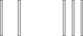
two surfaces that face each other. Let P0 be the energy lost per unit time by body 1. A new sheet of perfectly absorbing material is introduced between bodies 1 and 2, as shown in panel (b). It comes to equilibrium temperature T . Let
Pbe the net energy lost by surface 1 in this case. Find
P/P0 in terms of T1 and T2.
T1 |
T2 |
T1 |
T |
T2 |
|
(a) |
|
(b) |
|
Problem 26 The sun has a radius of 6.9 × 10⁸ m. The earth is 149.5 × 10⁹ m from the sun. Treat the sun as a thermal radiator at 5,800 K and calculate the energy from the sun per unit area per unit time striking the upper atmosphere of the earth (the solar constant). State the result in W m−2 and cal cm−2 min−1.
Problem 27 If all the energy received by the earth from the sun is lost as thermal radiation (a poor assumption because a significant amount is reflected from cloud cover), what is the equilibrium temperature of the earth?
Section 14.8
Problem 28 Show that an approximation to Eq. 14.40 for small temperature di erences is wtot = SKrad(T − Ts). Deduce the value of Krad at body temperature. Hint:
Factor T 4 − Ts4 = (T − Ts)(· · · ). You should get Krad = 6.76 W m−2 K−1.
Problem 29 What fractional change in Wλ(λ, T ) for thermal radiation from the human body results when there is a temperature change of 1 K at 5 µm? 9 µm? 15 µm?
Section 14.9
Problem 30 (a) Suppose that the threshold for erythema caused by sunlight with λ = 300 nm is 30 J m−2. Does this suggest that the result is thermal (an excessive temperature increase) or something else, like the photoelectric e ect or photodissociation? Make some reasonable assumptions to estimate the temperature rise.
(b) The energy in sunlight at all wavelengths reaching the earth is 2 cal cm−2 min−1. Suppose that the total body area exposed is 0.6 m2. What would be the temperature rise per minute for a 60-kg person if there were no heatloss mechanisms? Compare the rate of energy absorption to the basal metabolic rate, about 100 W.
Problem 31 Suppose that the energy fluence rate of a parallel beam of ultraviolet light that has passed through
Problems 395
thickness x of solution is given by ψ = ψ0e−µa x. (Scattering is ignored.) The absorption coe cient µa is related to the concentration C (molecules cm−3) of the absorbing molecules in the solution by µa = aC. Biophysicists working with ultraviolet light define the dose rate to be the power absorbed per molecule of absorber. (This is a di erent definition of dose than is used in Chapter 15.) Calculate the dose rate for a thin layer (µax 1).
Problem 32 A beam of photons passes through a monatomic gas of molecular weight A and absorption cross section σ. Ignore scattering. The gas obeys the ideal gas law, pV = N kB T .
(a)Find the attenuation coe cient in terms of σ, p, and any other necessary variables.
(b)Generalize the result to a mixture of several gases, each with cross section σi, partial pressure pi, and Ni molecules.
Problem 33 The attenuation of a beam of photons in a gas of pressure p is given by dΦ = −Φ(σp/kB T ) dx, where σ is the cross section, kB the Boltzmann constant, T the absolute temperature, and x the path length. Suppose that the pressure is given as a function of altitude y by p = p0e−mgy/kB T . What is the total attenuation by the entire atmosphere?
Problem 34 Consider a beam of photons incident on the atmosphere from directly overhead. The atmosphere
contains several species of molecules, each |
with par- |
tial pressure pi. The absorption coe cient |
is µa = |
(1/kB T ) i σipi. If each constituent of the atmosphere varies with height y as pi(y) = p0i exp(−migy/kB T ), show that the fluence rate striking the earth is given by an expression of the form e−α and find α.
Section 14.10
Problem 35 Consider a tissue with a heat capacity of 3.6 J kg−1 K−1, a density of 1,000 kg m−3, and a thermal conductivity of 0.5 W m−1 K−1. Assume the heat capacity of blood is the same, and that the tissue perfusion is 4.17× 10⁻⁶ m3 kg−1 s−1. Find the thermal di usivity, the time for the heat to flow 1 cm, and the thermal penetration depth.
Section 14.11
Problem 36 Suppose that a sphere radiates uniformly from its surface according to Lambert’s cosine law: L = L0. By considering area dS = 2πr2 sin θ dθ on the surface of a sphere, find the power radiated per steradian in the direction of the z axis and the total power radiated.
Problem 37 Show that the total power per unit area radiated from a surface obeying Lambert’s cosine law is Wr = πL0. This quantity is called the exitance.
396 14. Atoms and Light
Problem 38 How many photons per second correspond to 1 lm at 555 nm for photopic vision? At 510 nm for scotopic vision?
Section 14.12
Problem 39 A person is nearsighted, and the relaxed eye focuses at a distance of 50 cm. What is the strength of the desired corrective lens in diopters?
Problem 40 What is the distance of closest vision for an average person with normal vision at age 20? Age 40? Age 60?
Problem 41 A person of age 40 is fitted with bifocals with a +1 diopter strength bifocal lens. What are the closest and farthest distances of focus without the bifocal lens and with it? By the time the person is age 50, what are they with and without the same lens?
Problem 42 You can make a rough measurement of your own eye’s properties. Tape a piece of paper with some pattern on it on the wall. Cover one eye. Move away from the wall until the pattern starts to blur. Measure the distance to the wall in meters. Calculate the vergence of the object, U . Assume that the F of your relaxed eye is 59 diopters. Calculate V for your eye. Now find the closest distance at which you can see the paper. Calculate the accommodation of your eye.
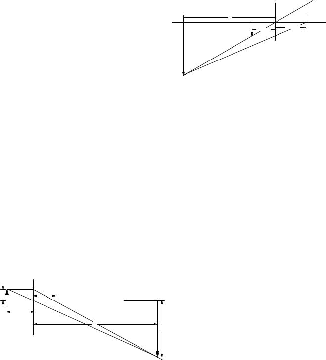
Problem 43 An object is placed 6 cm from a converging lens with a 5 cm focal length.
Plane of |
Ray through |
|
lens |
center of lens on |
|
axis is not |
||
|
||
|
deflected. |
Focal Point |
v |
f = 20 cm |
u = 15 cm |
Object |
Ray parallel to axis |
passes through |
focal point. |
Image |
(a)Use the thin-lens equation (Eq. 14.63) to calculate the image distance. Your value should be negative, corresponding to a “virtual image.”
(b)The magnification of the image is again given by m = −u/v. What is the magnification for the image in part (a)? This is how a magnifying glass works.
Problem 45 Combine the results of Problems 43 and 44. Consider two lenses, the first with focal length 5 cm and the second with focal length 20 cm, separated by 45 cm. The object is 6 cm in front of the first lens. The image from the first lens is the object for the second.
(a)Calculate the image distance and magnification of the image created by the first lens (called the objective).
(b)Use the first image as the object for the second lens (called the eyepiece), and calculate the image distance and magnification of the second image.
(c)The total magnification is the product of the magnifications of the objective and eyepiece. What is the total magnification? This is how the compound microscope works. The objective lens acts like a slide projector, and the eyepiece acts like a magnifying glass. Very large total magnifications can be obtained when the object is just to the left of the focal point of the objective, and the first image is just to the right of the focal point of the eyepiece.
|
|
|
|
|
|
|
Plane of lens |
|
|||||
|
|
|
|
|
|
|
|
|
|
|
|
||
h' |
|
|
|
|
|
|
f = 5cm |
|
|
|
Ray enters lens parallel |
||
|
|||||||||||||
|
|
|
|
|
|
||||||||
|
|
|
|
||||||||||
|
|
|
|
|
|
|
|
|
|
|
|
|
to axis and passes |
|
|
|
|
|
|
|
|
|
|
|
|
|
through focal point on |
|
|
|
|
|
|
|
|
|
|
|
|
|
other side. |
|
|
|
|
|
u = 6 cm |
|
|
|
|
|
|
|
|
|
|
|
|
|
|
|
|
|
|
|
|
||
|
|
|
|
|
|
|
|
|
|
|
|
|
|
v
h
Ray passing through lens plane on axis is undeflected.
(a)Use the thin-lens equation (Eq. 14.63) to calculate the image distance.
(b)The magnification of the image is given by m = −u/v. (A negative magnification implies an inverted image.) What is the magnification for the image in part (a)? A value |m| > 1 implies a“magnified” image. This is how a slide projector works.
Problem 44 An object is placed 15 cm from a converging lens with a focal length of 20 cm.
Problem 46 Snell’s law, n1 sin θ1 = n2 sin θ2, gives an interesting result if light passes from a medium with a higher index of refraction to one with a lower index of refraction, n1 > n2. Assume light passes from glass (n1 = 1.5) to air (n2 = 1.0).
(a)If θ1is 30 ◦, what is θ2?
(b)If θ1is 40 ◦, what is θ2?
(c)If θ1is 50 ◦, what is θ2?
This is really a trick question, because for θ1 greater than some critical angle, θc, θ2 exceeds 90 ◦, and light cannot pass into the second medium. Instead all the light is reflected and remains within the first medium.
(d) Calculate the critical angle for total internal reflection from glass to air.
Total internal reflection allows thin glass fibers to act as fiberoptic “light pipes,” which can be used to transmit signals. Bundles of such optical fibers are used in endoscopes to see inside the body.
Problem 47 Table 14.7 shows that most of the converging power of the eye occurs at the air-cornea interface. When a person is under water, this must be supplied by
the water-cornea surface. The index of refraction of the cornea is closer to that of water than to that of air. What are the implications for seeing under water? What are the implications for the vision of aquatic animals? [For more information on the di erence between the eyes of aquatic and terrestrial animals, see Denny (1993)].
Section 14.13
Problem 48 How many photons per 0.1 s enter the eye from a 100-W light bulb 1,000 ft away? Assume the pupil is 6 mm in diameter. How far away can a 100-W bulb be seen if there is no absorption in the atmosphere? Use a luminous e ciency of 17 lm W−1 and then assume an equivalent light source at 555 nm.
Problem 49 The table below shows the radiance of some extended sources. Without worrying about obliquity factors (assume that all the light is at normal incidence), calculate the number of photons entering a receptive field of 0.17 ◦ diameter when the pupil diameter is 6 mm and the integration time is 0.1 s. Assume a conversion e - ciency of 100 lm W−1 and then assume that all the photons are at 555 nm.
Source |
Radiance (lm m−2 sr−1) |
White paper in sunlight |
25,000 |
Clear sky |
3,200 |
Surface of the moon |
2.900 |
White paper in moonlight |
0.03 |
Problem 50 A piece of paper is illuminated by dim light so that its radiance is 0.01 lm m−2 sr−1 in the direction of a camera. A camera lens 1 cm in diameter is 0.6 m from the paper. The sheet of paper is 10 × 10 cm. The shutter of the camera is open for 1 ms. Assume all the light is at 555 nm. How many photons from the paper enter the lens of the camera while the shutter is open?
Problem 51 If three or more photons must be absorbed by a visual receptor field for the observer to see a flash, how often will the flash be seen if the average number of photons absorbed in a receptor field per flash is four?
Problem 52 Assume that an average of d photons are detected and that the photons are Poisson distributed. What must d be to detect a signal that is a 1% change in d, if the signal-to-noise ratio must be at least 5?
Problem 53 Suppose that the average number of photons striking a target during an exposure is m. The probability that x photons strike during a similar exposure is given by the Poisson distribution. What is the probability that an organism responds to an exposure of radiation in each of the following cases?
(a)The response of the organism requires that a single target within the organism be hit by two or more photons.
(b)The response of the organism requires that two targets within the organism each be struck by one or more photons during the exposure.
References 397
References
AAPM 57 (1996). Recommended Nomenclature for Physical Quantities in Medical Applications of Light. American Association of Physicists in Medicine (AAPM) Report No. 57. College Park, MD.
Armstrong, B. K., and A. Kricker (1995). Skin cancer.
Dermatolog. Clinics 13(3): 583–594.
Barlow, H. B. (1956). Retinal noise and absolute threshold. J. Opt. Soc. Am. 46: 634–639.
Barlow, H. B. (1957). Incremental thresholds at low intensities considered as signal/noise discriminations. J. Physiol. 136: 469–488.
Bergmanson, J. P. G., and P. G. S¨oderberg (1995). The significance of ultraviolet radiation for eye diseases. A review with comments on the e cacy of UVblocking contact lenses. Ophthalm. Physiol. Opt. 15(2): 83–91.
Berne, B. J., and R. Pecora (1976). Dynamic Light Scattering: With Applications to Chemistry, Biology, and Physics. New York, Wiley.
Blume, S. (1993). Social process and the assessment of a new imaging technique. Intl. J. Technol. Assess. Health Care 9(3): 335–345.
Bouma, B. E., and G. J. Tearney, eds. (2002). Handbook of Optical Coherence Tomography. New York, Marcel Dekker.
Bramson, M. A. (1968). Infrared Radiation. New York, Plenum.
Buka, R. L. (2004). Sunscreens and insect repellents.
Current Opinion in Pediatrics 16: 378–384.
Charman, W. N. (1995). Optics of the eye, Chap. 24 in M. Bass, editor in chief, Handbook of Optics, 2nd ed. Sponsored by the Optical Society of America. New York, McGraw-Hill.
Cornsweet, T. N. (1970). Visual Perception. New York, Academic.
Cotton, P. (1992). AMA’s council on scientific a airs to take a fresh look at thermography. J. Am. Med. Assoc. 267(14): 1885–1887.
De Boer, J. F., S. M. Srinivas, J. S. Nelson, T. E. Milner, and M. G. Ducros (2002). Polarization-sensitive optical coherence tomography. In Bouma, B. E. and G. J. Tearney, eds. Handbook of Optical Coherence Tomography. New York, Marcel Dekker, pp. 237–298.
Denny, M. W. (1993). Air and Water: The Biology and Physics of Life’s Media. Princeton, NJ, Princeton University Press.
Diem, M. (1993). Introduction to Modern Vibrational Spectroscopy. New York, Wiley.
Di ey, B. L. (1991). Solar ultraviolet radiation e ects on biological systems. Phys. Med. Biol. 36(3): 299–328.
Di ey, B. L., and J. Cameron (1984). A microcomputer program to predict sunburn exposure. Med. Phys. 11(6): 869–870.
Di ey, B. L., and J. Cheeseman (1992). Sun protection with hats. Brit. J. Dermatol. 127(1): 10–12.
398 14. Atoms and Light
Di ey, B. L., and P. M. Farr (1991). Quantitative aspects of ultraviolet erythema. Clin. Phys. Physiol. Meas. 12(4): 311–325.
Duderstadt, J. J., and L. J Hamilton (1976). Nuclear Reactor Analysis. New York, Wiley.
Esenaliev, R. O., K. V. Larin, I. V. Larina, and M. Motamedi (2001). Noninvasive monitoring of glucose concentration with optical coherence tomography. Optics Lett. 26(13): 992–994.
Farmer, J. (1997). Blood oxygen measurement. Chap. 3 in J. Webster. The Design of Pulse Oximeters. Bristol, Institute of Physics Publishers.
Fitzgerald, A. J., E. Berry, N. N. Zinonev, G. C. Walker, M. A. Smith and J. M. Chamberlain (2002). An introduction to medical imaging with coherent terahertz frequency radiation. Phys. Med. Biol. 47: R67–R84.
Fraden, J. (1991). Noncontact temperature measurements in medicine. In D. L. Wise, ed. Bioinstrumentation and Biosensors. New York, Dekker, pp. 511–549.
Gasiorowicz, S. (1974). Quantum Physics. New York, Wiley.
Giasson, C. J., N-M. Quesnel, and H. Boisjoly (2005). The ABCs of ultraviolet-blocking contact lenses: An ocular panacea for ozone loss? International Ophthalmologic Clinics 45(1): 117–139.
Gilchrist, B. A., M. S. Eller, A. C. Geller and M. Yaar (1999). The pathogenesis of melanoma induced by ultraviolet radiation. New Engl. J. Med. 340(17): 1341–1348.
Grievenkamp, J. E., J. Schwiegerling, J. M. Miller, and M. D. Mellinger (1995). Visual acuity modeling using optical raytracing of schematic eyes. Am. J. Ophthalmol. 120(2): 227–240.
Grossweiner, L. (1994). The Science of Phototherapy.
Boca Raton, FL, CRC Press.
Haddad, J. G. (1992). Vitamin D—Solar rays, the milky way, or both (Editorial). New Engl. J. Med. 326(18): 1213–1215.
Halliday, D., R. Resnick, and K. S. Krane (1992). Fundamentals of Physics, 4th ed. New York, Wiley.
Hanlon, E. B., R. Manoharan, T-W. Koo, K. E. Shafer, J. T. Motz, M. Fitzmaurice, J. R. Kramer, I. Itzkan, R. R. Dasari, and M. S. Feld (2000). Prospects for in vivo Raman spectroscopy. Phys. Med. Biol. 45: R1–R59.
Heald, M. A. (2003). Where is the “Wien peak”? Amer. J. Phys. 71 (12): 1322–1323.
Hecht, S., S. Schlaer, and M. H. Pirenne (1942). Energy, quanta, and vision. J. Gen. Physiol. 25: 819– 840.
Hielscher, A., S. L. Jacques, L. Wang, and F. K. Tittel (1995). The influence of boundary conditions on the accuracy of di usion theory in time-resolved reflectance spectroscopy of biological tissues. Phys. Med. Biol. 40: 1957–1975.
Huang, D., E. A. Swanson, C. P. Lin, J. S. Schuman, W. G. Stinson, W. Chang, M. R. Hee, T. Flotte, K. Gregory, C. A. Puliafito, and J. G. Fujimoto (1991). Optical coherence tomography. Science 254: 1178–1181.
Kevan, P. G., L. Chittka, and A. G. Dyer (2001). Limits to the salience of ultraviolet: Lessons from colour vision in bees and birds. J. Exp. Biol. 204: 2571–2580.
Kimlin, M. G., and A. V. Parisi (1999). Radiation penetrating vehicle glass: a field-based comparative study.
Phys. Med. Biol. 44: 917–926.
Kligman, L. H. (1989). Photoaging: manifestations, prevention, and treatment. Clin. Geriatr. Med. 5: 235– 251.
Knobler, E., I. Warmuth, C. Cocco, B. Miller, and J. Mackay (2002). Extracorporeal immunotherapy: the Columbia-Presbyterian experience. Photodermatology, Photoimmunololgy and Photomedicine 18 (5): 232– 237.
Kripke, M. L. (2003). The ABCs of sunscreen protective factors [Comment]. J. Invest. Dermatol. 121(1): vii– viii.
Lever, L. R., and C. M. Lawrence (1995). Nonmelanoma skin cancer associated with the use of a tanning bed. New Engl. J. Med. 332(21): 1450–1451.
Liu, H., D. A. Boas, Y. Zhang, A. G. Yodh, and B. Chance (1995). Determination of optical properties and blood oxygenation in tissue using continuous NIR light.
Phys. Med. Biol. 40: 1983–1993.
MacNeill, B. D., H. C. Lowe, M. Takano, V. Fuster and I-K. Jang (2003). Intravascular modalities for detection of vulnerable plaque. Arterioscler. Thromb. Vasc. Biol.
23: 1333–1342.
Madronich, S. (1993). The atmosphere and UV-B radiation at ground level. In A. R. Young et al., eds. Environmental UV Photobiology. New York, Plenum, pp. 1–39.
McDonagh, A. F. (1985). Light e ects on transport and excretion of bilirubin in newborns. In R. J. Wortman, M. J. Baum, and T. J. Potts, Jr., eds. The Medical and Biological E ects of Light. Ann. N.Y. Acad. Sci. 453: 65–72.
Messina, C., F. Locatelli, E. Lanino, C. Uderzo, G. Zacchello, S. Cesaro, M. Pillon, C. Perotti, C. Del Fante, M. Faraci, L. Rivabella, E. Calore, P. De Stefano, M. Zecca, G. Giorgiani, A. Bruglio, A. Balduzzi, G. Dini, L. Zanesco and R. Dall’Amico (2003). Extracorporeal photochemotherapy for paediatric patients with graft-versus- host disease after haematopoietic stem cell transplantation. Brit. J. Haematology. 122 (1):118–127.
Milonni, P. W. (1996). Answer to question on Snell’s law in quantum mechanics. Am. J. Phys. 64(7): 842.
Moseley, H. (1994). Ultraviolet and laser radiation safety. Phys. Med. Biol. 39: 1765–1799.
Moyal, D. D., and A. M. Fourtanier (2002). E ects of UVA radiation on an established immune response in humans and sunscreen e cacy. Exp. Dermatol. 11 (Suppl. 1): 28–32.
Murphy, M. R., and R. G. Oellrich (1990). A new method of phototherapy: Nursing perspectives. J. Perinatol. 10(3): 249–251.
Nickell, S., M. Hermann, M. Essenpreis, T. J. Farrell, U. Kr¨amer, and M. S. Patterson (2000). Anisotropy of
light propagation in human skin. Phys. Med. Biol. 45: 2873–2886.
Nijsten, T. E. and R. S. Stern (2003). The increased risk of skin cancer is persistent after discontinuation of psoralen+ultraviolet A: a cohort study. J. Invest. Dermatol. 121 (2): 252–258.
Patterson, M. S., B. Chance, and B. C. Wilson (1989). Time resolved reflectance and transmittance for the noninvasive measurement of tissue optical properties. Appl. Opt. 28(12): 2331–2336.
Paul, C. F. (2003). Treating severe psoriasis: How to navigate safely between Scylla and Charybdis? [Comment] J. Invest. Dermatol. 121 (2): ix–x.
Pirenne, M. H. (1962). Chaps. 5 and 6 of H. Davidson, ed., The Eye. Vol. 2. The Visual Process. New York, Academic.
Pogue, B. W., and M. S. Patterson (1994). Frequencydomain optical absorption spectroscopy of finite tissue volumes using di usion theory. Phys. Med. Biol. 39: 1157–1180.
Poon, T. S. C., R. StC. Barneston and G. M. Halliday (2003). Prevention of immunosuppression by sunscreen in humans is unrelated to protection from erythema and dependent on protection from ultraviolet A in the face of constant ultraviolet B protection. J. Invest. Dermatol. 121(1): 184–190.
Rieke, F., and D. A. Baylor (1998). Single-photon detection by rod cells of the retina. Revs. Mod. Phys. 70(3): 1027–1036.
Rose, A. (1973). Vision: Human and Electronic. New York, Plenum.
Saint-Jalmes, H., M. Lebec, E. Beaurepaire, A. Dubois and A. C. Boccara (2002). Full-field optical coherence microscopy. In Bouma, B. E. and G. J. Tearney, eds. Handbook of Optical Coherence Tomography. New York, Marcel Dekker, pp. 299–333.
Sakitt, B. (1972). Counting every quantum. J. Physiol. (London) 223: 131–150.
Schaefer, B. E. (1993). Suntan and the ozone layer. Sky and Telescope 86(1): 83–86, July.
Schmitt, J. M. (1999). Optical Coherence Tomography (OCT): A review. IEEE J. Select. Topics Quantum Electron. 5: 1205–1215.
Sevick, E. M., B. Chance, J. Leigh, S. Nioka, and M. Maris (1991). Quantitation of timeand frequencyresolved optical spectra for the determination of tissue oxygenation. Analyt. Biochem. 195: 330–351.
References 399
Sherwood, B. A. (1996). Answer to question on Snell’s law in quantum mechanics. Am. J. Phys. 64(7): 840–842.
Smye, S. W., J. M. Chamberlain, A. J. Fitzgerald and E. Berry (2001). The interaction between terahertz radiation and biological tissue. Phys. Med. Biol. 46: R101– R112.
So er, B. H., and D. K. Lynch (1999). Some paradoxes, errors, and resolutions concerning spectral organization of human vision. Amer. J. Phys. 67 (11):946–953.
Stark, L. C., and G. C. Theodoris (1973). Information theory in physiology, in J. H. U. Brown and D. S. Gans. eds. Engineering Principles in Physiology, New York, Academic, Vol. 1, pp. 13–31.
Steketee, J. (1973). Spectral emissivity of skin and pericardium. Phys. Med. Biol. 18: 686–694.
Studniberg, H. M., and P. Weller (1993). PUVA, UVB, psoriasis, and nonmelanoma skin cancer. J. Am. Acad. Dermatol. 29: 1013–1022.
Verheye, S., L. Diamantopoulos, P. W. Serruys, and G. Van Langenhove (2002). Intravascular imaging of the atherosclerotic plaque: Spotlight on temperature measurement. J. Cardiovasc. Risk 9: 247–254.
Webster, J. (1997). The Design of Pulse Oximeters.
Bristol, Institute of Physics Publishers.
Wieben, O. (1997). Light absorbance in pulse oximetry. Chap. 4 in J. Webster. The Design of Pulse Oximeters.
Bristol, Institute of Physics Publishers.
Yamashita, Y., A. Maki, and H. Koizumi (2001). Wavelength dependence of the precision of noninvasive optical measurement of oxy-, deoxy-, and total hemoglobin concentration. Med. Phys. 28(6): 1108–1114.
Yasuno, Y., S. Makita, Y. Sutoh, M. Itoh, and T. Yatagai (2002). Birefringence imaging of human skin by polarization-sensitive spectral interferometric optical coherence tomography. Optics Lett. 27(20): 1803–1805.
Yazdanfar, S., A. M. Rollins and J. A. Izatt (2000). Imaging and velocimetry of the human retinal circulation with color Doppler optical coherence tomography. Optics Lett. 25(19): 1448–1450.
Yodh, A., and B. Chance (1995). Spectroscopy and imaging with di using light. Phys. Today. March: 34–40.
Zalewski, E. F. (1995). Radiometry and photometry, Chap. 24 of M. Bass, editor in chief, Handbook of Optics, 2nd ed. Sponsored by the Optical Society of America. New York, McGraw-Hill.
Zhang, X-C. (2002). Terahertz wave imaging: horizons and hurdles. Phys. Med. Biol. 47: 3667–3677.