

Kluwer - Handbook of Biomedical Image Analysis Vol
.2.pdfA Knowledge-Based Scheme for Digital Mammography |
593 |
1.Deterministic component: This is the most common strategy for introducing adaptability into a CAD scheme. Typically the component method is fixed but parameters are adaptively determined on a per-image basis. The parameter setting is either performed in a deterministic manner, based directly on an observed feature of the image, e.g. variance of gray scales, or empirically through experimentation. In the past, this approach has been applied to each of the three CAD components, e.g., adaptive contrast enhancement methods to perform an optimal contrast enhancement on an image based on local neighborhoods [1, 2], adaptive segmentation techniques utilizing adaptive clustering [3–5], or thresholding techniques [6–8] to segment an image and adaptive classification methods in the reduction of false-positive regions.
2.Knowledge-based component: An alternative strategy for setting a given component’s parameters is to learn the optimal parameter settings for an individual or group of images using machine learning techniques. The mapping between the parameter settings and images is achieved using a global image characteristics.
3.Knowledge-based framework: An extension to the knowledge-based component is to use machine learning principles to learn the utility of a particular component technique for an individual or group of images. Such a knowledge-based framework would be capable of drawing on a variety of different techniques to meet the objectives of each CAD component. The framework would support the definition of an optimal pipeline through the CAD pyramid. To date, no research has been presented into the use of knowledge-based frameworks in medical imaging CAD schemes. We now highlight some past research into knowledge-based components used in CAD schemes to put our proposed model in context.
11.2.1A Review of Knowledge-Based Components in Medical Imaging CAD Schemes
Table 11.1 summarizes medical imaging CAD studies that have used a knowledge-based component. The studies are categorized according to their approach to representing the knowledge within the component. Each of the approaches is described in more detail below.

594 |
Singh and Bovis |
Table 11.1: Summary of studies utilizing knowledge-based components,
grouped according to their approach
|
|
|
Methodology |
|
|
|
|
|
|
|
|
Study |
Modality |
part |
ann |
usr |
cbr |
|
|
|
|
|
|
Zheng et al. [9] |
Mammography (x-ray) |
√ |
|
|
|
Matsubara et al. [10] |
Mammography (x-ray) |
√ |
√ |
|
|
Lai and Fang [11, 12] |
Angiography |
|
|
|
|
Pitiot et al. [13] |
MRI |
|
√ |
|
|
Sha and Sutton [14] |
MRI |
|
√ |
√ |
|
Fenster and Kender [15] |
CT |
|
|
√ |
|
Perner [16] |
CT |
|
|
|
|
|
|
|
|
|
|
part = image grouping; ann = multistage neural networks; usr = user interaction; cbr = case-based
reasoning.
11.2.1.1Knowledge Representation by Image Grouping on Various Criteria
This approach to implementing a knowledge-based component attempts to adaptively determine optimum parameter settings for groups of images on the basis of image feature vectors. The feature vector is used to group images accordingly, that in turn serve as a form of a priori knowledge for use in subsequent components. In this way components may be trained to operate on particular image groupings with different parameter settings.
In mammography, Zheng et al. [9] propose an adaptive computer-aided diagnosis scheme optimized on the basis of the characterization of the mammogram. The rule-based system proposes a difficulty index (DI). This is computed as the weighted sum of nine histogram-based features calculated from a separate training set. The computed DI score is used in conjunction with a banding scheme, based on empirically determined values corresponding to easy, moderately difficult and difficult groupings following human interpretation. An expert radiologist evaluates each mammogram and determines the group boundaries. The authors propose the use of a rule-based classification scheme such that different classification rules are independently set for the three different difficulty groups in training. On a locally defined dataset of 428 digitized mammograms (abnormal n = 220, normal n = 208), the authors report the simple adaptive
A Knowledge-Based Scheme for Digital Mammography |
595 |
scheme reduced the average number of false-positive detections from 0.85 to 0.53 per image.
Matsubara et al. [10] proposed the use of an image grouping scheme for digitzed mammograms. In their study, images are assigned to one of four categories based on histogram analysis of the image gray scales. Subsequent imageprocessing operations, such as threshold-based segmentation and region classification operate on parameters defined empirically and independently within each category. The authors use this scheme to ignore high-density mammograms. On a small dataset of 30 images, the authors report a sensitivity of 93%.
11.2.1.2Knowledge Representation with Multistage Neural Networks
An alternative to a hard bounded grouping scheme such as those proposed above is in the use of soft decision boundaries. These knowledge-based components utilize a mixture-of-experts paradigm. Lai and Fang [11, 12] proposed the use of a hierarchical neural network to model the optical transformation of a 12-bit magnetic resonance image (MRI) into an 8-bit representation for display on a computer monitor. This optimal optical transformation is crucial if the expert radiologist is to effectively interpret the displayed image. The authors cite a major obstacle to the implementation of a simple linear solution as the difference in optimal parameters for different types of MR images. For example, 3D angiographic images and T1or T2-weighted images should be parameterized differently. To account for such differences, the authors propose a hierarchical arrangement of neural networks trained to provide accurate estimation for a wide range of images. To achieve the mapping of optimal transformation parameter values with individual images, a variety of histogram, wavelet and spatial features are used to characterize each image. The decision of each network module in the hierarchy is combined using a weighted averaged fusion scheme. Evaluating their framework on a dataset of more than 2400 images the authors report that their methodology gives satisfactory results and it is robust to unknown images.
Similarly, Pitiot et al. [13] proposed an automated method for extracting anatomical structures in MRI based on textural classification. The authors hypothesize that performing a pretextural classification prior to segmentation will
596 |
Singh and Bovis |
lead to a more accurate definition of the anatomical boundary. The pretextural classification is based on a mixture-of-experts paradigm, such that each expert is trained on a particular grouping of textural features extracted from a moving window within the image. A second-stage multiscale neural network is trained on equally drawn numbers of random samples from correctly and incorrectly classified pixels from the first stage. The network arrangement of stage two is trained on local morphology and texture features from a wider pixel neighborhood in the task of detecting anatomical structures. Evaluating their framework on a small dataset of 10 testing images, the authors report an increase in classification rates as a result of the two-stage hybrid neural classification.
Sha and Sutton [14] proposed the use of a network-of-networks paradigm first discussed by Guan et al. [17] for dynamically reconfiguring a test image for enhancement and segmentation. Under the proposed framework, the image is connected on a pixel-by-pixel basis by weights in a manner analogous to an attractor neural network. Pixels are updated based on local variances obtained from weight connections with neighbours in an iterative manner until convergence of the network architecture. In their study, the authors present only qualitative results on the enhancement and segmentation of MRI images.
11.2.1.3Knowledge Representation Learnt from User Interactions
A novel method of implementing an adaptable characteristic in a knowledgebased component has been suggested by capturing user interactions with a CAD tool.
Fenster and Kender [15] proposed the use of a diagnostic tool for the interpretation of computed tomography (CT) images. The authors utilize a boundarybased segmentation technique termed the live wire paradigm. The motivation for the scheme is based on an attempt to utilize the interaction with an expert clinician during segmentation, thereby learning from the users feedback for use in subsequent segmentations. Under the proposed framework, a 2D boundary is constructed around a ROI based in part on image properties and knowledge acquired in training when manual segmentation is performed. The tool utilizes an optimal feature selection process to determine the best boundary position from the available information.
A Knowledge-Based Scheme for Digital Mammography |
597 |
11.2.1.4Knowledge Representation Using Case-Based Reasoning
Case-based reasoning (CBR) approaches have been used extensively as a means of directly utilizing image properties. CBR is an approach to computerbased cognition that involves reasoning from prior experiences. It solves new problems by adapting solutions that were used to solve old problems. The knowledge base of a CBR system consists of cases indexed by their pertinent features.
Perner [16] proposes a novel method of image segmentation based on CBR. The CBR unit for image segmentation consists of a case base in which formerly processed cases are stored. Accompanying each case is information regarding the parameters used in the segmentation of the image. On test, a similarity measure is used to select the most similar case in the archive from which the segmentation parameters are selected and used to segment the test image. The author hypothesizes that images having similar characteristics will show good segmentation results when the same segmentation parameters are applied. The image information comprises a vector of statistical features extracted from the gray scale histogram of the matching image. In the author’s implementation, nonimage information is obtained from CT image headers, such as sex, age, CT-slice thickness, etc. The final similarity measure comprises both image and nonimage information. The segmentation of the image is performed using histogram analysis. The parameters for this process comprise those defining a smoothing function for the histogram and a set of thresholds used for histogram analysis. The system has been evaluated using a 130 image case base and by comparing the automatic CBR segmentation with that of an expert clinician on 600 CT images of the brain. The author reports a linear correlation coefficient of 0.85 between the CBR segmentation and those manually drawn by an expert clinician.
11.2.2 An Adaptive Knowledge-Based Model
As we can see from the survey in the previous section, little research has been undertaken in attempting to optimize a hierarchy of image processing operators. In this chapter an adaptive knowledge-based model is proposed. The model comprises deterministic and knowledge-based components for the detection of
A Knowledge-Based Scheme for Digital Mammography |
599 |
Mammogram grouping: By grouping mammograms on a predefined criteria, subsequent CAD components may be engineered to operate on specific mammograms types. Our study hypothesizes that a mammogram can be grouped on the basis of its parenchymal patterns. The aim of this component is to predict a mammogram’s group by utilizing supervised learning techniques in conjunction with a training set of example images.
Optimal contrast enhancement: A range of contrast enhancement techniques previously used in mammographic CAD research are surveyed in [18]. The adaptive knowledge-based model aims to accommodate many of these methods in the form of enhancement experts and learn, on the basis of feature vectors from training mammograms, the optimal contrast enhancement expert for a given mammogram on test. We propose machine learning techniques such as artificial neural networks (ANN) for learning this mapping.
Optimal image segmentation: A variety of different image segmentation methods have been identified for mammographic CAD in [18, 19]. Adopting a similar strategy to that of the knowledge-based contrast enhancement experts, a set of segmentation experts are proposed. As opposed to different contrast enhancement experts, each segmentation expert is functionally identical. The adaptability property in the segmentation component is achieved by learning the saliency of input features used to perform the segmentation. This chapter hypothesizes that different segmentation experts operating on different input feature spaces will have a greater utility in the segmentation of different mammograms. Input features for expert construction will be drawn from a subset commonly utilized in mammographic CAD. For example, the subset may include image gray scales, contrast enhanced gray scales, textures, and edgegradient information possibly at different scales of resolution. Each segmentation expert operates on a predefined set of features for a predefined group of mammograms. The implementation of an optimal segmentation is achieved by predicting the best blend of segmentation decisions given by the collection of experts for an individual mammogram.
Reduction of false-positive regions: The final component is the reduction of false-positive regions. This component operates by discriminating between normal and abnormal regions based on a feature vector
600 |
Singh and Bovis |
extracted from a suspicious ROI. This component is implemented within the adaptive knowledge-based model as a modular arrangement of ANNs trained to specialize in particular groupings of mammograms.
11.2.2.2The Complete Adaptive Knowledge-Based Model Framework
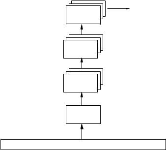
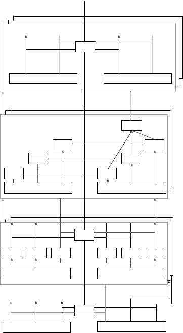
A schematic low-level representation of the adaptive knowledge-based model is shown in Fig. 11.2. Each component identified in Fig. 11.2 is discussed now to show how the complete model comprising a knowledge-based framework is to be implemented. Each blocked level in Fig. 11.2 is described in further detail in each part of this chapter and summarized below:
Image preprocessing component: The following image preprocessing is performed. Firstly, mammograms are grouped on the basis of their breast density. The Digital Database of Screening Mammograms (DDSM) database used in our study (http://marathon.csee.usf.edu/Mammography /Database.html) comes with the complete ground-truth definitions of breast cancer lesions specified according to the American College of Radiology (ACR) Breast Imaging Reporting and Data System (BI-RADS) lexicon. We develop a classification scheme that indexes the texture features of training data with their ground truth breast density information. This classification scheme is used to predict the correct density of a test image and hence used the correct algorithms for application.
Secondly, we redefine the boundaries (ground truth) supplied with the DDSM database to accurately represent the location of the lesion. This is done using an active contour model (full details are available in [18]). The reason for using this redefined ground truth is to improve the learning in the contrast enhancement and image segmentation components.
Expert image contrast enhancement: The main aim of this component is to select the optimal image enhancement method per image. The aim is to choose the enhancement method that optimizes the following image segmentation performance.
Expert image segmentation: The aim of image segmentation is to label pixels within an image as corresponding to real world objects. For a
602 |
Singh and Bovis |
mammographic CAD scheme, this involves labeling pixels in the image as being normal or suspicious. In this way suspicious pixels may be combined into suspicious regions. By utilizing machine learning principles, a segmentation expert can be constructed from a set of training images drawn from a particular mammographic breast type. Our study evaluates the knowledge-based segmentation component using 10 different input feature spaces, including the original image, contrast-enhanced image, and a textural representation at different scales. To segment a mammogram, of a given breast type, the 10 trained segmentation experts each give an estimate of the segmentation based on their input feature spaces. These decisions are then combined such that the optimal blend of segmentation experts is determined thereby resulting in an optimal segmentation. From the segmented image, region boundaries are identified and the regions passed onto the final component for the reduction of false-positive regions.
Reduction of false-positive regions: We select a set of region-based features that can be used in conjunction with a separate training set of regions for learning component knowledge. By training an ANN for each breast type, a modular arrangement of ANNs can be used to specialize in decisionmaking. The aim on test is to reduce the average number of false-positive regions per image while maintaining a high level of sensitivity to lesion detection.
We now detail the individual components of the model in much greater detail.
11.3 Image Contrast Enhancement Layer
In order to construct a scheme for the optimal selection of image enhancement, some quantitative indices are needed that measure the amount of enhancement. Not enough research has been conducted to tackle this difficult issue. In our previous work [19, 20] we introduced three new quantitative measures of image enhancement based on the change in contrast between the target (mass) and the backgound (a border 20 pixels wide around the target). We cover these measures for the sake of completeness here in section 11.3.1. In addition, we also discuss