

Obstetrics_by_Ten_Teachers_19E_-_Kenny_Louise
.pdf126 Pre-eclampsia and other disorders of placentation
as diabetes and hypertension) are more common in adults who were born with FGR.
Definition and incidence
FGR is defined as a failure of a fetus to achieve its genetic growth potential. This usually results in a fetus that is small for gestational age (SGA). SGA means that the weight of the fetus is less than the tenth centile for its gestation. Other cut-off points (e.g. the third centile) can be used. The terms SGA and FGR are not synonymous. It is important to remember that most SGA fetuses are constitutionally small and are not compromised. Intrauterine growth restriction (IUGR) indicates that there is a pathological process operating to restrict the growth rate of the fetus. Consequently, some FGR fetuses may not actually be SGA, but nevertheless will have failed to fulfil their growth potential.
Aetiology
The common causes of FGR are listed in Table 10.1. They are grouped into two main categories: factors that directly affect the intrinsic growth potential of the fetus and external influences that reduce the support for fetal growth. Chromosome abnormalities, genetic syndromes and fetal infections can alter intrinsic fetal growth potential. External influences that affect fetal growth can be subdivided into maternal systemic factors and placental insufficiency.
Maternal under-nutrition is globally the major cause of FGR. Low maternal oxygen saturation,
Table 10.1 Causes of fetal growth restriction
which can occur with cyanotic heart disease or at high altitude, will reduce fetal PO2 levels and fetal metabolism. Smoking, by increasing the amount of carboxyhaemoglobin in the maternal circulation, effectively reduces the amount of oxygen available to the fetus, thus causing FGR. A wide variety of drugs other than tobacco can affect fetal growth including alcohol and cocaine, probably through multiple mechanisms affecting fetal enzyme systems, placental blood flow and maternal substrate levels.
In developed countries, the most common cause of FGR is poor placental function secondary to inadequate trophoblast invasion of the spiral arteries. This results in reduced perfusion of the intracotyledon space which in turn leads to abnormal development of the terminal villi and impaired transfer of oxygen and nutrients to the fetus. Less frequently, reduced perfusion can occur from other conditions such as maternal sickle cell disease and the antiphospholipid syndrome (see Chapter 12, Medical diseases complicating pregnancy). Multiple pregnancy usually results in a sharing of the uterine vascularity, which causes a relative reduction in the blood flow to each placenta. On the fetal side of the placental circulation, abnormalities of the umbilical cord, such as a single umbilical artery, are associated with FGR as are the intraplacental vascular connections found in monochorionic twinning.
Pathophysiology
FGR is frequently classified as symmetrical or asymmetrical. Symmetrically small fetuses are
Reduced fetal growth potential |
Aneuploidies, e.g. trisomy 18 |
|
Single gene defects, e.g. Seckel’s syndrome |
|
Structural abnormalities, e.g. renal agenesis |
|
Intrauterine infections, e.g. cytomegalovirus, toxoplasmosis |
|
|
Reduced fetal growth support |
|
|
|
Maternal factors |
Under-nutrition, e.g. poverty, eating disorders |
|
Maternal hypoxia, e.g. living at altitude, cyanotic heart disease |
|
Drugs, e.g. alcohol, cigarettes, cocaine |
|
|
Placental factors |
Reduced uteroplacental perfusion, e.g. inadequate trophoblast |
|
invasion, sickle cell disease, multiple gestation |
|
Reduced fetoplacental perfusion, e.g. single umbilical artery, |
|
twin–twin transfusion syndrome |

normally associated with factors that directly impair fetal growth, such as chromosomal disorders and fetal infections. Asymmetrical growth restriction is classically associated with uteroplacental insufficiency which leads to reduced oxygen transfer to the fetus and impaired excretion of carbon dioxide by the placenta. A fall in PO2 and a rise in pCO2 in the fetal blood induces a chemoreceptor response in the fetal carotid bodies with resulting vasodilatation in the fetal brain, myocardium and adrenal glands, and vasoconstriction in the kidneys, splanchnic vessels, limbs and subcutaneous tissues. The liver circulation is also severely reduced. Normally, 50 per cent of the well-oxygenated blood in the umbilical vein passes to the right atrium through the ductus venosus, eventually to reach the fetal brain, with the remainder going to the portal circulation in the liver. When there is fetal hypoxia, more of the well-oxygenated blood from the umbilical vein is diverted through the ductus venosus, which means that the liver receives less. The result of all these circulatory changes is an asymmetrical fetus with relative brain sparing, reduced abdominal girth and skin thickness. The vasoconstriction in the fetal kidneys results in impaired urine production and oligohydramnios. The fetal hypoxaemia also leads to severe metabolic changes in the fetus reflecting intrauterine starvation. Antenatal fetal blood sampling has shown reduced levels of nutrients such as glucose and amino acids (especially essential amino acids) and of hormones such as thyroxine and insulin. There are increased levels of corticosteroids and catecholamines, which reflect the increased perfusion of the adrenal gland. Haematological changes also reflect the chronic hypoxia, with increased levels of erythropoietin and nucleated red blood cells.
Chronic fetal hypoxia in FGR may eventually lead to fetal acidaemia, both respiratory and metabolic, which if prolonged can lead to intrauterine death if the fetus is not removed from its hostile environment. FGR fetuses are especially at risk from profound asphyxia in labour due to further compromise of the uteroplacental circulation by uterine contractions.
Management
The assessment of fetal well-being is described in detail in Chapter 6, Antenatal imaging and assessment of fetal well-being.
Definition and incidence |
127 |
In brief, the detection of an SGA infant contains two elements: first, the accurate assessment of gestational age and second, the recognition of fetal smallness.
Early measurement of the fetal crown–rump length before 13 weeks plus 6 days gestation or head circumference between 13 6 and 20 weeks remains the method of choice for confirming gestational age. Thereafter, the most precise way of assessing fetal growth is by ultrasound biometry (biparietal diameter, head circumference, abdominal circumference and femur length) serially at set time intervals (usually of 4 weeks and no less than 2 weeks). As resources in most units do not permit comprehensive serial ultrasound in all pregnancies, serial ultrasound biometry is usually performed in ‘at risk’ pregnancies (see box below).
Pregnancies at risk of FGR
•Multiple pregnancies (see Chapter 9, Twins and higher multiple gestations)
•History of FGR in previous pregnancy
•Current heavy smokers
•Current drug users
•Women with underlying medical disorders:
•hypertension
•diabetes
•cyanotic heart disease
•antiphospholipid syndrome
•Pregnancies where the symphysis-fundal height is less than expected
When a diagnosis of SGA has been made, the next step is to clarify whether the baby is normal and simply constitutionally small or whether it is FGR. A comprehensive ultrasound examination of the fetal anatomy should be made looking for fetal abnormalities that may explain the size. Even if the anatomy appears normal, the presence of symmetrical growth restriction in the presence of a normal amniotic fluid volume raises the suspicion of a fetal genetic defect and the parents should be counselled accordingly. Amniocentesis and rapid fetal karyotype should be offered. Features suspicious of uteroplacental insufficiency are an asymmetrically growth restricted fetus with a relatively small

128Pre-eclampsia and other disorders of placentation
abdominal circumference, oligohydramnios and a high umbilical artery resistance (see Chapter 6, Antenatal imaging and assessment of fetal well-being, Figures 6.8 and 6.16).
At present, there are no widely accepted treatments available for FGR related to uteroplacental insufficiency. Obvious contributing factors, such as smoking, alcohol and drug abuse, should be stopped and the health of the women should be optimized. Low-dose aspirin may have a role in the prevention of FGR in high-risk pregnancies but is not effective in the treatment of established cases.
When growth restriction is severe and the fetus is too immature to be delivered safely, bed rest in hospital is usually advised in an effort to maximize placental blood flow although the evidence supporting this practice is limited. The aim of these interventions is to gain as much maturity as possible before delivering the fetus, thereby reducing the morbidity associated with prematurity. However, timing the delivery in such a way that maximizes gestation without risking the baby dying in utero demands intensive fetal surveillance. The most widely accepted methods of monitoring the fetus are discussed in detail in Chapter 6, Antenatal imaging and assessment of fetal well-being, and summarized briefly in the following box.
Surveillance of the FGR fetus
•Serial biometry and amniotic fluid volume measurement performed at no less than 2-weekly intervals
•In the FGR fetus dynamic tests of fetal well-being including:
•Umbilical artery Doppler wave form analysis
•Absence or reversed flow of blood in the umbilical artery during fetal diastole requires delivery in the near future
•In extremely pre-term or pre-viable infants with absent or reversed end diastolic flow in the umbilical artery, other fetal arterial and venous Doppler studies can be performed although their use has not yet been proven by large prospective trials.
•Fetal cardiotocography
Prognosis
The prognosis of FGR is highly dependent upon the cause, severity and the gestation at delivery. When FGR is related to a congenital infection or chromosomal
abnormality, subsequent development of the child will be determined by the precise abnormality.
Of babies with FGR secondary to uteroplacental insufficiency, some babies will suffer morbidity or mortality as a result of prematurity. For the survivors, the long-term prognosis is good with low incidences of mental and physical disability and most infants demonstrate ‘catch-up growth’ after delivery when feeding is established.
A link between FGR and the adult onset of hypertension and diabetes has been established. It remains to be seen whether other associations will be found in the future.
Placental abruption
Definition
This is the premature separation of a normally sited placenta from the uterine wall. In more than twothirds of cases, the separation is at the edge of the placenta and the blood tracks down to the cervix and is revealed as vaginal bleeding. The remaining cases are concealed and present as uterine pain and potentially maternal shock, fetal distress or fetal death without obvious or with minimal bleeding.
Incidence
This has been documented as between 0.4 and 2.0 per cent of pregnancies, but varies depending upon the criteria used for diagnosis. Histological examination of the placenta suggest that the actual incidence is much higher (4 per cent), but obviously many of these cases are clinically silent.
Aetiology
This is unknown in the majority of cases, although there is an association with defective trophoblastic invasion, as with pre-eclampsia and FGR. Other associations include direct abdominal trauma, high parity, uterine over-distension (polyhydramnios and multiple gestation), sudden decompression of the uterus (e.g. after delivery of the first twin or after rupture of the membrane in polyhydramnios). The association with hypertension may reflect a direct cause, or may be a manifestation of poor trophoblast invasion.

Risk factors for placental abruption
•Hypertension
•Smoking
•Trauma to abdomen
•Cocaine use
•Anticoagulant therapy
•Polyhydramnios
•FGR
Clinical presentation and diagnosis
The classical presentation is that of abdominal pain, vaginal bleeding and uterine contractions, often close to term or in established labour. Bleeding may be concealed so its absence does not preclude the diagnosis. Large abruptions may present with maternal shock and/or collapse. Abdominal palpation typically reveals a tender, tense uterus that is often described as being ‘woody hard’. The fetus is often difficult to palpate. Depending on the size and location of the abruption, the fetus may be dead, in distress or unaffected. The diagnosis is usually made on clinical grounds. Occasionally, ultrasound demonstrates the presence of retroplacental clot, but this is not a reliable diagnostic tool.
Effects of placental abruption
Hypovolaemic shock
There is often a tendency to underestimate the blood loss, particularly if the abruption is concealed. In addition, some patients will have underlying hypertension and this can mask subsequent hypotension. Central venous pressure monitoring may be helpful both in assessing the degree of blood loss and in accurate fluid replacement.
Disseminated intravascular coagulation
Disseminated intravascular coagulation (DIC) is a secondary phenomenon following generalized activation of the coagulation systems. The triggers known to precipitate DIC include tissue thromboplastin release, endothelial damage to small vessels and pro-coagulant phospholipid production secondary to intravascular coagulation.
Acute renal failure
This is a consequence of poor renal perfusion, secondary to hypovolaemia, hypotension and DIC. The patient initially becomes oliguric and may develop acute tubular necrosis.
Although dialysis may be required, the long-term prognosis for
Placental abruption |
129 |
acute renal failure after placental abruption in women who are adequately resuscitated is excellent.
Fetomaternal haemorrhage
This is particularly important for mothers who are rhesus negative, who should have a Kleihauer test to quantify the size of the fetomaternal haemorrhage and an appropriate dose of anti-D immunoglobulin.
Perinatal mortality
Abruption is a significant cause of fetal and neonatal loss. Perinatal mortality rates are influenced by the size of the abruption, interval to delivery, gestational age at which the abruption and delivery occurred and other associated factors, such as growth restriction, related to poor placentation.
FGR
When abruption is chronic or recurrent, the area of placenta available for nutrient and waste exchange between the fetus and the mother is reduced. This may contribute to or exacerbate pre-existing FGR.
Management
A large placental abruption is a life-threatening emergency for both mother and baby. The management is described in detail in Chapter 16, Obstetric emergencies. Where smaller degrees of abruption have occurred and there is no fetal distress, particularly where gestational age favours delaying the delivery to allow greater fetal maturity, conservative management may be instituted. This will require close monitoring of fetal well-being, using ultrasound scans of fetal growth, amniotic fluid volume, umbilical artery Doppler and cardiotocography. As with many complicated obstetric problems, the timing of delivery will be based on when the perceived risks of leaving the fetus undelivered outweigh the risks of premature delivery and the decision is best taken in conjunction with paediatricians.
Key points
•Abnormal trophoblast invasion in the first trimester of pregnancy will not enable a low-resistance uteroplacental circulation to develop.
•The consequences of abnormal trophoblast invasion are pre-eclampsia, FGR, placental abruption and intrauterine death.
•There is currently no clinically useful screening test for these conditions in a low-risk population.

130 Pre-eclampsia and other disorders of placentation
C A S E H I S T O R Y
Mrs B was a 34-year-old Caucasian primigravid teacher. At a gestation of 11 weeks, she was seen in the hospital antenatal clinic for the first time. She was noted to be a non-smoker. There was no relevant past history, but family history revealed that her mother has had hypertension since her late forties. Mrs B was 1.56 m tall and weighed 83 kg. Her booking blood pressure was 110/74 mmHg and urinalysis was normal.
The antenatal period was uneventful until 37 weeks. At 37 weeks gestation, a community midwife noted that Mrs B’s blood pressure had risen to 150/100 mmHg and that there was 1 of protein in the urine. Mrs B was referred to the hospital as an emergency admission.
On arrival at the hospital, Mrs B’s blood pressure was 160/110 mmHg and there was 3 of protein in the urine. She was complaining of some upper abdominal pain and there was hyperreflexia. The fetal heart rate was normal.
What are the risks in this case?
Mrs B was hypertensive and had marked proteinuria, having previously been normotensive. The diagnosis is pre-eclampsia. The level of the blood pressure denotes severe disease. The pregnancy is at term.
Mrs B is at risk of developing a worsening condition. A further rise in her blood pressure will put her at risk of intracranial
haemorrhage. She may have an eclamptic fit, develop a coagulopathy and HELLP syndrome, and possibly renal failure. There is a further risk of placental abruption and severe haemorrhage. The fetus is at risk secondary to the mother’s condition.
Plan of action
•The patient does not require resuscitation.
•The fetus does not require emergency delivery.
•Call for help.
•Establish an intravenous line with a wide-bore cannula.
•Take blood for clotting studies, full blood count and blood biochemistry and save serum.
•Prevent an eclamptic fit from occurring.
•Give magnesium sulphate intravenously 4 g bolus over 20 minutes. Continue with 1 g/hour.
•At these doses, monitoring blood levels is not necessary unless the urine output falls to less than 20 mL/hour (magnesium sulphate is excreted via the kidneys).
•Lower the blood pressure. The aim is to achieve a diastolic blood pressure of 90–100 mmHg and the
systolic blood pressure should be treated if above
160 mmHg. Check the blood pressure every 5 minutes. Oral labetalol or nifedipine can be used to treat blood pressure. If unsuccessful, intravenous hydrallazine or labetalol, as a bolus followed by an infusion will be needed.
•Measure input and output of fluids.
•Put a Foley catheter into the bladder.
•Restrict input from all sources to 80 mL/hour (or 1 mL/kg/hour).
•A CVP line may be needed.
•If the clotting becomes deranged (platelets <50 109/L), contact a consultant haematologist for advice.
Management of the case
In this case, the blood pressure fell to 145/96 mmHg on treatment with labetalol. Treatment with magnesium sulphate was started and the urine output averaged 35 mL/hour. Clotting studies, full blood count and biochemistry remained normal. The cardiotocograph showed a normal fetal heart pattern.
Once stabilization had been achieved, delivery was planned. Because the clotting studies were normal, an epidural was put in place. Vaginal examination showed that the cervix was favourable, with the fetus presenting by the head. Therefore, induction of labour was commenced, and after a rapid labour a 3.2 kg boy was delivered, with normal Apgar scores. The estimated blood loss was 600 mL.
After delivery, Mrs B was nursed in the delivery suite for 36 hours. The magnesium sulphate infusion was continued for 24 hours after delivery. The labetalol infusion was stopped and labetalol was continued orally to control blood pressure.
There was initial concern with regard to the urine output, which remained at 25 mL/hour for the first 6 hours after delivery.
The position was watched, but no active steps were taken to redress the issue and, between 6 and 12 hours after delivery, the patient began to have a marked diuresis. Seven days after delivery, Mrs B’s blood pressure had returned to normal without medication.
Conclusion
This case demonstrates appropriate management of moderate to severe pre-eclampsia at term. Major problems were prevented by swift action. Mrs B is at risk of pre-eclampsia in her next
pregnancy, although it is likely to be less severe. She is also at risk of developing hypertension later in life.

Additional reading
Confidential Enquiry into Maternal and Child Health. Saving Mothers’ Lives: Reviewing maternal deaths to make motherhood safer – 2003–2005: The Seventh Report of the Confidential Enquiries into Maternal Deaths in the United Kingdom. London: CEMACH.
Additional reading |
131 |
Milne F, Redman C, Walker J et al. The pre-eclampsia community guideline (PRECOG): how to screen for and detect onset of pre-eclampsia in the community. BMJ 2005; 330: 576–80.

|
|
|
|
|
|
|
|
|
|
|
|
|
|
|
|
|
C H A P T E R 1 1 |
L A T E M I S C A R R I A G E A N D |
|
|
|||||||||||
|
|
|
E A R L Y B I R T H |
|
|
||||||||||
|
|
|
Griffith Jones |
|
|
|
|
|
|
|
|
|
|||
|
|
|
|
|
|
|
|||||||||
|
Definition....................................................................................................... |
132 |
|
Clinical features of preterm pre-labour rupture of the |
|
|
|||||||||
|
Prevalence ................................................................................................... |
132 |
|
|
membranes ....................................................................................... |
137 |
|
||||||||
|
Classification .............................................................................................. |
134 |
|
Clinical features of late miscarriage ........................................... |
137 |
|
|||||||||
|
Aetiology........................................................................................................ |
134 |
|
Management of symptomatic women....................................... |
138 |
|
|||||||||
|
Clinical features of preterm labour |
.............................................. 135 |
|
Management of high-risk asymptomatic women.............. |
140 |
|
|||||||||
|
|
|
|
|
|
|
|
|
|
|
|
|
|
|
|
O V E R V I E W
Preterm or early delivery occurs after viability but before 37 weeks gestation. Spontaneous preterm labour and preterm pre-labour rupture of membranes account for approximately two-thirds of preterm births with the remainder resulting from medical or obstetric complications. Second trimester or late miscarriage occurs prior to viability. It is particularly distressing to the woman and her family because the pregnancy has become obvious abdominally and the mother may have started to notice fetal movements. Currently, the ‘grey zone’ for viability is around 23 weeks. Early births remain the predominant cause of perinatal mortality and morbidity, particularly those occurring between viability and 32 weeks gestation. The aetiology underlying late miscarriage and spontaneous preterm delivery varies with gestational age.
Definition
In pregnancy, term refers to the gestational period from 37 0 to 41 6 weeks. Preterm births occur between 24 0 and 36 6 weeks. Although births earlier than this are referred to as miscarriages, occasional survivors are seen after delivery at 23 weeks, which has become the ‘grey zone’ for viability. A ‘late’ or second trimester miscarriage occurs between 12 and 23 weeks gestation. The predominant causes of losses at 12–16 weeks are those of first trimester miscarriage, namely fetal chromosomal and structural anomalies and some implantation abnormalities. A more practical definition of late miscarriage is one occurring between 17 and 23 weeks.
Early births occur either because delivery is felt to be in the best interests of the mother or baby (indicated deliveries) or because the mother develops spontaneous contractions or membrane rupture earlier than normal (spontaneous deliveries). After viability, the latter group has two subdivisions: spontaneous preterm labour (PTL) and preterm prelabour rupture of membranes (PPROM). Indicated deliveries, PTL and PPROM each account for approximately one-third of early births (Figure 11.1).
1/3 1/3
1/3
|
PTL |
|
PPROM |
|
Indicated |
Figure 11.1 Origin of preterm births. (PTL, preterm labour; PPROM, preterm pre-labour rupture of membranes.)
Prevalence
In 2005, there were just over 645000 live births in the UK. Of these 7.4 per cent were preterm, delivering between 24 and 36 weeks. Significantly higher rates of preterm birth of 12 per cent are reported from the USA. Conversely, many Nordic countries with very reliable data collection
Prevalence 133
quote rates around 5 per cent. This must reflect, at least in part, differing socioeconomic and cultural factors. There is no evidence that the incidence of preterm birth is declining. In fact, the rate appears to be slowly increasing, in part due to an increasing incidence of multiple pregnancy.
In most countries, including the UK, there are no formal records of miscarriages. The exact incidence of late miscarriages is unknown, but is estimated to be approximately 1 per cent.
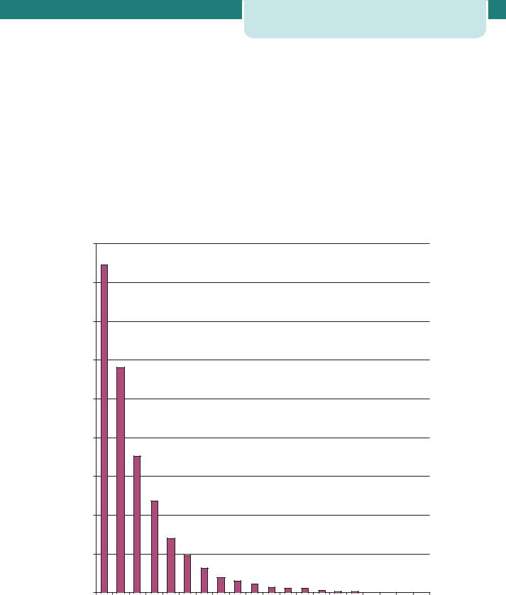
Preterm births contribute significantly to perinatal mortality, half of which results from babies born before 32 weeks. UK infant mortality figures in 2005 for live births at each gestational age are shown in Figure 11.2.
Predicted survival can be modified if accurate information concerning fetal sex, weight and well-being is available. Parents are particularly anxious about the risks of later disability and handicap. These risks are especially significant below 26 weeks gestation. When assessed at six years of age, nearly half the survivors at 23–25 weeks gestation have a moderate or severe disability. Furthermore, many of these disabilities only become apparent after two to three years of age. Survival with no disability is only seen in 1, 3 and 8 per cent of live births at 24, 24 and 25 weeks, respectively. There are other long-term worries after very preterm births, including subsequent growth, educational needs and social behaviour. There may also be influences on later
Infant mortality
|
90 |
|
|
|
|
|
|
|
|
|
|
|
|
|
|
|
|
|
|
|
|
|
80 |
|
|
|
|
|
|
|
|
|
|
|
|
|
|
|
|
|
|
|
|
|
70 |
|
|
|
|
|
|
|
|
|
|
|
|
|
|
|
|
|
|
|
|
|
60 |
|
|
|
|
|
|
|
|
|
|
|
|
|
|
|
|
|
|
|
|
of Live births |
50 |
|
|
|
|
|
|
|
|
|
|
|
|
|
|
|
|
|
|
|
|
40 |
|
|
|
|
|
|
|
|
|
|
|
|
|
|
|
|
|
|
|
|
|
% |
|
|
|
|
|
|
|
|
|
|
|
|
|
|
|
|
|
|
|
|
|
|
|
|
|
|
|
|
|
|
|
|
|
|
|
|
|
|
|
|
|
|
|
|
30 |
|
|
|
|
|
|
|
|
|
|
|
|
|
|
|
|
|
|
|
|
|
20 |
|
|
|
|
|
|
|
|
|
|
|
|
|
|
|
|
|
|
|
|
|
10 |
|
|
|
|
|
|
|
|
|
|
|
|
|
|
|
|
|
|
|
|
|
0 |
23 |
24 |
25 |
26 |
27 |
28 |
29 |
30 |
31 |
32 |
33 |
34 |
35 |
36 |
37 |
38 |
39 |
40 |
41 |
42 |
|
|
||||||||||||||||||||
|
|
|
|
|
|
|
|
|
|
|
Gestation |
|
|
|
|
|
|
|
|
|
|
Figure 11.2 |
Infant mortality of live born infants in the UK, 2005 |
|
|
|
|
|
|
|
|
|
|
||||||||||

134Late miscarriage and early birth
adult health. Fortunately, both morbidity and mortality fall dramatically with increasing gestation.
It is important to recognize the effect of denominator differences in survival figures, especially at the earliest gestations. If the figures are based on fetuses alive at the start of labour, the survival rates will be lower, as there is an inherent risk of intrapartum death. If the figures are based on NICU admissions, the figures will be higher, as some live births will succumb during initial resuscitation in the maternity unit. At 25 weeks or less, such statistical manipulations may lead to a change of nearly 10 per cent in the quoted survival figures.
Classification
For reasons related to aetiology, outcome and recurrence risk, preterm births should be divided into three gestational periods: mildly preterm births at 32 0 to 36 6 weeks (incidence 6.1 per cent), very preterm births at 28 0 to 31 6 weeks (incidence 0.9 per cent) and extremely preterm births at 24 0 to 27 6 weeks (incidence 0.4 per cent).
Late miscarriages occur across a broad gestational age range. At the latter part of the second trimester, between 17 and 23 weeks, the commonest factors underlying such losses will be those linked to extremely preterm births. Indeed, research shows that women who have such a late second trimester delivery are at increased risk of a very preterm birth in their next pregnancy.
As the interval between symptom onset and delivery can occasionally be measured in weeks, some ‘inevitable’ or ‘threatened’ second trimester miscarriages may result in preterm deliveries. Pre-viable membrane rupture represents the most common precedent for this scenario. Before 22 weeks gestation, membrane rupture with severe oligohydramnios carries a significant risk of lethal fetal lung hypoplasia. Unfortunately, this complication cannot be reliably predicted antenatally. The parents should be counselled as to the poor prognosis but if there are no signs of infection, there is no urgency to interfere. Some may want to wait for nature to take its course; others may want more immediate action. In both cases, continuing support is important. No harm will come from conservative management as long as careful observation is made for signs of infection. Established chorioamnionitis usually declares itself with contractions and delivery. Rarely do the membranes seal and fluid reaccumulate. This is usually only seen when membrane rupture follows amniocentesis.
Aetiology
Labour at term and prior to it share a common pathway involving uterine contractility, cervical effacement and dilatation and membrane rupture. At term, the activation of this pathway is physiological. However, a variety of pathologies underlie labour remote from term. It has been suggested by some authors that preterm labour be considered a syndrome, in order to emphasize its multifactorial nature.
Infection
Subclinical intrauterine infection of the choriodecidual space and amniotic fluid is the most widely studied aetiological factor underlying spontaneous preterm births. The uterine cavity is normally sterile but the vagina contains commensal bacteria. Depending on the bacterial load and cervical resistance, the bacteria may ascend through the cervix and reach the fetal membranes. This may activate the decidua, increase prostaglandin release and trigger contractions. Alternatively, it may weaken the membranes, leading to rupture. Early-onset neonatal sepsis, maternal postpartum endometritis and histological chorioamnionitis are all significantly more common after preterm birth, particularly those very early deliveries before 32 weeks.
Over-distension
The commonest cause of uterine over-distension is multiple gestation. Polyhydramnios has a similar effect. Overstretching of the myometrium (and possibly the membranes) leads to increased contractile activity and premature shortening and opening of the cervix.
Vascular
Disturbance at the uteroplacental interface may lead to intrauterine bleeding. The blood can track down behind the membranes to the cervix and be revealed. Alternatively, it may track away from the cervix and be concealed. Either way, the blood irritates the uterus, leading to contractions, and damages the membranes, leading to early rupture.
Surgical procedures and intercurrent illness
Serious maternal infective illnesses such as pyelonephritis, appendicitis and pneumonia are associated with preterm labour. In these cases,

preterm labour is presumed to be due either to direct blood-borne spread of infection to the uterine cavity or indirectly to chemical triggers, such as endotoxins or cytokines. Many other illnesses, such as cholestasis of pregnancy, and non-obstetric surgical procedures are associated with preterm labour, although the mechanisms for this remain obscure.
Amniocentesis is a pregnancy-specific procedure associated with an increased risk of late miscarriage and early birth. It is most commonly performed at 15–18 weeks gestation. It is associated with a 0.5 per cent chance of subsequent pregnancy loss before viability. This may happen in the days after the procedure but many losses occur several weeks later and a small increased chance of preterm delivery persists after reaching viability.
Abnormal uterine cavity
A uterine cavity that is distorted by congenital malformation may be less able to accommodate the developing pregnancy. Associated abnormal placentation and cervical weakness may also contribute. Fibroids in a low position may also lead to complications. However, fibroids are common and most pregnancies are successful despite their presence.
Cervical weakness
Due to previous surgical damage or a congenital defect, the cervix may shorten and open prematurely. The membranes then prolapse and may be damaged by stretching or by direct contact with vaginal pathogens. These same pathogens may ascend and trigger contractions. Often referred to as ‘cervical incompetence’, weakness may be a better term. The evidence suggests that gradations of deficiency exist, rather than an ‘all-or-nothing’ phenomenon.
This remains a notoriously difficult diagnosis to make, as dilatation of the cervix remains the final common pathway for all late miscarriages and early births. Reliably distinguishing between such dilatation being the primary event or secondary to other pathologies is challenging.
Idiopathic
In many cases, especially mildly preterm births between 34 and 36 weeks, no cause will be found. In these cases, the physiological pathways to parturition may simply have been turned on too early.
Clinical features of preterm labour |
135 |
Risk factors for preterm labour/PPROM
Non-modifiable, major
•Last birth preterm: 20 per cent risk
•Last two births preterm: 40 per cent risk
•Twin pregnancy: 50 per cent risk
•Uterine abnormalities
•Cervical anomalies:
•cervical damage (cone biopsy, repeated dilatation)
•fibroids (cervical)
•Factors in current pregnancy:
•recurrent antepartum haemorrhage
•intercurrent illness (e.g. sepsis)
•any invasive procedure or surgery
Non-modifiable, minor
•Teenagers having second or subsequent babies
•Parity (0 or 5)
•Ethnicity (black women)
•Poor socioeconomic status
•Education (not beyond secondary)
Modifiable
•Smoking: two-fold increase of PPROM
•Drugs of abuse: especially cocaine
•Body mass index (BMI) 20: underweight women
•Inter-pregnancy interval 1 year
Clinical features of preterm labour
History
Always check the dating of the pregnancy by reviewing the menstrual history and, if possible, any prior ultrasound examinations. This is critical at gestations near viability.
Less than 50 per cent of all women presenting with symptoms suggesting a risk of early delivery will deliver within 7 days. Too much emphasis is often placed on the contraction frequency. In isolation, it correlates poorly with the risk of preterm birth. Markers of intensity, such as analgesic requirements or simple bedside clinical impression, may add refinement. Vague complaints such as increased discharge, pelvic pressure or low backache are sometimes reported, with the latter two often showing a cyclical pattern. Nonetheless, the diagnosis of