

Obstetrics_by_Ten_Teachers_19E_-_Kenny_Louise
.pdf116 Twins and higher multiple gestations
At present, transvaginal cervical ultrasound shows the most promise as a predictor of very preterm delivery (see Chapter 11, Late miscarriage and early birth). As regular ultrasound examination is already part of the care of multiple pregnancy, there is little impact on health-care resources. Once preterm labour is diagnosed, neonatal unit staff must be promptly involved. The use of tocolytic drugs in this situation, particularly the beta-agonists, carries risks of serious maternal morbidity.
Multiple pregnancy support groups
Twin pregnancies are associated with a number of financial, personal and social costs for families, that continue long beyond the neonatal period. A significant contribution to these costs comes from the increased incidence of handicap, largely secondary to preterm delivery. Several specialized support groups for multiple pregnancy exist. In the UK, these include the Twins and Multiple Birth Association (TAMBA) and the Multiple Birth Foundation. All parents expecting twins should be given contact details for such resources locally.
Intrapartum management
Complications in labour are more common with twin gestations. These include premature birth, abnormal presentations, prolapsed cord, premature separation of the placenta and postpartum haemorrhage. Judiciously managed, labour is generally considered to be safe. It may require considerable expertise and is the only situation in which internal podalic version is still practised in obstetrics.
keep this separate, not run through a giving-set until needed and clearly labelled ‘for postpartum use only’. It is essential that two neonatal resuscitation trolleys, two obstetricians and two paediatricians are available and that the special care baby unit and anaesthetist are informed well in advance of the delivery.
Analgesia during labour
Epidural analgesia is recommended. Indeed, if the presentation of twins is anything other than vertex– vertex, the use of an epidural can be justified in terms of analgesia for possible intrauterine manipulations required in the second stage for delivery of the second twin. Contrary to recommendations for the management of a singleton labour, the epidural should be kept running throughout the second stage, as it is most likely to be required after the delivery of the first twin. Having an anaesthetist present and ready to administer anaesthesia if complications arise is a satisfactory alternative. However, many obstetric units now opt to deliver twins in the operating theatre.
Fetal well-being in labour
Fetal heart rate monitoring should be continuous throughout labour, ideally using a specialized twin monitor. An abnormal fetal heart rate pattern in the first twin may be assessed using fetal scalp sampling, as for a singleton pregnancy. However, a non-reassuring pattern in the second twin will usually necessitate delivery by Caesarean section. The condition of the second twin must be carefully monitored after the delivery of the first twin, as acute complications such as cord prolapse and placental separation are well recognized.
Preparation
This should begin long before labour, with antenatal education and an intrapartum care plan. A twin CTG machine should be used for fetal monitoring and a portable ultrasound machine should be available during the delivery. A standard oxytocin solution for augmentation should be prepared, run through an intravenous giving-set and clearly labelled ‘for augmentation’, for use for delivery of the second twin, if required. A second high-dose oxytocin infusion should also be available for the management of postpartum haemorrhage. However, it is advisable to
Vaginal delivery of vertex–vertex
Although this combination is considered low risk, an obstetrician should be present, as complications with delivery of the second twin can occur. Delivery of the first twin is undertaken in the usual manner and thereafter the majority of second twins will be delivered within 15 minutes. However, there is no urgency to deliver the second twin within a set time period, providing both mother and baby remain well.
After the delivery of the first twin, abdominal palpation should be performed to assess the lie of the second twin. It is helpful to use ultrasound for

|
|
Intrapartum management |
117 |
confirmation, which is also useful for checking the fetal heart rate. If the lie is longitudinal with a cephalic presentation, one should wait until the head is descending and then perform amniotomy with a contraction. If contractions do not ensue within 5–10 minutes after delivery of the first twin, an oxytocin infusion should be started. The indications for instrumental delivery of the second twin are as for singletons (see Chapter 15, Operative intervention in obstetrics).
Delivery of vertex–non-vertex
If the second twin is non-vertex, which occurs in about 40 per cent of twins, numerous studies have shown that vaginal delivery can be safely considered.
If the second twin is a breech, the membranes can be ruptured once the breech is fixed in the birth canal. A total breech extraction may be performed if fetal distress occurs or if a footling breech is encountered, but this requires considerable expertise. Complications are less likely if the membranes are not ruptured until the feet are held by the operator. Where the fetus is transverse, external cephalic version can be successful in more than 70 per cent of cases. The fetal heart rate should be closely monitored, and ultrasound can be helpful to demonstrate the final position of the baby. If external cephalic version is unsuccessful, and assuming that the operator is experienced, an internal podalic version can be undertaken (Figure 9.4).
A fetal foot is identified by recognizing a heel through intact membranes. The foot is grasped and pulled gently and continuously into the birth canal. The membranes are ruptured as late as possible. This procedure is easiest when the transverse lie is with the back superior or posterior. If the back is inferior or if the limbs are not immediately palpable, ultrasound may help to show the operator where they would be found. This will minimize the unwanted experience of bringing down a fetal hand in the mistaken belief that it is a foot.
Non-vertex first twin
When the first twin presents as a breech, clinicians usually recommend delivery by elective Caesarean section. This is largely because of the increased risks associated with singleton breech vaginal delivery. Other factors include dwindling experience of breech delivery and the rarely seen phenomenon of ‘locked twins’.
Figure 9.4 Internal podalic version
118 Twins and higher multiple gestations
In this latter case, the chin of the first (breech) baby locks against the chin of the second (cephalic) twin.
Preterm twins
Even in low birth weight twin gestations, the method of delivery in relation to fetal presentation will have little or no effect on neonatal mortality and subsequent neonatal developmental outcome. No significant differences in perinatal outcome exist when comparing breech-extracted second twins to those delivered by Caesarean section.
Requirements for twin delivery
•Large delivery room
•Operating theatre and staff ready
•Anaesthetist present
•Senior obstetrician present
•At least two midwives present
•Twin resuscitaires
•Ventouse/forceps to hand
•Blood grouped and saved
•Intravenous access
•Neonatologists present
•Pre-mixed oxytocin infusion ready
Postpartum haemorrhage
The risk of postpartum haemorrhage is increased in twin pregnancies due to the larger placental site and uterine over-distension. For that reason, all multiple gestations should have an intravenous line and blood grouped and saved during labour.
Management is generally no different from that of postpartum haemorrhage complicating singleton delivery (see Chapter 16, Obstetric emergencies). However, ideally, the third stage should be actively managed and a high-dose oxytocin infusion commenced following delivery as prophylaxis.
Higher multiples
A consequence of the widespread introduction of assisted reproductive techniques has been an exponential increase in the incidence of higher
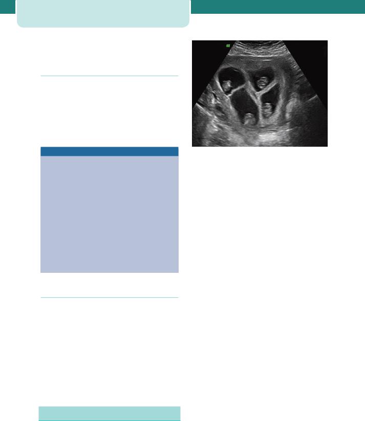
Figure 9.5 First trimester ultrasound showing quadruplet pregnancy
multiple gestations (Figure 9.5), mostly triplets. At least 75 per cent of triplet pregnancies are secondary to assisted conception. They are associated with increased risks of miscarriage, perinatal death and handicap. The median gestational age at birth is 33 weeks and long-term complications are primarily a consequence of extremely preterm delivery. Although the demands on maternal physiology are greater still, antenatal care is essentially no different from that for a twin gestation. Caesarean section is usually advocated for delivery due to the difficulties of intrapartum fetal monitoring; however, the evidence to support this strategy is weak, and several case series of vaginal birth have reported comparable neonatal outcomes.
In an attempt to reduce the morbidity and mortality associated with extremely preterm delivery, the procedure of multi-fetal reduction was introduced. Iatrogenic fetal death is achieved by the ultrasound-guided puncture of the fetal heart and injection of potassium chloride. Although it is technically feasible to perform reduction from as early as 7 weeks gestation, it is usually delayed until around 11–12 weeks. This allows for spontaneous reduction to occur and for the screening and diagnosis of major fetal abnormalities and chromosomal defects. Following reduction, there is gradual resorption of the dead fetuses and their placentae.
A triplet pregnancy managed expectantly has a 4 per cent chance of loss prior to 24 weeks and a

|
|
Higher multiples |
119 |
25 per cent risk of extremely preterm delivery between 24 and 32 weeks. After multi-fetal reduction, usually to twins, the risk of loss before viability is increased to 8 per cent. However, the chance of a
very preterm birth drops to 10 per cent. The risks of subsequent miscarriage and extremely preterm delivery have been found to increase with the number of fetuses reduced.
C A S E H I S T O R Y
Miss C is a 19-year-old woman who is single. She is unemployed. She has no active medical problems. She was discovered to have a twin pregnancy at 12 weeks gestation, after presenting to the A&E deptartment with an episode of bleeding. The pregnancy was classified as DCDA. She had no further prenatal care until she presented to the Maternity Unit with reduced fetal movements at 28 weeks.
An ultrasound was organized and showed one fetus (twin A) to be growth restricted (with an estimated weight on the fifth centile) and have oligohydramnios. The other fetus was on the 40th centile.
Could this be TTTS?
One-third of monozygotic twins will be dichorionic. However, it is the monochorionic placentation that carries a risk of unbalanced vascular anastamoses between the two fetuses. Therefore, TTTS is unlikely.
What other useful information should you seek from this
ultrasound?
Miss C must have missed her review of fetal anatomy at
19–20 weeks. As anomalies can be associated with intrauterine growth restriction (IUGR), a careful anatomy review should be performed. Doppler studies of the fetal circulation may also aid in diagnosis, with increased resistance in the umbilical arteries and a ‘brain-sparing’ effect in the fetal circulation seen in true IUGR. As the patient presented with reduced fetal movement, some
assessment of individual fetal activity could be undertaken, such as
a biophysical profile.
If fetal activity was not assessed during the ultrasound,
which test is indicated?
A CTG should be performed. Although there may be only slight differences in the heart rate patterns of the two fetuses, a special twin monitor allows them to be clearly identified.
Although the umbilical Doppler for twin A is abnormal,
the CTG is reactive with accelerations. Should delivery be
organized?
Delivery would subject the healthy and well-grown twin B to the risks of iatrogenic prematurity. The risk of death or serious morbidity to twin A by remaining in utero has to be balanced against this.
With a normal CTG and no evidence of acute compromise, delivery would not be appropriate at this time.
What other antenatal measures should be taken?
Close follow-up of fetal well-being should be organized, at least on a weekly basis. Steroids should be considered for fetal lung maturity, as early delivery is a possibility. All the routine antenatal tests should now be organized, as Miss C has missed a large component of
her prenatal care. Miss C may be at risk for anaemia. Social work involvement may be appropriate, to discover the underlying factors behind her failure to obtain antenatal care.
New developments
•Transvaginal ultrasound measurement of cervical length at 20–24 weeks may be used to assess the risk of very preterm delivery.
•The interrelated phenomena of delayed childbearing and increased use of assisted conception techniques have dramatically increased the incidence of multiple pregnancy.
•The optimal way to screen for Down’s syndrome in multiple pregnancy is by nuchal translucency at 12 weeks gestation. Chorionicity can also be reliably determined at this time.
•Multi-fetal reduction is increasingly accepted as improving overall outcomes in higher multiples.
Key points
•Twins account for about 1.5 per cent of pregnancies but up to 25 per cent of special care baby unit admissions.
•Perinatal mortality rate in twins is nearly six times higher than in singletons, primarily due to spontaneous preterm births.
•Both serious maternal complications and minor discomforts are increased in multiple gestation.
•The determination of chorionicity is very important – the highest risks are seen in monochorionic twins.

|
C H A P T E R 1 0 |
|
|
|
|
|
|
|
|
|
|
|
|
|
PRE - ECLAMPSIA AND OTHER |
|
|
||||||||||
|
|
DISORDERS OF PLACENTATION |
|||||||||||
|
|
Louise C Kenny |
|
|
|||||||||
|
|
|
|
|
|
||||||||
|
The placenta............................................................................................... |
120 |
Definition and incidence ..................................................................... |
126 |
|
||||||||
|
Pre-eclampsia ........................................................................................... |
121 |
Placental abruption ................................................................................ |
128 |
|
||||||||
|
Fetal growth restriction ....................................................................... |
125 |
Additional reading................................................................................... |
131 |
|
||||||||
|
|
|
|
|
|
|
|
|
|
|
|
|
|
O V E R V I E W
Pre-eclampsia is a leading cause of maternal death. The World Health Organization estimates that globally between 50 000 and 75 000 women die of this condition each year. Furthermore, pre-eclampsia is frequently accompanied by fetal growth restriction (FGR), which is responsible for considerable perinatal morbidity and mortality. Although fetal growth is controlled by a number of factors including genetic predisposition and maternal nutritional status, it is now apparent that the origins of both pre-eclampsia and much of FGR seen in clinical practice lie in defective placentation. A further condition frequently related to impaired placentation is abruptio placentae or placental abruption. This is the premature separation of a normally sited placenta, which is usually of sudden onset and associated with a high fetal mortality and substantial maternal morbidity and mortality. Knowledge of the early events in the invasion of the maternal uterine wall by placental trophoblast cells is therefore helpful in understanding the aetiology of these important clinical conditions.
The placenta
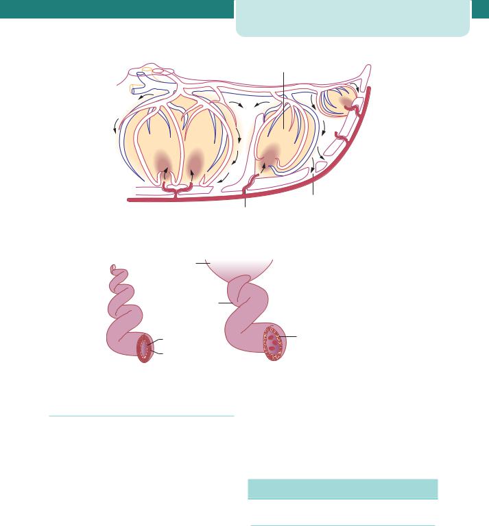
The placenta is a fetomaternal organ. The functional unit of the placenta is the fetal cotyledon. The mature human placenta has about 120 fetal cotyledons grouped into visible lobes (frequently and somewhat confusingly termed ‘maternal cotyledons’). Each cotyledon contains a primary villus stem arising from the chorionic plate and supplied by primary branches of fetal vessels. The primary stems divide to form secondary and tertiary stems from which arise the terminal villi, where maternal–fetal exchange takes place. The fetal cotyledons appear to develop around the entries of the maternal spiral arteries from the decidual plate. The centre of each cotyledon is hollow and during maternal systole, blood spurts from the spiral arteries and enters the intercotyledon space. Blood rises high to the chorionic plate then disperses laterally between and over the surface of the terminal villi, becoming increasingly desaturated of oxygen and nutrients and picking up carbon dioxide and waste products. The blood then filters into narrow venous channels between the cotyledons, before falling back to the maternal decidual plate, where the maternal veins
return the desaturated blood to the maternal circulation (Figure 10.1). Maternal and fetal blood is separated by three microscopic tissue layers: trophoblastic tissue, connective tissue and the endothelium of the fetal capillaries. However, microscopic examination of the terminal villi surrounding the intracotyledon space shows numerous vasculosyncytial membranes where the fetal capillaries and trophoblast fuse to form a very thin membrane, where most of the transfer of nutrients and blood gases takes place.
Normal placentation
The maternal blood flow to the placenta increases throughout pregnancy from 50 mL/min in the first trimester to 500–750 mL/min at terms. This increase in perfusion is accomplished by anatomical conversion of the maternal spiral arteries by trophoblast. Trophoblast cells invade the spiral arterioles within the first 12 weeks of pregnancy and replace the smooth muscle of the wall of the vessels, thus converting them to wide bore, low resistance, large capacitance vessels (Figure 10.2). This process is normally complete by 20 weeks gestation.
Pre-eclampsia 121
Intracotyledonary
space
Maternal
veins
Spiral artery
Figure 10.1 Anatomy and distribution of blood flow through the intracotyledonary space
Invading |
Creation of a high-flow, |
|
trophoblasts |
low resistance zone |
|
Loss of |
|
|
spirality |
|
|
Endothelium |
Replacement of smooth |
|
muscle and endothelium |
||
|
||
Smooth muscle |
with trophoblasts |
|
(tunica media) |
|
Figure 10.2 Physiological change of spiral arteries by invading trophoblast
Abnormal placentation
In pregnancies destined to be complicated by preeclampsia, FGR and/or abruptio placentae, there is a complete or partial failure of trophoblast invasion of the myometrial segments of the spiral arteries. Hence, spiral arteries retain some of their pre-pregnancy characteristics being relatively narrow bore and of low capacitance and high resistance and resulting in impaired perfusion of the fetoplacental unit. The mechanism underlying decreased trophoblast invasion in complicated pregnancies is poorly understood but it may reflect an ‘immune intolerance’ of the mother to the invading trophoblast. Affected placentae have gross morphological changes, which include infarcts and basal haematomas. An infarct represents an area of ischaemic necrosis of a cotyledon resulting from a spiral artery occlusion, usually by thrombosis. A placenta
with multiple infarcts is significantly associated with intrauterine fetal death and growth restriction. Basal haematomas consist of a mass of blood in the centre of the fetal cotyledon due to the rupture of a damaged spiral artery. This lesion is associated with maternal hypertension and increased perinatal mortality.
Pre-eclampsia
Definition
In the past, the definition of pre-eclampsia has been inconsistent and this has led to difficulty in comparing studies on treatments or outcomes. There is now a widely accepted classification system of hypertensive disorders in pregnancy (see Chapter 12, Medical diseases complicating pregnancy), which defines

122Pre-eclampsia and other disorders of placentation
pre-eclampsia as hypertension of at least 140/90 mmHg recorded on at least two separate occasions and at least 4 hours apart and in the presence of at least 300 mg protein in a 24 hour collection of urine, arising de novo after the 20th week of pregnancy in a previously normotensive woman and resolving completely by the sixth postpartum week.
Chronic hypertension (with or without renal disease) existing prior to pregnancy can predispose to the later development of superimposed pre-eclampsia. Even in the absence of superimposed pre-eclampsia, chronic hypertension is associated with increased maternal and fetal morbidity (see Chapter 12, Medical diseases complicating pregnancy) and pregnancies complicated by chronic hypertension should therefore be regarded as high risk.
Non-proteinuric gestational hypertension, i.e. hypertension arising for the first time in the second half of pregnancy and in the absence of proteinuria, is not associated with adverse pregnancy outcome. Every effort therefore should be made to clearly distinguish it from pre-eclampsia.
Incidence
Pre-eclampsia complicates approximately 2–3 per cent of pregnancies, but the incidence varies depending on the exact definition used and the population studied. In the most recent Confidential Enquiry (2003–2005), there were 18 deaths due to pre-eclampsia, making this the second most common cause of direct death in pregnancy and the puerperium in the UK.
Epidemiology
Pre-eclampsia is more common in primigravid women. It is thought that the normal fetal–maternal transfusion that occurs during pregnancy and particularly during delivery exposes the mother to products of the fetal (and hence paternal) genome, protecting her in subsequent pregnancies. In line with this, the protective effect of first pregnancy seems to be lost if a woman has a child with a new partner. Overall the recurrence risk in a subsequent pregnancy is 20 per cent, but is much higher if severe pre-eclampsia developed at an extremely early gestation in the first pregnancy. There also appears to be a maternal genetic predisposition to pre-eclampsia as there is a threeto four-fold increase in the incidence of pre-eclampsia in the first degree relatives of affected women. Finally,
there are a number of general medical conditions and pregnancy-specific factors that predispose to the development of pre-eclampsia.
Risk factors for pre-eclampsia
•First pregnancy
•Multiparous with:
•pre-eclampsia in any previous pregnancy
•ten years or more since last baby
•Age 40 years or more
•Body mass index of 35 or more
•Family history of pre-eclampsia (in mother or sister)
•Booking diastolic blood pressure of 80 mmHg or more
•Booking proteinuria (of 1 on more than one occasion or quantified at 0.3 g/24 hour)
•Multiple pregnancy
•Certain underlying medical conditions:
•pre-existing hypertension
•pre-existing renal disease
•pre-existing diabetes
•antiphospholipid antibodies
Aetiology and pathophysiology
Pre-eclampsia only occurs in pregnancy, but has been described in pregnancies lacking a fetus (molar pregnancies) and in the absence of a uterus (abdominal pregnancies) (see Chapter 9, Problems in early pregnancy, in Gynaecology by Ten Teachers, 19th edn), suggesting that it is the presence of trophoblast tissue that provides the stimulus for the disorder. Placental bed biopsies have demonstrated that trophoblast invasion is patchy in pre-eclampsia and the spiral arteries retain their muscular walls. This is thought to prevent the development of a high flow, low impedance uteroplacental circulation. The reason why trophoblast invades less effectively in these pregnancies is not known but may reflect an abnormal adaptation of the maternal immune system.
It is widely believed that defective trophoblast invasion results in relative under-perfusion of the placenta and that this releases a factor(s) into the maternal circulation that targets the vascular endothelium (Figure 10.3). The nature of this factor
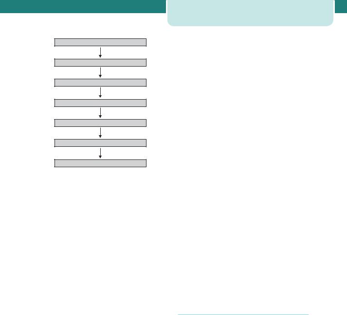
Genetic predisposition
Abnormal immunological response
Deficient trophoblast invasion
Hypoperfused placenta
Circulating factor(s)
Vascular endothelial cell activation
Clinical manifestations of disease
Figure 10.3 The proposed aetiology of pre-eclampsia
has not been identified, although numerous candidates have been proposed including a variety of growth factors, cytokines and products of oxidative stress caused by hypoxic-reperfusion injury in the placenta.
As the target cell of the disease process, the vascular endothelial cell, is so ubiquitous, pre-eclampsia is a truly multisystem disease, affecting multiple organ systems, often concurrently.
Cardiovascular system
Normal pregnancy is characterized by marked peripheral vasodilatation resulting in a fall in total peripheral resistance despite an increase in plasma volume and cardiac rate. Pre-eclampsia is characterized by marked peripheral vasoconstriction, resulting in hypertension. The intravascular high pressure and loss of endothelial cell integrity results in greater vascular permeability and contributes to the formation of generalized oedema.
Renal system
In the kidney, a highly characteristic lesion called ‘glomeruloendotheliosis’ is seen. This is relatively specific for pre-eclampsia (it is not seen with other hypertensive disorders) and is associated with impaired glomerular filtration and selective loss of intermediate weight proteins, such as albumin and transferrin, leading to proteinuria. This, in turn, causes a reduction in plasma oncotic pressure and exacerbates the development of oedema.
Pre-eclampsia 123
Haematological system
In the event of endothelial damage, platelets adhere to the damaged area. Furthermore, diffuse vascular damage is associated with the laying down of fibrin. Pre-eclampsia in association with increased fibrin deposition and a reduction in the platelet count may accompany and occasionally predate the onset of disease.
The liver
In the liver, subendothelial fibrin deposition is associated with elevation of liver enzymes. This can be associated with haemolysis and a low platelet count due to platelet consumption (and subsequent widespread activation of the coagulation system). The presence of these findings is called HELLP syndrome (haemolysis, elevation of liver enzymes and low platelets). HELLP syndrome is a particularly severe form of pre-eclampsia, occurring in just 2–4 per cent of women with the disease. It is associated with a high fetal loss rate (of up to 60 per cent). The management of HELLP syndrome is discussed further in Chapter 16, Obstetric emergencies.
Neurological system
The development of convulsions in a woman with pre-eclampsia is defined as eclampsia. Vasospasm and cerebral oedema have both been implicated in the pathogenesis of eclampsia. Retinal haemorrhages, exudates and papilloedema are characteristic of hypertensive encephalopathy and are rare in preeclampsia, suggesting that hypertension alone is not responsible for the cerebral pathology.
Clinical presentation
The classic symptoms of pre-eclampsia include a frontal headache, visual disturbance and epigastric pain. However, the majority of women with pre-eclampsia are asymptomatic or merely complain of general, vague ‘flu-like’ symptoms.
Clinical examination should include a complete obstetric and neurological examination. Hypertension is usually the first sign, but occasionally is absent or transient until the late stages of the disease. Dependent oedema of the feet is very common in healthy pregnant women. However, rapidly progressive oedema of the face and hands may suggest pre-eclampsia. Epigastric tenderness is a worrying sign and suggests liver involvement. Neurological examination may reveal

124 Pre-eclampsia and other disorders of placentation
hyperreflexia and clonus in severe cases. Urine testing for protein should be considered part of the clinical examination.
Testing for proteinuria
Dipstick urinalysis
•Instant result but quantitatively inaccurate
•Results: trace: seldom significant; 1 : possible significant proteinuria, warrants quantifying; 2 : probable significant proteinuria, warrants quantifying
Protein:creatinine ratio
•Fast (within an hour)
•Results semi-quantitative: 30 mg/mol – probable significant proteinuria
24 hour collection
•Slow
•Results: 0.3 g/24 hour represents confirmed significant proteinuria
Management and treatment
There is no cure for pre-eclampsia other than to end the pregnancy by delivering the baby (and placenta). This can be a significant problem if pre-eclampsia occurs early in pregnancy, particularly at gestations below 34 weeks. Therefore, management strategies are aimed at minimizing risk to the mother in order to permit continued fetal growth. In severe cases, this is often not possible.
The principles of management of pre-eclampsia are:
•early recognition of the symptomless syndrome;
•awareness of the serious nature of the condition in its severest form;
•adherence to agreed guidelines for admission to hospital, investigation and the use of
antihypertensive and anticonvulsant therapy;
•well-timed delivery to pre-empt serious maternal or fetal complications;
•post-natal follow up and counselling for future pregnancies.
A diagnosis of pre-eclampsia usually requires admission. Patients with mild hypertension, minimal protein and normal haematological and biochemical parameters may be monitored as outpatients but will
require frequent attendance for fetal and maternal assessment. Women with moderate or severe hypertension, significant proteinuria or abnormal haematological or biochemical parameters require admission and inpatient management. Investigations indicated in the ongoing management of preeclampsia are listed in the following box.
Investigations for pre-eclampsia
To monitor maternal complications:
•Full blood count (with particular emphasis on falling platelet count and rising haematocrit)
•If platelet values are normal, additional clotting studies are not indicated
•Serum renal profile (including serum uric acid levels)
•Serum liver profile
•Frequent repeat proteinuria quantification is probably unhelpful once a diagnosis of pre-eclampsia has been made
To monitor fetal complications
•Ultrasound assessment of:
•fetal size
•amniotic fluid volume
•maternal and fetal Dopplers
•Antenatal cardiotocography used in conjunction with ultrasound surveillance, provides a useful but by no means infallible indication of fetal well-being. A loss of baseline variability or decelerations may indicate fetal hypoxia
The aim of antihypertensive therapy is to lower the blood pressure and reduce the risk of maternal cerebrovascular accident without reducing uterine blood flow and compromising the fetus. There are a variety of antihypertensives used in the management of pre-eclampsia. Methyldopa is a centrally acting antihypertensive agent. It has a long established safety record in pregnancy. However, it can only be given orally, it takes upwards of 24 hours to take effect and has a range of unpleasant side effects, including sedation and depression. These properties limit its usefulness. Labetalol is an alpha-blocking and betablocking agent. It too has a good safety record in pregnancy and can be given orally and intravenously. Nifedipine is a calcium-channel blocker with a rapid onset of action. It can, however, cause severe headache that may mimic worsening disease.

In severe cases of fulminating disease, an intravenous infusion of hydralazine or labetalol can be titrated rapidly against changes in the blood pressure. The drug of choice for the treatment of eclampsia is magnesium sulphate. This is given intravenously and has been shown to reduce the incidence of further convulsions in women with eclampsia. Magnesium sulphate should also be used in women with severe pre-eclampsia to prevent the onset of convulsions.
In the UK, over half of all women who died of pre-eclampsia or eclampsia in the last Confidential Enquiry into Maternal and Child Health (2003–2005) died from intracranial haemorrhage. This illustrates the importance of obtaining adequate blood pressure control in women with pre-eclampsia. In women who develop serious multisystem complications, the importance of a multidisciplinary approach involving clinicians from other specialties (e.g. intensive care, haematology, nephrology) cannot be overstated.
Screening and prevention
The accurate prediction of women at risk of developing pre-eclampsia will facilitate targeting of increased antenatal surveillance while allowing women at low risk to participate in community-based antenatal care. In addition, a predictive test would in turn facilitate the development of novel therapeutic preventative interventions. Unfortunately, there is currently no screening test for pre-eclampsia. Despite intensive research in this area, no single blood biomarker has emerged that either alone or in combination with other biomarkers or clinical data possesses sufficient sensitivity and specificity to be clinically useful.
The ability of Doppler ultrasound uterine artery waveform analysis to identify women at risk of pre-eclampsia (and other adverse pregnancy outcomes) has been investigated with varying success. In pregnancies with incomplete trophoblast remodelling of the spiral arteries, a characteristic ‘notch’ can often be seen in the waveform pattern which frequently also demonstrates high resistance (see Chapter 6, Antenatal imaging and assessment of fetal well-being, and Figure 6.20). This screening test may have a role in the women who have already been identified as being at risk of the disease because of their medical or past obstetric history. However, it is not of value in screening low risk women.
Established preventative interventions include low-dose aspirin (typically 75 mg daily), which
Fetal growth restriction |
125 |
modestly reduces the risk of pre-eclampsia in high-risk women, and calcium supplementation may also reduce risk, but only in women with dietary intake. Despite encouraging preliminary studies, its now appears certain that vitamins C and E do not lower the risk of pre-eclampsia.
Additional points in management
Iatrogenic premature delivery of the fetus is often required in severe pre-eclampsia. If her condition allows, the mother should be transferred to a centre with adequate facilities to care for her baby and prior to 34 weeks gestation steroids should be given intramuscularly to the mother to reduce the chance of neonatal respiratory distress syndrome. Delivery before term is often by Caesarean section. Such patients are at particularly high risk for thromboembolism and should be given prophylactic subcutaneous heparin and issued with antithromboembolic stockings. In the case of spontaneous or induced labour and if clotting studies are normal, epidural anaesthesia is indicated as it helps control blood pressure. Ergometrine is avoided in the management of the third stage as it can significantly increase blood pressure.
Post-natally, blood pressure and proteinuria will resolve; however, in a minority of cases one or both persist beyond 6 weeks and this suggests the presence of underlying chronic hypertension or renal disease. Additionally, a careful search should be made post-natally for underlying medical disorders in women who present with severe pre-eclampsia before 34 weeks gestation.
Fetal growth restriction
There are a wide variety of reasons why a baby may be born small including congenital anomalies, fetal infections and chromosomal abnormalities. However, most babies that are born small are either constitutionally small (i.e. healthy, but born to small parents and fulfilling their genetic growth potential) or are small secondary to abnormal placenta function and have FGR.
FGR is a major cause of neonatal and infant morbidity and mortality. There is a significant cost associated with providing adequate facilities to look after these babies. In addition, there is an increasing body of evidence that certain adult diseases (such