Добавил:
Upload Опубликованный материал нарушает ваши авторские права? Сообщите нам.
Вуз: Предмет: Файл:

Obstetrics_by_Ten_Teachers_19E_-_Kenny_Louise

.pdf
Скачиваний:
59
Добавлен:
27.02.2016
Размер:
5.6 Mб
Скачать

196Labour

possible from home or a midwifery-run unit. Women with issues which increase the chances of problems occurring during labour should be recommended to deliver in an obstetric unit and NICE have published a list of obstetric, fetal and medical factors to help guide midwives and obstetricians when they are counselling women. Also, a variety of indications are listed for intrapartum transfer into an obstetric unit, including high maternal pyrexia in labour, delay in labour, concerns regarding fetal well-being, hypertension, retained placenta and complicated perineal trauma requiring suturing.

Management of normal labour

Women are told to contact their local labour suite or their community midwife if they think their waters may have broken (rupture of membranes) or when their contractions are occurring every 5 minutes or more. It is important to recognize that women have very different thresholds for seeking advice and reassurance. The need for pain relief may result in admission to hospital before either of these two criteria is reached. Whether at home or in hospital, the attending midwife will then make an assessment of the situation based on the history and on clinical examination.

History

The following are important points to note in the admission history:

Details of previous births and the size of previous babies (the uneventful birth of normal or large

babies is encouraging). A previous Caesarean section, for example, is an adverse feature, especially if it was performed because of a mechanical problem.

The frequency, duration and perception of strength of the contractions and when they began.

Whether the membranes have ruptured and, if so, the colour and amount of amniotic fluid lost.

The presence of abnormal vaginal discharge or bleeding.

The recent activity of the fetus.

Any medical issues of note that may influence the labour and delivery, e.g. pregnancy-induced

hypertension, fetal growth restriction.

Does she have any special requirements, e.g. an interpreter, or particular emotional/psychological

needs?

What are her expectations of the labour and delivery? Has she made a birth plan? What did she

hope to use for pain relief?

General examination

It is important to recognize women who have a raised body mass index, as this may complicate the management of labour. The temperature, pulse and blood pressure must be recorded and a sample of urine tested for protein, blood, ketones, glucose and nitrates.

Abdominal examination

After the initial inspection for scars indicating previous surgery, it is important to determine the lie of the fetus (longitudinal, transverse or oblique) and the nature of the presenting part (cephalic or breech). If it is a cephalic presentation, the degree of engagement must be determined. A head that remains high and unengaged is a poor prognostic sign for successful delivery. If there is any doubt as to the presentation or if the head is high (five-fifths palpable), an ultrasound scan will determine the presenting part or the reason for the high head (e.g. OP position, deflexed head, placenta praevia, fibroid, etc.) An attempt to estimate the size of the fetus should be made.

Abdominal examination also includes an assessment of the contractions; this takes time (at least 10 minutes) and is done by palpating the uterus directly, not by looking at the tocograph, which provides information only on the frequency and duration of contractions, not the strength.

Vaginal examination

A full explanation of the purpose and technique of vaginal examination is given to the woman and her consent must be obtained. Many women find vaginal examinations extremely distressing and every effort should be made to maintain the woman’s dignity and privacy. The index and middle fingers are passed to the top of the vagina and the cervix. The cervix is examined for dilatation, effacement and application to the presenting part. The dilatation is estimated digitally in centimetres. When no cervix can be felt,

this means the cervix is fully dilated (10 cm). The length of the cervix should be recorded. The cervix at 36 weeks is about 3 cm long. It gradually shortens by the process of effacement. In early labour, it may still be uneffaced. At about 4 cm of dilatation, the cervix should be fully effaced. Providing the cervix is at least 4 cm dilated, it should be possible to determine both the position and the station of the presenting part.

In a normal labour, the vertex will be presenting and the position can be determined by locating the occiput. The occiput is identified by feeling for the triangular posterior fontanelle. Failure to feel the posterior fontanelle may be because the head is deflexed, the occiput is posterior or there is so much caput that the sutures cannot be felt. All of these indicate the possibility of a prolonged labour. Normally, the occiput will be transverse (OT position) or anterior (OA). Relating the lowest part of the head to the ischial spines will give an estimation of the station. This vaginal assessment of station should always be taken together with assessment of the degree of engagement by abdominal palpation. If the head is at or below the ischial spines (0 to 1 or more) and the occiput is anterior (OA), the outlook is favourable for vaginal delivery.

The condition of the membranes should also be noted. If they have ruptured, the colour and amount of fluid draining should be noted. Copious amounts of clear fluid are a good prognostic feature; scanty, heavily blood-stained or meconium-stained fluid is a warning sign for fetal compromise.

Women who are found not to be in established labour should be offered appropriate analgesia and support. Most can safely go home, to return when the contractions increase in strength and frequency.

The admission history and examination act as an initial screen for abnormal labour and increased maternal/fetal risk. If all features are normal and reassuring, the woman will remain under midwifery care. If there are risk factors identified, medical involvement in the form of the on-call obstetric team may be appropriate.

Women in labour should have their pulse measured hourly and their temperature and blood pressure every 4 hours. The frequency of contractions should be recorded every 30 minutes and a vaginal examination performed every 4 hours (unless other factors suggest it needs to be repeated on a different time-frame). It should be noted when the woman voids urine, and this should be tested for ketones and

Management of normal labour

197

protein. Once the second stage is reached, the blood pressure and pulse should be performed hourly, and vaginal examinations offered every hour also.

Fetal assessment in labour

A healthy term fetus is usually able to withstand the rigours of a normal labour. However, with each contraction, placental blood flow and oxygen transfer are temporarily interrupted and a fetus that is already compromised before labour will become increasingly so. Insufficient oxygen delivery to the fetus causes a switch to anaerobic metabolism and results in the generation of lactic acid and hydrogen ions. In excess, these saturate the buffering systems of the fetus and cause a metabolic acidosis which, in the extreme, can cause neuronal damage and permanent neurological injury, even intrapartum fetal death. Hypoxia and acidosis cause a characteristic change in the fetal heart rate pattern, which can be detected by ausculatation and the cardiotocograph (CTG). Meconium is often passed by a healthy fetus at or after term as a result of maturation of gastrointestinal physiology; in this scenario, it is usually thin and a very dark green or brown colour. However, it may also be expelled from a fetus exposed to marked intrauterine hypoxia or acidosis; in this scenario, it is often thicker and much brighter green in colour.

Fetal assessment in labour takes four forms:

1.observation of the colour of the liquor – fresh meconium staining and heavy bleeding are markers of potential fetal compromise

2.intermittent auscultation of the fetal heart using a Pinard stethoscope or a hand-held Doppler ultrasound,

3.continuous external fetal monitoring (EFM) using CTG,

4.fetal scalp blood sampling (FBS).

The fetal heart rate (FHR) should be auscultated with a Pinard stethoscope, or by using a hand-held Doppler device, early on in the initial assessment. It should be listened to for at least a minute, immediately after a contraction. This should be repeated every 15 minutes during first stage, and at least every 5 minutes in second stage. The practice of performing an ‘admission cardiotocograph’ on all women is no longer recommended, however a CTG should be performed if there are issues which might

198Labour

complicate labour and delivery. Most of these women will also be advised to have continuous electronic fetal monitoring (EFM) throughout labour, using the CTG. This may negatively impact on their mobility. Women who begin labour with intermittent auscultation may be advised to change to continuous EFM if any of the following events occur during their labour:

significant meconium staining to the liquor;

abnormal fetal heart rate detected by intermittent auscultation;

maternal pyrexia;

fresh vaginal bleeding;

augmentation of contractions with oxytocin;

at the request of the woman.

The quality of a CTG recording is sometimes poor because of fetal position or maternal obesity. A fetal scalp electrode may overcome this problem. It is fixed into the skin of the fetal scalp and picks up the fetal heart rate directly. It rarely causes any harm to the fetus but requires a certain degree of cervical dilatation to be fitted, and for the membranes to be ruptured if they have remained intact.

The interpretation of the fetal heart rate pattern on a CTG is discussed in Chapter 6, Antenatal imaging and assessment of fetal well-being. In brief,

features of a normal fetal heart rate pattern include a baseline rate of between 110 and 160 bpm (beats per minute), a baseline variability of between 5 and 25 bpm, and the absence of decelerations. Interpreting the CTG in labour is somewhat different to that of an antenatal CTG, particularly in second stage. The absence of accelerations is of uncertain significance during labour, and the presence of simple variable decelarations, or early decelerations, later on in labour is extremely common and not usually a sign of significant fetal compromise.

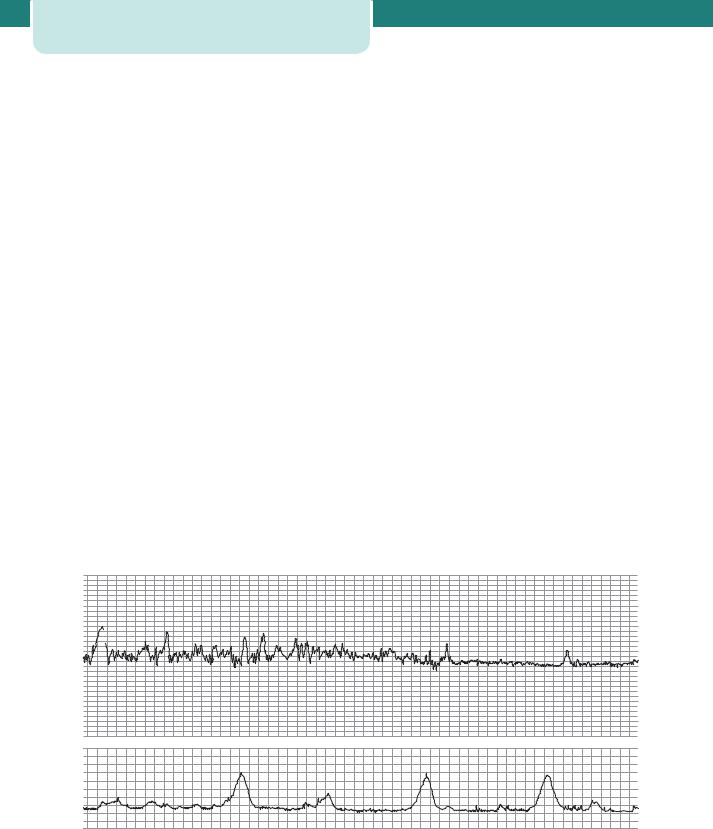
Each feature of the CTG (baseline rate, variability, decelerations and accelerations) should be assessed each time a CTG is reviewed. Each feature can be described as ‘reassuring’, ‘non-reassuring’ or ‘abnormal’ according to certain strict nationally agreed definitions outlined in the NICE guideline on intrapartum management. If all four features are reassuring then the CTG is considered ‘normal’ (see Figure 14.18). If one feature is non-reassuring (and the other three are reassuring) then the CTG is described as ‘suspicious’. If there are two or more nonreassuring features, or any abnormal features, then the CTG is ‘pathological’. Any reversible causes must be considered and addressed and further assessment of the fetus made with fetal blood sampling. If this is not possible or safe then the baby should be delivered without delay.


200


200


180


180


160


160


140


140


120


120


100


100


80


80


60


60


09.05.07 1cm/min

12

100

09.05.07 1cm/min


12

100

09.05.07 1cm/min

12


80


80


10


10


10


8

60


8

60


8


6

40


6

40


6


4


4


4


20


20


2


2


2


013

0

0


014

0

0

015

0

kPa


kPa


kPa


Figure 14.18 A normal cardiotocograph (CTG), showing a baseline fetal heart rate of approximately 120 bpm, frequent accelerations, baseline variability of 10–15 bpm and no decelerations. The uterus is contracting approximately once every 5 minutes

Unfortunately, the CTG can be difficult to interpret and it carries a significant false-positive rate, i.e. it often raises the possibility of fetal compromise when in truth the fetus is still in good condition. In order that the use of the CTG does not lead to unnecessary intervention, a fetal blood sampling may be performed during labour to measure fetal pH and base excess directly (see below under Management of possible fetal compromise). Often, these results are normal even when the CTG is abnormal.

The use of electronic fetal monitors, only introduced in the 1970s, has been controversial and their value in ‘low-risk’ labours is doubtful. Education and training are crucial in the proper use of all equipment. Unfortunately, these devices were introduced before CTG recordings and their outcomes were fully understood. There is little doubt that babies’ lives have been saved by the use of electronic fetal monitors, but they have also contributed to the rise in the Caesarean section and instrumental delivery rates and they lead to reduced mobility in labour and increased parental anxiety. This remains a challenge.
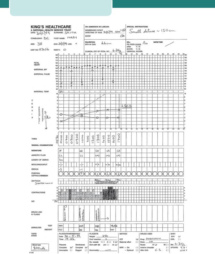
The partogram

The introduction of a graphic record of labour in the form of a partogram has been an important development. This record allows an instant visual assessment of the rate of cervical dilatation and comparison with an expected norm, according to the parity of the woman, so that slow progress can be recognized early and appropriate actions taken to correct it where possible. Other key observations are entered on to the chart, including the frequency and strength of contractions, the descent of the head in fifths palpable, the amount and colour of the amniotic fluid draining, and basic observations of maternal well-being, such as blood pressure, pulse rate and temperature (Figure 14.19).

A line can be drawn on the partogram at the end of the latent phase demonstrating progress of 1 cm dilatation per hour. Another line (‘the action line’) can be drawn parallel and 4 hours to the right of it. If the plot of actual cervical dilatation reaches the action line, indicating slow progress, then consideration should be given to a number of different measures which aim to improve progress (see below). Progress can also be considered slow if the cervix dilates at less than 1 cm every 2 hours.

Management of normal labour

199

Management during first stage

Key management principles of first stage of

labour

The first stage of labour is timed from the diagnosis of onset of labour to full dilatation of the cervix.

Provision of continuity of care and emotional support to the mother.

Observation of the progress of labour with timely intervention if it becomes abnormal.

Monitoring of fetal well-being.

Adequate and appropriate pain relief consistent with the woman’s wishes.

Adequate hydration to prevent ketosis.

Women who are in the latent phase of labour should be encouraged to mobilize and should be managed away from the labour suite where possible. Indeed, they may well go home, to return later when the contractions are stronger or more frequent. Encouragement and reassurance are extremely important. Intervention during this phase is best avoided unless there are identified risk factors. Simple analgesics are preferred over nitrous oxide and epidurals. There is no reason to restrict eating and drinking, although lighter foods and clear fluids may be better tolerated. Vaginal examinations are usually performed every 4 hours to determine when the active phase has been reached (approximately 4 cm dilatation and full effacement). Thereafter, the timing of examinations should be decided by the midwife. Fourhourly is standard practice, however, this frequency may be increased if the midwife thinks that progress is unusually slow or fast. The lower limit of normal progress is 1 cm dilatation every 2 hours once the active phase has been reached. Descent of the presenting part through the pelvis is another crucial component of progress and should be recorded at each vaginal examination. Full dilatation may be reached, but if descent is inadequate, vaginal delivery will not occur.

During the first stage, the membranes may be intact, may have ruptured spontaneously or may have been ruptured artificially. Generally speaking, if the membranes are intact, it is not necessary to rupture them if the progress of labour is satisfactory.

Maternal and fetal observations are carried out as described previously, and recorded on the partogram. Women should receive one-to-one care (i.e. from

200 Labour

Figure 14.19 A typical partogram. This is a partogram of a nulliparous woman of short stature with a big baby and an augmented labour. The labour culminates in an emergency Caesarean section for cephalopelvic disproportion

a dedicated midwife) and should not be left alone for any significant period of time once labour has established. They should be able to choose birth partners themselves and should be able to adopt whatever positions they find most comfortable. Mobility during labour is encouraged and it is likely that standing upright encourages progress. Unfortunately, many women adopt a supine position, especially if there is a need for continuous EFM (i.e. the CTG). Women may drink during established labour and those who are becoming dehydrated may benefit from intravenous fluids to prevent ketosis, which can impair uterine contractility. Light diet is acceptable if they have no obvious risk factors for needing a general anaesthetic and if they have not had pethidine or diamorphine for pain relief. Shaving and enemas are unnecessary and antacids need only be given to women with risk factors for complications, or to those who have had opioid analgesia. A variety of methods of pain relief are available, depending on the location of the birth, and these are discussed below under Pain relief in labour.

‘Active management of labour’ was a collection of interventions which was routinely recommended to nulliparous women to maximize the chances of a normal birth. It included one-to-one midwifery care, 2-hourly vaginal examinations and early artificial rupture of membranes and use of oxytocin augmentation if progress fell more than 2 hours behind the schedule of 1 cm dilatation per hour. A variety of studies failed to show any obvious benefit of active management, except that derived from one- to-one care, the only component now recommended for all women in normal labour.

Second stage

If the labour has been normal, the first sign of the second phase of the second stage is likely to be an urge to push experienced by the mother. Full dilatation of the cervix should be confirmed by a vaginal examination if the head is not visible. The woman will get an expulsive reflex with each contraction, and will generally take a deep breath, hold it, and strain down (the Valsalva manoeuvre). Women will be guided by their own urge to push, however, the midwife has an important role to play, with advice, support and reassurance if progress is poor. Women should be discouraged from lying supine, or semisupine, and should adopt any other position that they

Management of normal labour

201

find comfortable. Lying in the left lateral position and squatting are particularly effective options. There is insufficient good quality evidence to either support or discourage water-birth. Maternal and fetal surveillance intensifies in second stage, as described previously. The development of fetal acidaemia may accelerate, and maternal exhaustion and ketosis increase in line with the duration of active pushing. Use of regional analgesia may interfere with the normal urge to push, and second stage is more often diagnosed on a routine scheduled vaginal examination. Pushing is usually delayed for at least an hour if an epidural is in situ (the ‘passive stage’), however, the baby should be delivered within 4 hours of reaching full dilatation.

Descent and delivery of the head

The progress of the descent of the head can be judged by watching the perineum. At first, there is a slight general bulge as the woman strains. When the head stretches the perineum, the anus will begin to open, and soon after this the baby’s head will be seen at the vulva at the height of each contraction. Between contractions, the elastic tone of the perineal muscles will push the head back into the pelvic cavity. The perineal body and vulval outlet will become more and more stretched, until eventually the head is low enough to pass forwards under the subpubic arch. When the head no longer recedes between contractions (crowning), this indicates that it has passed through the pelvic floor, and delivery is imminent. Vaginal and perineal tears are common consequences of vaginal birth, particularly during first deliveries. The ‘hands-on’ approach has been very popular. As crowning occurs, the hands of the accoucheur are used to flex the fetal head and guard the perineum. The belief is that controlling the speed of delivery of the fetal head will limit maternal damage, however, there is little evidence to support this practice over the alternative ‘hands-off’ approach. Once the head has crowned, the woman should be discouraged from bearing down by telling her to take rapid, shallow breaths (‘panting’). An episiotomy is a surgical cut, performed with scissors, which extends from the vaginal fourchette in a mediolateral direction, usually to the right, through the perineum and incorporating the lower vaginal wall (see Chapter 15, Operative intervention in obstetrics). It is performed during instrumental birth (ventouse or forceps), or to hasten delivery if there is suspected fetal compromise. It will only assist the birth if the head has passed through

202Labour

the pelvic floor, so should not be performed too early. It does not help prevent more severe perineal injury and its routine use in normal spontaneous labour was abandoned some time ago. Effective analgesia is required, and this will usually be with infiltration of local anaesthetic if the woman does not have an epidural.

Delivery of the shoulders and rest of the body

Once the fetal head is born, a check is made to see whether the cord is wound tightly around the neck, thereby making delivery of the body difficult. If this is the case, the cord may need to be clamped and divided before delivery of the rest of the body. With the next contraction, there is external rotation of the head and the shoulders can be delivered. To aid delivery of the shoulders, the head should be pulled gently downwards and forwards until the anterior shoulder appears beneath the pubis. The head is then lifted gradually until the posterior shoulder appears over the perineum and the baby is then swept upwards to deliver the body and legs. If the infant is large and traction is necessary to deliver the body, it should be applied to the shoulders only, and not to the head. Shoulder dystocia (difficulty in delivering the shoulders) is discussed in Chapter 16, Obstetric emergencies.

Immediate care of the neonate

After the infant is born, it lies between the mother’s legs or is delivered directly on to the maternal upper abdomen. The baby will usually take its first breath within seconds. There is no need for immediate clamping of the cord, and indeed about 80 mL of blood will be transferred from the placenta to the baby before cord pulsations cease, reducing the chances of neonatal anaemia and iron deficiency. The baby’s head should be kept dependent to allow mucus in the respiratory tract to drain, and oropharyngeal suction should only be applied if really necessary. After clamping and cutting the cord, the baby should have an Apgar score calculated at 1 minute of age (see Chapter 19, Neonatology) which is then repeated at 5 minutes. Immediate skin-to-skin contact between mother and baby will help bonding, and promote the further release of oxytocin, which will encourage uterine contractions. The baby should be dried and covered with a warm blanket or towel, maintaining this contact. Initiation of breastfeeding should be encouraged within the first hour of life, and routine

newborn measurements of head circumference, birthweight and temperature are usually performed soon after this hour has elapsed. Before being taken from the delivery room, the first dose of vitamin K should be given (if parental consent has been given) and the infant should have a general examination for abnormalities and a wrist label attached for identification.

Third stage

This is timed from the delivery of the baby to the expulsion of the placenta and membranes. This normally takes between 5 and 10 minutes.

Separation of the placenta occurs because of the reduction of volume of the uterus due to uterine contraction and the retraction (shortening) of the lattice-like arrangement of the myometrial muscle fibres. A cleavage plane develops within the decidua basalis and the separated placenta lies free in the lower segment of the uterine cavity. Signs of separation are:

lengthening of the cord protruding from the vulva;

a small gush of blood from the placental bed, which normally stops quickly due to a retraction

of the myometrial fibres;

rising of the uterine fundus to above the umbilicus (Figure 14.20);

the fundus becomes hard and globular compared to the broad, softer fundus prior to separation.

Figure 14.20 Signs of separation and descent of the placenta. After separation, the uterine upper segment rises up and feels more rounded

Management of the third stage can be described as ‘active’ or ‘physiological’. Modern active management of the third stage is a package of care which includes the following:

intramuscular injection of 10 IU of oxytocin, given as the anterior shoulder of the baby is

delivered, or immediately after delivery of the baby;

early clamping and cutting of the umbilical cord;

controlled cord traction (see Figure 14.21).

Figure 14.21 Delivering the placenta by controlled cord traction

Active management of the third stage should be recommended to all women because high quality evidence shows that it reduces the incidence of postpartum haemorrhage from 15 to 5 per cent. When the signs of placental separation are recognized, controlled cord traction is used to expedite delivery of the placenta. When a contraction is felt, the left hand should be moved suprapubically and the fundus elevated with the palm facing towards the mother. At the same time, the right hand should grasp the cord and exert steady traction so that the placenta separates and is delivered gently, care being taken to peel off all the membranes, usually with a twisting motion. Uterine inversion is a rare complication which may occur if the uterus is not adequately controlled with the left hand and excessive traction is exerted on the cord in the absence of a uterine contraction (see Chapter 16, Obstetric emergencies).

In approximately 2 per cent of cases, the placenta will not be expelled by this method. If no bleeding occurs, a further attempt at controlled cord traction should be made after 10 minutes. If this fails, the placenta is ‘retained’ and will require manual removal under general or regional anaesthesia in the operating theatre. Direct injection of oxytocin into the umbilical

Pain relief in labour

203

vein may bring about delivery of the placenta while preparations are being made for theatre.

Physiological management of the third stage is where the placenta is delivered by maternal effort, and no uterotonic drugs are given to assist this process. It is associated with heavier bleeding, but women who are not at undue risk of postpartum haemorrhage should be supported if they choose this option. In the event of haemorrhage, or if the placenta remains undelivered after 60 minutes of physiological management, active management should be recommended.

After completion of the third stage, the placenta should be inspected for missing cotyledons or a succenturiate lobe. If these are suspected, manual removal of the placenta (possibly under ultrasound guidance) should be arranged, because in this situation the risk of postpartum haemorrhage is high.

Finally, the vulva of the mother should be inspected for any tears or lacerations. Minor tears do not require suturing, but tears extending into the perineal muscles (or, indeed, an episiotomy) will require careful repair (see Chapter 15, Operative intervention in obstetrics).

Key points

The key features of normal labour are:

spontaneous onset,

single cephalic presentation,

37–42 weeks gestation,

no artificial interventions,

unassisted spontaneous vaginal delivery,

dilatation of at least 1 cm every 2 hours in the active phase of first stage,

an active second stage of no more than 2 hours in a primiparous woman, and no more than 60 minutes in multiparous women,

a third stage lasting no more than 30 minutes with active management.

Pain relief in labour

The provision of analgesia in childbirth varies between cultures. Some women and their carers believe that there is an advantage in avoiding analgesia, whereas other women will use all methods on offer to limit their pain. Professionals who are knowledgeable about

204Labour

labour and are sympathetic to the labouring woman should give advice regarding pain relief in labour. The method of pain relief is to some extent dependent on the previous obstetric record of the woman, the course of labour and also the estimated length of labour. Just as one woman’s labour can be made into an unhappy experience by unsolicited and unnecessary analgesia, pain relief that is inadequate, or offered too late, can ruin another’s. It should be remembered that the final decision rests with the woman, although there are certain circumstances in which particular forms of analgesia are contraindicated and should not be offered.

Non-pharmacological methods

One-to-one care in labour from a midwife or effective birth partner has been shown to reduce the need for analgesia. Relaxation and breathing exercises may help the woman to manage her pain. Prolonged hyperventilation can make the woman dizzy and can cause alkalosis. Homeopathy, acupuncture and hypnosis are sometimes employed, but their use has not been associated with a significant reduction in pain scores or with a reduced need for conventional methods of analgesia, and they are probably not widely applicable.

Relaxation in warm water during the first stage of labour often leads to a sense of well-being and allows women to cope much better with the pain. The temperature of the water should not exceed 37.5ºC. Clearly, a woman in labour cannot use an opiate or have an epidural sited while in water. Transcutaneous electrical nerve stimulation (TENS) works on the principle of blocking pain fibres in the posterior ganglia of the spinal cord by stimulation of small afferent fibres (the ‘gate’ theory). It has been shown to be ineffective in reducing pain scores or the need for other forms of analgesia and should no longer be offered to women in established labour. It does not have any adverse effects, but is often disappointing. It may still be of use in the latent phase of labour.

Pharmacological methods

Opiates, such as pethidine and diamorphine, are still used in most obstetric units and indeed can be administered by midwives without the involvement of the medical staff. This may be one of the reasons for their popularity. They should be available in all

birth settings but they provide only limited pain relief during labour and furthermore may have significant side effects including:

nausea and vomiting (they should always been given with an anti-emetic);

maternal drowsiness and sedation;

delayed gastric emptying (increasing the risks if general anaesthesia is subsequently required);

short-term respiratory depression of the baby;

possible interference with breastfeeding.

Opiates tend to be given as intramuscular injections, however, an alternative is a subcutaneous or intravenous infusion by a patient-controlled analgesic device (PCA). This allows the woman, by pressing a dispenser button, to determine the level of analgesia that she requires. If a very short-acting opiate is used, the opiate doses can be timed with the contractions. This method of pain relief is particularly popular among women who cannot have an epidural and find non-pharmacological options insufficient.

Inhalational analgesia

Nitrous oxide (NO) in the form of Entonox (an equal mixture of NO and oxygen) is used on most labour wards. It has a quick onset, a short duration of effect, and is more effective than pethidine. It may cause light-headedness and nausea. It is not suitable for prolonged use from early labour because hyperventilation may result in hypocapnoea, dizziness and ultimately tetany and fetal hypoxia. It is most suitable later on in labour or while awaiting epidural analgesia.

Epidural analgesia

Epidural (extradural) analgesia is the most reliable means of providing effective analgesia in labour. Failure to provide an epidural is one of the most frequent causes of anxiety and disappointment among labouring women. The epidural service must be well organized to be effective, and fortunately resources are now in place so that a significant delay in the placement of an epidural is unusual.

Indications

The decision to have an epidural sited should be a combined one between the woman, her midwife,

the obstetric team and the anaesthetist. The woman must be informed about the risks and benefits and the final decision in most cases rests with the woman unless there is a definite contraindication. It is important to warn the woman that she may temporarily lose sensation and movement in her legs, and that intravenous access and a more intensive level of maternal and fetal monitoring will be necessary, for example with continuous electronic fetal monitoring (the CTG). The effect of epidural analgesia on labour and the operative delivery rate has been a controversial issue. The evidence is now clear that epidural analgesia does not increase Caesarean section rates. However, second stage is longer and there is a greater chance of instrumental delivery, which may be lessened by a more liberal use of oxytocin infusions during second stage in primiparous women with an epidural. In certain clinical situations, an epidural in the second stage of labour may assist a vaginal delivery by relaxing the woman and allowing time for the head to descend and rotate.

The main indication is for effective pain relief. There are other maternal and fetal conditions for which epidural analgesia would be advantageous in labour. These are:

prolonged labour,

maternal hypertensive disorders,

multiple gestation,

certain maternal medical conditions,

a high risk of operative intervention.

The main contraindications are:

coagulation disorders,

local or systemic sepsis,

hypovolaemia,

insufficient numbers of trained staff (both anaesthetic and midwifery).

An epidural will limit mobility and for this reason is not ideal for women in early labour. However, women in severe pain, even in the latent phase of labour, should not be denied regional anaesthesia. Neither is advanced cervical dilatation necessarily a contraindication to an epidural. It is more important to assess the rate of progress, the anticipated length of time to delivery and the type of delivery expected.

Pain relief in labour

205

Complications of regional analgesia

Accidental dural puncture during the search for the epidural space should occur in no more

than 1 per cent of cases. The needle used for an epidural is wider bore than that used for a spinal. If the subarachnoid space is accidentally reached with an epidural needle, there is a risk that the hole left afterwards in the dura will be

large enough to allow the leakage of cerebrospinal fluid. This results in a ‘spinal headache’. This is characteristically experienced on the top of the head and is relieved by lying flat and exacerbated by sitting upright. If the headache is severe or persistent, a blood patch may be necessary. This involves injecting a small volume of the woman’s blood into the epidural space at the level of

the accidental dural puncture. The resulting blood clot is thought to block off the leak of cerebrospinal fluid.

Accidental total spinal anaesthesia (injection of epidural doses of local anaesthetic into the

subarachnoid space) causes severe hypotension, respiratory failure, unconsciousness and death if not recognized and treated immediately.

The mother requires intubation, ventilation and circulatory support. Hypotension must be treated with intravenous fluids, vasopressors and left uterine displacement, although urgent

delivery of the baby may be required to overcome aorto-caval compression and so permit maternal resuscitation.

Spinal haematomata and neurological complications are rare, and are usually associated

with other factors such as bleeding disorders.

Drug toxicity can occur with accidental placement of a catheter within a blood vessel. This is

normally noticed by aspiration prior to injection.

Bladder dysfunction can occur if the bladder is allowed to overfill because the woman is

unaware of the need to micturate, particularly after the birth while the spinal or epidural is wearing off. Over-distension of the detrusor muscle of the bladder can permanently damage it and leave long-term voiding problems. To avoid this, catheterization of the bladder should be carried out during labour if the woman

does not void significant volumes of urine spontaneously.

Соседние файлы в предмете [НЕСОРТИРОВАННОЕ]